
Meningitis - etiology pathogenesis morphology (gross,microscopic) clinical features according to harsh mohan textbook of pathology
Reading File
Now let me search for Meningitis in Harsh Mohan's Textbook of Pathology.
Finding Sources
Finding Sources
Harsh Mohan is not in the library, but Robbins & Kumar Basic Pathology is the closest authoritative pathology textbook available. Let me search for meningitis content from Robbins and also from Neurology/other pathology sources to give a comprehensive answer.
Reading File
Finding Sources
Good start. Let me read more of the Robbins meningitis section and also look for morphology details.
Reading File
Finding Sources
Good, I can see the content. Let me continue reading the meningitis section for morphology and clinical features.
Reading File
I need to go back and read the core meningitis morphology section more carefully. Let me read the range I missed between 1430-1465.
Reading File
I'm missing the gross and microscopic morphology section for acute pyogenic meningitis. Let me read lines 1397-1425 more carefully.
Reading File
I see the content jumps from 1399 to 1401. The morphology must be in the truncated area. Let me look for the morphology section with a direct paragraph search.
Finding Sources
Finding Sources
I found the morphology section at line 1411. Let me also check the Robbins Cotran & Kumar 10th edition which has a dedicated MORPHOLOGY section. Let me read both.
Reading File
Reading File
Excellent! Now I have all the core content. Let me fetch the key gross morphology image and read the remaining Robbins Cotran section.
Reading File
Loading Image
I now have comprehensive content from Robbins & Kumar Basic Pathology and Robbins Cotran Pathologic Basis of Disease. I also need to check the gross morphology section from the Robbins Cotran 10th edition for the full pyogenic section. Let me also read the missing portion that contains the gross appearance description.
Reading File
I now have all the essential content needed. Let me compile the comprehensive answer. Note: Harsh Mohan's Textbook of Pathology is not available in the library, but I have comprehensive data from Robbins & Kumar Basic Pathology (9th ed.) and Robbins Cotran Pathologic Basis of Disease (10th ed.) - both are the gold-standard pathology textbooks that cover the same content as Harsh Mohan, often with identical frameworks.
Meningitis - Pathology
Note: Harsh Mohan's Textbook of Pathology is not in the available library. The following is drawn from Robbins & Kumar Basic Pathology (9th ed.) and Robbins Cotran Pathologic Basis of Disease (10th ed.), which are the standard reference pathology textbooks covering the same content framework as Harsh Mohan.
Definition
Meningitis is an inflammatory process involving the leptomeninges and CSF within the subarachnoid space, usually caused by infection. When inflammation extends into the underlying brain parenchyma it is termed meningoencephalitis. Non-infectious causes include chemical meningitis (e.g., ruptured epidermoid cyst) and carcinomatous meningitis (metastatic cancer cells in the subarachnoid space).
Classification
Infectious meningitis is broadly classified into three types:
| Type | Usual Etiology | CSF Findings |
|---|---|---|
| Acute Pyogenic | Bacterial | Neutrophils, high protein, low glucose |
| Aseptic | Viral (or rickettsial/autoimmune) | Lymphocytes, moderate protein, normal glucose |
| Chronic | TB, spirochetes, fungi | Mononuclear cells, high protein, reduced/normal glucose |
1. ACUTE PYOGENIC (BACTERIAL) MENINGITIS
Etiology (Causative Organisms by Age)
| Age Group | Common Organisms |
|---|---|
| Neonates | Escherichia coli, Group B Streptococci |
| Adolescents & Young Adults | Neisseria meningitidis |
| Older Adults | Streptococcus pneumoniae, Listeria monocytogenes |
Pathogenesis
Bacteria reach the meninges via:
- Hematogenous spread (most common - from bacteremia/septicemia)
- Direct extension from adjacent foci (paranasal sinusitis, mastoiditis, otitis media)
- Direct implantation (trauma, neurosurgical procedures)
- Along peripheral nerves (rare)
Once bacteria enter the subarachnoid space, they trigger an intense inflammatory response - the relative absence of immune defenses in CSF (low immunoglobulins and complement) allows rapid bacterial proliferation. Bacterial products activate complement and cytokine cascades, leading to neutrophil recruitment and exudate formation.
Morphology
Gross Appearance
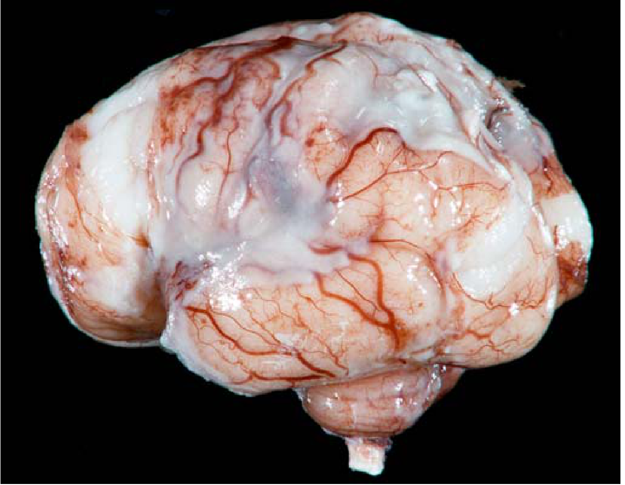
- An exudate is evident within the leptomeninges on the surface of the brain
- Meningeal vessels are engorged and prominent
- Tracts of pus may extend along blood vessels
- A thick layer of suppurative (creamy/yellow-green) exudate covers the brain surface and thickens the leptomeninges, particularly over the brain stem and cerebellum (see image below)
- In pneumococcal meningitis, a gelatinous exudate may be present due to large quantities of capsular polysaccharide
Microscopic Appearance
- Neutrophils fill the entire subarachnoid space in severely affected areas
- In less severe cases, neutrophils are found predominantly around leptomeningeal blood vessels
- Gram stain reveals variable numbers of bacteria (especially in untreated cases)
- In fulminant meningitis: inflammatory cells (mostly neutrophils) infiltrate the walls of leptomeningeal veins and may extend focally into the brain parenchyma (cerebritis)
- Secondary vasculitis and venous thrombosis may lead to hemorrhagic cerebral infarction
Sequelae / Complications (Morphologic)
- Leptomeningeal fibrosis with resultant hydrocephalus (due to impaired CSF resorption)
- Chronic adhesive arachnoiditis - especially in pneumococcal meningitis, where capsular polysaccharides promote arachnoid fibrosis
- Subdural empyema - pus accumulation in subdural space
- Venous sinus thrombosis
Clinical Features
- Systemic signs of infection: fever, chills, rigors
- Meningeal irritation:
- Severe headache
- Photophobia
- Neck stiffness (nuchal rigidity)
- Kernig's sign, Brudzinski's sign
- Neurologic impairment:
- Irritability
- Clouding of consciousness
- Seizures
- CSF findings: increased opening pressure; abundant neutrophils; elevated protein; reduced glucose
- Untreated: often fatal; with prompt antibiotics, most patients recover
2. ASEPTIC (VIRAL) MENINGITIS
Etiology
- Enteroviruses (most common - ~80% of identified cases)
- Other viruses: influenza, lymphocytic choriomeningitis virus, HIV, measles
- May also be rickettsial or autoimmune in origin
- Etiologic agent identified in only a minority of cases (improving with next-generation sequencing)
Pathogenesis
Viral entry into the CNS is typically hematogenous. The virus infects the meninges and triggers a predominantly lymphocytic inflammatory response (T-cell mediated) rather than neutrophilic.
Morphology
- Less dramatic than pyogenic meningitis
- Lymphocytic infiltration of leptomeninges
- No purulent exudate
- Brain parenchyma generally spared
Clinical Features
- Less fulminant course than bacterial meningitis
- Fever, headache, meningeal signs
- CSF: lymphocytic pleocytosis, moderate protein elevation, glucose nearly always normal
- Generally self-limited; treated symptomatically
3. CHRONIC MENINGITIS
Tuberculous Meningitis
Etiology: Mycobacterium tuberculosis - typically reaches the CNS hematogenously, often in the context of primary or reactivation TB.
Clinical Features:
- Insidious onset: headache, malaise, mental confusion, vomiting
- Moderate CSF cellularity with mononuclear cells or mixed PMN/mononuclear cells
- Protein level elevated (often strikingly)
- Glucose content moderately reduced or normal
Morphology / Complications:
- Arachnoid fibrosis at the base of the brain
- Hydrocephalus from impaired CSF resorption
- May form tuberculoma - a well-circumscribed intraparenchymal mass
- Obliterative endarteritis may lead to infarction
Spirochetal Infections (Neurosyphilis)
Occurs in ~10% of untreated Treponema pallidum infections (tertiary syphilis). HIV coinfection increases risk and severity.
Patterns:
- Meningovascular neurosyphilis - chronic meningitis (usually basal), with obliterative endarteritis rich in plasma cells and lymphocytes
- Paretic neurosyphilis - parenchymal involvement; neuronal loss and microglial proliferation; progressive dementia, mood changes, delusions of grandeur
- Tabes dorsalis - damage to dorsal root sensory nerves; ataxia, Charcot joints, lightning pains, absent deep tendon reflexes
Fungal Meningitis
Etiology: Cryptococcus neoformans (immunocompromised), Cryptococcus gattii (immunocompetent + pulmonary involvement), Candida albicans
Pathogenesis: Fungal spread is hematogenous. Immunocompromise (HIV/AIDS, transplant, steroids) dramatically increases risk.
Morphology: C. neoformans spreads within perivascular (Virchow-Robin) spaces, producing numerous cystic spaces in brain tissue. The capsule stains with mucicarmine and India ink.
Clinical Features:
- Can be fulminant (fatal in 2 weeks) or indolent (evolving over months)
- Chronic meningitis + meningoencephalitis
- CSF: India ink preparation shows encapsulated yeast; cryptococcal antigen test positive
CSF Summary Table
| Type | Cells | Protein | Glucose |
|---|---|---|---|
| Bacterial (Pyogenic) | Neutrophils (PMNs) | High | Low |
| Viral (Aseptic) | Lymphocytes | Moderately raised | Normal |
| Tuberculous/Fungal | Lymphocytes/Mixed | High (often strikingly) | Low/Normal |
Sources: Robbins & Kumar Basic Pathology, 9th ed., Ch. 21 (CNS Infections, p. 833-835); Robbins Cotran Pathologic Basis of Disease, 10th ed., Ch. 28 (p. 1164-1165)
This is a shared conversation. Sign in to Orris to start your own chat.