HCL poisoning from the context of forensic medicine
Reading File
Finding Sources
Finding Sources
Finding Sources
Finding Sources
Reading File
Finding Sources
Reading File
Finding Sources
Finding Sources
Reading File
Reading File
Reading File
Finding Sources
Finding Sources
Finding Sources
I now have comprehensive information from two major forensic medicine textbooks. Let me compile the full answer:
HCl (Hydrochloric Acid) Poisoning - Forensic Medicine
Physical & Chemical Properties
Hydrochloric acid (HCl) is also called Muriatic Acid or Spirit of Salt. Key properties:
- Pure form: colorless, fuming liquid with a strong pungent, irritating odour
- Strong solution fumes even at ordinary temperatures in damp air
- When ingested, the liberated gas is simultaneously inhaled
- Natural constituent of gastric and intestinal juice (physiologically present)
- Commercially used for: preparing chlorine, dissolving metals, cleaning drains, removing scale from kettles, and as medicine
- Muriatic acid (commercial form): yellow-colored solution, fumes strongly in damp air - less destructive than sulphuric or nitric acid
- Does not stain skin or mucous membranes, but stains dark clothing reddish-brown
- Being volatile, it readily affects respiratory tract mucosa
Classification in Toxicology
HCl belongs to the group of Corrosive Poisons (specifically, mineral/inorganic acids). These are agents that, besides being strong irritants, actually produce ulceration and necrosis of tissues on contact. The mineral acid group includes H2SO4, HNO3, and HCl.
Mechanism of Action
Corrosive acids act by:
- Coagulation of cellular proteins - forming a firm eschar
- Conversion of haemoglobin to acid haematin (accounts for dark brown/black discoloration)
- Extraction of water from tissues (dehydration)
- HCl is less corrosive than sulphuric acid in local action - it does not char or blacken tissues as severely
- It readily destroys mucous membranes but does not usually cause serious skin damage (unlike H2SO4)
- Color change of mucous membrane: initially grey or grey-white, later becomes brown or black (due to acid haematin formation)
Signs and Symptoms
Acute (Ingestion)
- Burning pain in mouth, throat, oesophagus, and stomach
- Mucous membrane: initially grey/grey-white turning brown/black
- Nausea, vomiting (brownish acidic vomit)
- Less skin corrosion than H2SO4 (does not cause burns/blistering on skin)
Inhalation of Fumes
- Intense irritation of throat and air passages
- Spasm of glottis
- Symptoms of suffocation
- Cough, dyspnoea, and cyanosis
- Pulmonary oedema in severe cases
Chronic Poisoning (Occupational exposure to fumes)
Constant exposure produces a distinct chronic syndrome:
- Coryza
- Conjunctivitis
- Corneal ulcer
- Pharyngitis
- Bronchitis
- Inflammation of gums
- Loosening of teeth (dental erosion)
Fatal Dose & Fatal Period
| Parameter | Value |
|---|---|
| Fatal dose | 15-20 mL (of concentrated acid) |
| Fatal period | 18-36 hours (Essentials of Forensic Medicine, 36th ed.); 18-24 hours (Dikshit) |
Treatment
Treatment is the same as for sulphuric acid poisoning:
- Do NOT perform gastric lavage (risk of perforation; also contraindicated in all corrosive poisonings)
- Do NOT induce emesis
- Neutralize with alkali (dilute sodium bicarbonate, milk of magnesia, lime water, or soap solution)
- Give milk, egg white, or water to dilute
- Analgesics for pain
- Treat shock, oedema of larynx (may need tracheostomy)
- IV fluids and supportive care
Postmortem (Autopsy) Appearances
Findings are similar to sulphuric acid poisoning but with less tendency to charring and tissue destruction:
External:
- No staining of skin (unlike H2SO4 which leaves grey/black stains)
- Dark clothing may show reddish-brown stains
- Skin around mouth/chin: grey becoming darker from blood
Internal:
- Oral cavity and oesophageal mucosa: ashy grey, may become blackened, swollen, and hardened if acid was concentrated
- Stomach: contains brownish acidic fluid; mucosa is corroded and greyish-white; folds of the whole stomach mucosa are brownish
- Perforation: rare (unlike H2SO4 where perforation is more common)
- Respiratory tract: acute inflammation and oedema of respiratory passages and lung tissue are common (due to vapour inhalation)
- "Blotting paper stomach" appearance (dryness/brittleness) may be seen (characteristic of concentrated acid poisoning generally)
Chemical/Laboratory Test
Silver nitrate test: Adding a solution of silver nitrate to the suspected material produces a heavy, curdy, white precipitate of silver chloride (AgCl) - this is the diagnostic confirmatory test for chloride/HCl.
AgNO₃ + HCl → AgCl↓ (white, curdy precipitate) + HNO₃
Medico-Legal Aspects
| Mode | Frequency |
|---|---|
| Suicidal | Most common - majority of cases |
| Accidental | A few cases (industrial exposure, improper storage) |
| Homicidal | Very rare (HCl is not an ideal homicidal poison due to its pungent odour and local action) |
Special medico-legal points:
- Abortifacient use: HCl has been rarely injected into the vagina to produce abortion. This causes atresia of the vagina as a sequel.
- Forgery/Obliteration: Used for erasing writing or signatures from documents - forensic document examination may reveal attempts at forgery.
- Occupational exposure must be considered when evaluating chronic presentations.
- The distinctive volatile fume characteristic means that even without direct ingestion, inhalation can be fatal in confined spaces (industrial/occupational poisonings).
Comparison with Other Mineral Acids (Quick Reference)
| Feature | H2SO4 (Sulphuric) | HNO3 (Nitric) | HCl (Hydrochloric) |
|---|---|---|---|
| Colour of stain | Grey → black | Yellow (xanthoproteic) | No skin stain; reddish-brown on dark cloth |
| Corrosive action | Most severe | Moderate | Least severe of the three |
| Skin burns | Severe charring | Yellow staining | Usually none |
| Stomach perforation | Common | Common | Rare |
| Fatal dose | 5 mL | 5 mL | 15-20 mL |
| Fatal period | 12-18 hrs | 12-24 hrs | 18-36 hrs |
Sources:
- The Essentials of Forensic Medicine and Toxicology, 36th edition (2026), Section: HYDROCHLORIC ACID (HCL), p. 1281-1315
- P.C. Dikshit Textbook of Forensic Medicine and Toxicology, Section: HYDROCHLORIC ACID (HCL), pp. 454-455
- Parikh's Textbook of Medical Jurisprudence, Forensic Medicine and Toxicology, Classification of Corrosive Poisons section
How does sulphuric acid blackens or Charr the skin and mucus membrane
Finding Sources
Finding Sources
Finding Sources
Reading File
Finding Sources
Finding Sources
Reading File
Reading File
Reading File
I now have everything needed for a complete, mechanism-focused answer. Here it is:
How Sulphuric Acid Blackens and Chars Skin & Mucous Membranes
This is the result of four simultaneous, overlapping mechanisms acting together:
1. Powerful Dehydration (Primary mechanism of charring)
Concentrated H2SO4 is an extremely hygroscopic (water-absorbing) substance. It has an overwhelming chemical affinity for water molecules.
When it contacts biological tissue (which is ~70% water), it violently strips water from organic molecules - particularly carbohydrates and proteins. The dehydration reaction:
Organic tissue (contains C, H, O in carbohydrate/protein chains) H2SO4 removes H and O as H2O Carbon skeleton is left behind → appears black
This is the same principle seen in a chemistry demonstration where H2SO4 poured on sucrose (sugar, C12H22O11) rapidly turns it into a black carbon column:
C12H22O11 → 12C (black) + 11H2O
The same reaction occurs in the tissue's carbohydrate-rich mucous layer and the carbon-containing proteins - leaving a blackened carbonaceous residue. This is true charring - chemical, not thermal.
2. Exothermic Reaction Generating Heat
When concentrated H2SO4 contacts moist tissue (any surface with water), an intense exothermic reaction occurs:
H2SO4 + H2O → H3O⁺ + HSO4⁻ + HEAT
This reaction generates a large amount of heat locally. The Essentials of Forensic Medicine and Toxicology (36th ed.) states directly:
"An exothermic reaction occurs when strong mineral acids, e.g., sulphuric acid, comes into contact with moist skin. The heat together with corrosion causes coagulation necrosis."
This heat component compounds the chemical damage, contributing to thermal burning on top of chemical destruction - hence sulphuric acid causes superficial burns after just 1 second of contact, and full-thickness burns after 30 seconds.
3. Protein Denaturation and Coagulative Necrosis
H2SO4 causes precipitation and coagulation of cellular proteins. The hydrogen ions (H⁺) released by the acid denature structural proteins and enzymes by:
- Breaking hydrogen bonds in protein secondary/tertiary structure
- Precipitating proteins out of solution
This is documented in Dikshit's Forensic Toxicology as the mechanism:
"Causes coagulation necrosis by precipitation of proteins"
The coagulated, denatured protein forms a firm, leathery, dark eschar (coagulative necrosis). Rosen's Emergency Medicine confirms:
"Sulphuric acid burns result in a black or brown eschar."
This eschar formation is actually a self-limiting feature - the crust theoretically prevents further penetration of acid to deeper layers. However, in practice with concentrated H2SO4, the damage is already severe before the eschar forms.
4. Conversion of Haemoglobin to Acid Haematin
This is the specific mechanism that produces the brownish-black colour on mucous membranes (as opposed to the external charred skin):
H2SO4 + Haemoglobin → Acid Haematin
Acid haematin is a dark brown-to-black degradation product of haemoglobin. It forms when strong acids react with the haem component of haemoglobin in the superficial blood vessels of the mucosa. This is why:
- Mucous membranes of the mouth, oesophagus and stomach appear grey → brown → black
- Vomit is described as "brown or black, mucoid, and strongly acidic"
- Stomach mucosa appears brownish-black at autopsy
- Tongue becomes "swollen, sodden, and black"
Summary: Why Black Specifically?
| Substrate | What H2SO4 does | Result |
|---|---|---|
| Carbohydrates (mucus, glycoproteins) | Dehydration → removes H2O | Black carbon residue |
| Proteins | Denaturation + coagulation | Black-brown leathery eschar |
| Haemoglobin | Converts to acid haematin | Brown-black pigment |
| Water in tissue | Exothermic reaction → heat | Thermal burn adds to charring |
All four processes happen simultaneously and reinforce each other. The final result is a dry, hard, blackened, brittle eschar - characteristically described in forensic pathology as:
- Skin: brownish-black streaks/burns running from corners of the mouth down the chin/neck (from dripping acid)
- Mucous membrane of mouth/tongue: initially white/grey necrotic membrane → brownish-black
- Stomach: blackened, corroded mucosa; may become paper-thin ("blotting paper stomach"); perforation is common
Why HCl Does NOT Char
This mechanism explains why HCl does not produce the same charring:
- HCl is volatile and does not have the same dehydrating (hygroscopic) property as H2SO4
- It lacks the extreme heat of dilution
- No comparable carbonization of organic molecules occurs
- HCl produces a grey-white eschar (via acid haematin alone, without the carbon deposition)
Sources:
- Essentials of Forensic Medicine and Toxicology, 36th ed. (2026) - Mechanism of Action: Mineral Acids; Sulphuric Acid section
- P.C. Dikshit Textbook of Forensic Medicine and Toxicology - Mode of Action; Properties of Sulphuric Acid; Signs & Symptoms
- Rosen's Emergency Medicine - Acid burns, coagulative necrosis, eschar formation
Doesn't mucosa of git get burnt by H2SO4 , mucosa has carb and protein too from which H2O can be taken out?
Finding Sources
Finding Sources
Reading File
Reading File
Reading File
Reading File
Loading Image
Loading Image
Now I have everything - the textbook text and the actual autopsy images. Here is the complete answer to your excellent question:
Yes - the GIT Mucosa Absolutely Gets Charred by H2SO4
Your reasoning is completely correct. The GIT mucosa is rich in carbohydrates (mucins, glycoproteins) and proteins (epithelial cells, basement membrane, glandular secretions) - all of which undergo the same dehydration and carbonization reactions. The stomach is actually the most severely affected organ of all.
What Actually Happens in the GIT - Region by Region
Oesophagus - Relatively Spared (and here's why)
This is the one apparent exception - and the reason is anatomical/physiological, not chemical:
- The oesophagus is lined by squamous epithelium, which is physically tougher and more resistant than the columnar epithelium lower down
- H2SO4 swallowed rapidly causes rapid transit through the oesophagus - contact time is short
- The coagulum (eschar) forms quickly on the squamous mucosa and limits further penetration
- Perforation of the oesophagus is therefore rare
But even so - the oesophageal mucosa shows inflammation, oedema, and severe interstitial haemorrhage even when gross corrosion is not obvious.
Stomach - The Primary Target and Site of Maximum Destruction
This is where all four mechanisms (dehydration, exothermic heat, protein coagulation, acid haematin formation) operate at full force - and your question is entirely validated here:
"The greater part of the stomach may be converted into a soft, spongy, black mass which readily disintegrates when touched."
- Essentials of Forensic Medicine & Toxicology, 36th ed.
"The mucous membrane of the stomach is inflamed, oedematous, blackened with a peppery feel."
- Dikshit's Forensic Medicine
Why the stomach bears the brunt:
- Acid pools here - the pyloric sphincter goes into spasm on acid contact, trapping the H2SO4 in the stomach for a prolonged time. Contact time is far longer than in the oesophagus.
- Columnar epithelium (gastric mucosa cells) is far more vulnerable than the squamous epithelium of the oesophagus
- Rich blood supply = more haemoglobin = more acid haematin formation = darker black colouration
- Mucin-rich surface = abundant carbohydrate substrate for dehydration/carbonization
- Protein-dense glandular cells (chief cells, parietal cells, mucous cells) = extensive coagulative necrosis
The specific autopsy finding that results:
| Finding | Mechanism |
|---|---|
| Soft, boggy, black mass | Dehydration + carbonization of carbs/proteins + acid haematin |
| Mucosal ridges more damaged than furrows | Acid contacts the ridges first and more directly |
| Brown/black stomach contents | Acid haematin from digested blood + carbonized tissue debris |
| Wall studded with acute erosions | Protein coagulation/necrosis through the mucosal layers |
| Perforation (common with H2SO4) | Full-thickness destruction when contact time is long enough |
| "Peppery feel" of mucosa | Granular texture from carbonized tissue residue |
Here are the actual autopsy images from the textbooks:
From Dikshit's Forensic Medicine - Stomach mucosa in H2SO4 poisoning (gross specimen):
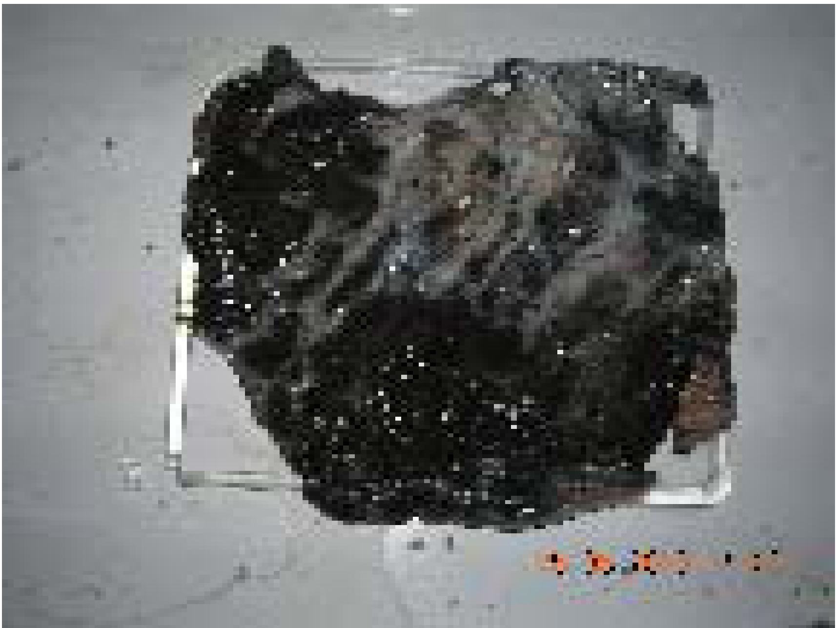
From Essentials of Forensic Medicine (36th ed.) - Stomach in sulphuric acid poisoning:
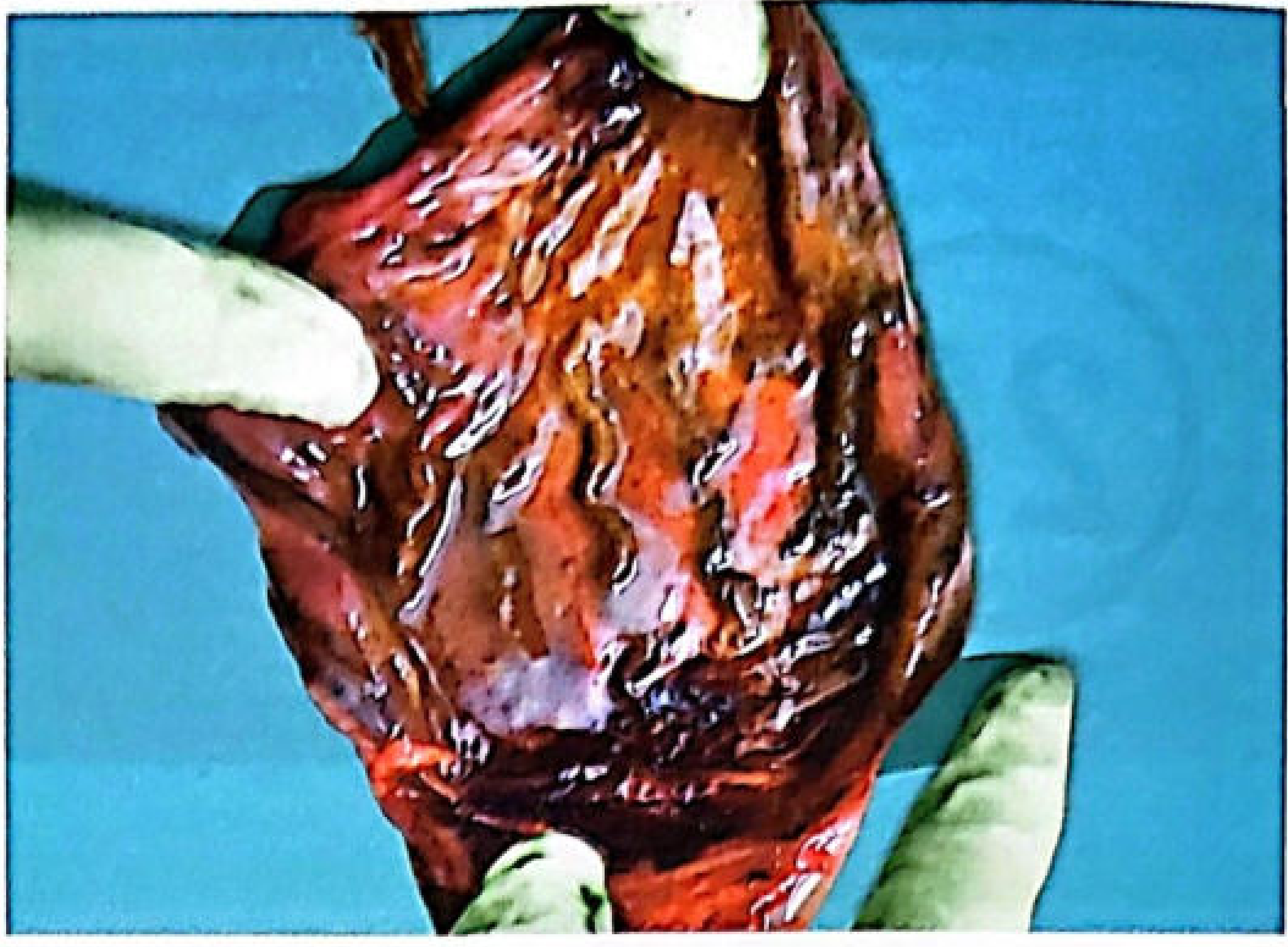
Both images show the extensively blackened, destroyed gastric mucosa - precisely what your chemical reasoning predicts.
Small Intestine & Beyond
- Duodenum - shows similar but less intense changes (acid reaches here after dilution/partial neutralization)
- Small intestine - signs of irritation only
- Peritoneal cavity - if perforation occurs, the spilled acid causes chemical peritonitis, and the viscera in the peritoneal cavity also become blackened and corroded
The Key Insight Your Question Reveals
The reason textbooks say "skin is charred" more prominently is because charring on skin is visible externally and is a diagnostic sign seen before autopsy. But internally, the GIT mucosa - especially the stomach - undergoes equal or worse destruction by the exact same mechanisms. The stomach is actually the forensic pathologist's primary target organ at autopsy.
The sequence in the stomach is:
- H2SO4 contacts mucin layer → strips water from mucopolysaccharides → black carbonized residue
- Contacts columnar epithelial cells → denatures and precipitates proteins → firm, then disintegrating, brown-black necrotic mass
- Reacts with haemoglobin from ruptured mucosal capillaries → acid haematin → adds to the black colour
- Exothermic reaction with tissue water → local heat generation compounds necrosis
- Pyloric spasm traps acid → reaction continues until the stomach wall is fully destroyed
Sources:
- Essentials of Forensic Medicine & Toxicology, 36th ed. (2026), Sulphuric Acid - Postmortem Appearances, pp. 1123-1172
- P.C. Dikshit Textbook of Forensic Medicine & Toxicology, Internal Findings in H2SO4 Poisoning, pp. 451-455
- Cummings Otolaryngology - Caustic Ingestions: coagulative necrosis and eschar formation at mucosal surfaces
Why can stomach mucosa be dark brown innitric acid consumption
Finding Sources
Finding Sources
Reading File
Reading File
Reading File
I now have the full picture from both textbooks. Here's the precise answer:
Why the Stomach Mucosa is Brown/Brown-Black in Nitric Acid Poisoning (Not Yellow)
This is a classic forensic medicine paradox that trips people up. Nitric acid's signature is yellow (xanthoproteic), yet the oesophagus and stomach are brown or brownish-black. Both textbooks state this explicitly:
"In the esophagus and stomach, corrosion of mucous membrane may not be accompanied by yellow discoloration, which may appear brown or brown-black due to acid haematin."
- Essentials of Forensic Medicine & Toxicology, 36th ed.
"Oesophagus and stomach are not stained yellow but is brown or brownish-black due to the formation of acid haematin."
- Dikshit's Forensic Medicine
The Core Reason: Two Competing Reactions
When nitric acid is ingested, two reactions happen simultaneously in the GIT, but they produce different coloured products - and one dominates over the other inside the gut:
Reaction 1: Xanthoproteic Reaction → YELLOW
(Dominates on skin, lips, teeth, clothing)
Concentrated HNO3 reacts with the aromatic amino acid rings (tyrosine, phenylalanine, tryptophan) in proteins:
HNO3 + aromatic ring of protein → nitration → nitro-compounds (picric acid derivatives)
Picric acid and its relatives are intensely yellow. This is why:
- Skin, lips, face, hands turn yellow
- Teeth turn yellow
- Clothing is yellow stained
- Urine turns brown-yellow
This reaction works beautifully on keratinised/squamous surfaces (skin, buccal mucosa, teeth) because the aromatic amino acids in keratin are accessible and the reaction proceeds without competition.
Reaction 2: Acid Haematin Formation → BROWN-BLACK
(Dominates in the oesophagus and stomach)
Inside the GIT, the mucosa is richly vascular - the submucosal capillaries rupture immediately on acid contact, releasing large quantities of haemoglobin into the tissue.
HNO3 (strong acid, H⁺) + Haemoglobin → Acid Haematin
Acid haematin is a dark brown to black compound, formed when H⁺ ions displace the iron from the haem ring, producing a denatured ferric porphyrin complex. This is the same molecule produced by H2SO4 and HCl - it is not unique to nitric acid.
Why Does Acid Haematin Win Inside the Stomach?
The stomach and oesophagus have specific conditions that favour acid haematin over xanthoproteic colouring:
| Factor | Effect |
|---|---|
| Abundant submucosal blood vessels | Massive release of haemoglobin on mucosal disruption |
| High haemoglobin concentration | Acid haematin produced in very large quantities, its dark colour overwhelms the yellow |
| Moist, vascular columnar epithelium | Direct contact of acid with blood, not just surface keratin |
| Pooling of acid in stomach | Prolonged contact ensures complete haemoglobin conversion |
| No keratin barrier | The gastric mucosa lacks the thick keratin layer of skin, so the xanthoproteic reaction (which works best on keratin-rich surfaces) is less prominent |
The brown-black of acid haematin is simply a stronger, more abundant pigment in this environment than the yellow xanthoproteic products. It visually overrides the yellow staining.
Summary: Colour Rule for Nitric Acid
| Location | Colour | Reason |
|---|---|---|
| Skin, lips, face, hands | Yellow | Xanthoproteic reaction (nitration of aromatic amino acids in keratin) |
| Teeth | Yellow | Xanthoproteic reaction on enamel proteins |
| Clothing | Yellow | Nitration of fabric organic compounds |
| Urine | Brown-yellow | Excretion of nitrated compounds |
| Oesophagus & stomach mucosa | Brown/brown-black | Acid haematin formation dominates (blood-rich environment) |
| Vomitus | Yellowish-brown | Mix of both - yellow xanthoproteic + brown acid haematin |
This is also why the vomit is described as "yellowish-brown containing altered blood" - it contains both products mixed together.
Sources:
- Essentials of Forensic Medicine & Toxicology, 36th ed. (2026) - Nitric Acid, Postmortem Appearances
- P.C. Dikshit Textbook of Forensic Medicine & Toxicology - Nitric Acid, Autopsy Findings
What is meant by acid corrosion
Finding Sources
Finding Sources
Finding Sources
Finding Sources
Finding Sources
Finding Sources
Reading File
Reading File
Reading File
All the key definitions and mechanisms are now in hand. Here is the complete answer:
What is Acid Corrosion?
Definition
The word comes from the Latin corrodere - meaning "to gnaw through" flesh. A corrosive acid is one that does not merely irritate tissue on contact, but physically destroys and eats it away - dissolving structure, killing cells, and leaving behind a zone of dead, denatured, often disintegrating tissue.
The standard forensic definition from the textbooks:
"A corrosive poison fixes, destroys and erodes the surface with which it comes in contact."
- Essentials of Forensic Medicine & Toxicology, 36th ed.
"A corrosive poison is simply a highly active irritant and not only produces inflammation but also actual ulceration of the tissues."
- Parikh's Textbook of Medical Jurisprudence
Corrosion vs. Irritation - The Critical Distinction
This is the key conceptual difference:
| Irritant | Corrosive | |
|---|---|---|
| What it does | Inflames tissue, causes redness, swelling, pain | Destroys and dissolves tissue structurally |
| Depth | Superficial inflammation | Actual ulceration, necrosis, perforation |
| Reversibility | Generally reversible inflammation | Irreversible structural destruction |
| Concentration-dependent | Yes - same corrosive in dilute form acts as an irritant | In concentrated form causes full corrosion |
| Example | Dilute HCl (0.1%) | Concentrated HCl, H2SO4, HNO3 |
Note: Corrosives in dilute solution act as irritants - this is a classic exam point. The same H2SO4 that corrodes tissue in concentrated form merely irritates when well diluted.
The Three Mechanisms That Constitute "Acid Corrosion"
When a strong acid corrodes tissue, it does so via three simultaneous chemical actions:
1. Extraction of Water from Tissues (Dehydration)
Concentrated acids, especially H2SO4, are powerfully hygroscopic. They strip water molecules from carbohydrates, glycoproteins, and proteins in the tissue. The dehydrated organic residue collapses structurally and carbonizes - the tissue literally loses its molecular architecture. This is the "gnawing" at the molecular level.
2. Coagulation of Cellular Proteins
H⁺ ions denature and precipitate proteins by:
- Breaking hydrogen bonds in protein structure
- Unfolding and clumping structural proteins, enzymes, and cell membrane proteins
- Forming a firm coagulum (eschar) of denatured protein on the surface
This produces coagulative necrosis - the cells die in a fixed, firm, structurally preserved but non-functional state, like cooked egg white. The eschar this creates is hard and leathery.
"Ingestion of corrosives causes coagulation necrosis, a denaturation of proteins with formation of a gel-like substance on the mucosa called the coagulum."
- Cummings Otolaryngology
3. Conversion of Haemoglobin to Acid Haematin
Strong acids react with haemoglobin released from disrupted mucosal blood vessels, converting it to dark-brown acid haematin - visually manifesting as the brown-black discolouration seen in corroded mucosae.
What "Corrosion" Looks Like Macroscopically
The sequential stages of acid corrosion on tissue:
| Stage | Appearance | What's happening |
|---|---|---|
| Immediate | Blanching, grayish-white area | Protein coagulation, dehydration begins |
| Minutes | White → brown → black | Acid haematin forming, carbonization progressing |
| Established | Hard, leathery, black eschar | Full coagulative necrosis, structural destruction |
| Advanced | Soft, boggy, disintegrating | Eschar breaks down, wall thinning, perforation risk |
The mucosal ridges corrode faster than the furrows because the acid contacts the raised ridges first and in greater concentration - another hallmark finding at autopsy.
Why Corrosion is Dangerous Beyond the Local Burn
Even though corrosion is described as primarily a "local action" (no remote effects), the consequences radiate outward:
- Perforation - when corrosion breaches the full thickness of the stomach/oesophageal wall, acid spills into the peritoneal or thoracic cavity → chemical peritonitis or mediastinitis
- Systemic acidosis - absorbed H⁺ ions enter the bloodstream → pH falls → metabolic acidosis, haemolysis, cardiovascular collapse
- Oedema of glottis - corrosive fumes or splash on the larynx → swelling → acute asphyxia (can kill within minutes)
- Late stricture - as the corroded area heals (3 weeks to 3 months), dense scarring causes pyloric stenosis or oesophageal stricture
Corrosion vs. Caustic - Are They the Same?
Often used interchangeably, but technically:
- Corrosive = any agent (acid or alkali) that destroys tissue on contact
- Caustic = specifically refers to strong alkalis (caustic soda = NaOH; caustic potash = KOH)
- Both cause tissue destruction but by different mechanisms:
- Acids → coagulative necrosis (firm eschar limits penetration)
- Alkalis → liquefactive necrosis (no eschar, penetration goes deeper and is often more severe)
Sources:
- Essentials of Forensic Medicine & Toxicology, 36th ed. (2026) - Corrosive Poisons: Action and Classification
- Parikh's Textbook of Medical Jurisprudence, Forensic Medicine & Toxicology - Classification by Mode of Action
- Cummings Otolaryngology Head & Neck Surgery - Caustic Ingestions: definition and mechanism
Treatment of sulphuric acid poisoning
Finding Sources
Finding Sources
Reading File
Reading File
I have all the detail needed from both textbooks. Here is the complete treatment outline:
Treatment of Sulphuric Acid Poisoning
ABSOLUTE CONTRAINDICATIONS (Do First, Remember Always)
These two are the most important and most exam-tested points:
| NEVER do | Reason |
|---|---|
| Gastric lavage / stomach wash | Acid has already thinned and corroded the stomach wall - the tube or pressure will perforate it |
| Emetics / induced vomiting | Brings acid back up through the already-damaged oesophagus, causing repeat corrosion and risk of aspiration |
| Alkaline carbonates or bicarbonates (NaHCO3, Na2CO3) | Neutralization releases CO2 gas rapidly → gastric distension → rupture/perforation of the weakened stomach wall |
STEP-BY-STEP TREATMENT
Step 1 - Immediate Dilution and Neutralization (within 30 minutes)
The acid must be diluted and neutralized in situ - where it sits - not removed.
Give one-fourth litre (250 mL) of any of the following immediately:
- Water (plain, large quantity)
- Milk (preferred - also a demulcent)
- Milk of magnesia (MgO suspension - mild alkali, safe)
- Lime water (Ca(OH)2 solution - mild alkali, safe)
- Soap suds (mild alkali)
- Aluminium hydroxide gel (Al(OH)3 - antacid, safe)
- Calcium oxide or magnesium oxide dissolved in water (one tablespoonful)
Why these are safe neutralizers: They are weak/mild alkalis - they neutralize H⁺ slowly without producing explosive CO2 gas, unlike strong carbonates.
Step 2 - Demulcents (Coat and Protect the Mucosa)
Give substances that form a protective coating over the corroded mucosa:
- Olive oil
- Egg whites (raw)
- Starch water
- Mineral oil
- Melted butter
- Barley water
- Vegetable oil
These coat the mucosal surface, reduce further acid contact, and provide some pain relief.
Step 3 - Pain Relief
Severe burning pain must be treated aggressively:
- Morphine 15 mg IM or IV (Dikshit)
- Meperidine (Pethidine) HCl 50-150 mg orally or IV
Step 4 - Airway Management
Acid fumes and oedema of the glottis/larynx are a life-threatening emergency:
- Tracheostomy - if oedema of glottis develops (stridor, dyspnoea)
- Oxygen inhalation if respiratory distress
- Assisted/artificial respiration if needed
Step 5 - IV Support
- Nothing by mouth for approximately 1 week
- IV fluids and nutrition for 5-7 days, then gradual reintroduction of liquids → soft food → normal diet
- Blood transfusion if haemorrhage has been significant
- IV Calcium gluconate 10 mL of 10% solution (for systemic effects)
Step 6 - Corticosteroids (to Prevent Stricture)
- Prednisolone 60 mg/day in divided doses
- Purpose: reduce inflammatory fibrosis that leads to oesophageal/pyloric stricture
- Note: generally not recommended now due to increased risk of perforation - use with caution and clinical judgment
Step 7 - Endoscopy (within 24-48 hours)
- Flexible fibre-optic endoscopy to assess extent of damage to oesophagus and stomach
- If circumferential 2nd or 3rd degree burns are found → proceed to exploratory laparotomy
- If gastric necrosis is present → oesophagogastrostomy may be required
Step 8 - Prevention of Stricture Formation (Late Treatment)
Once acute phase is over, prevent scarring/stricture:
- Mercury-filled bougie (4 cm diameter) passed daily through the oesophagus to prevent it from narrowing as it heals
- If stricture already established: repeat dilatation
Step 9 - External Burns (Skin and Eyes)
Skin burns:
- Wash immediately with large quantities of water
- Apply paste of magnesium oxide or sodium bicarbonate to neutralize residual acid
Eye burns:
- Irrigate immediately with water or sodium bicarbonate solution for 10-15 minutes
- A suspended IV bag giving low-pressure continuous irrigation is ideal
CAUSES OF DEATH (Timing)
| Time | Cause |
|---|---|
| Within hours | Shock, spasm/oedema of glottis → asphyxia |
| Within 24 hours | Perforation of stomach → peritonitis + shock |
| Within 1st week | Septicaemia from septic absorption |
| Months to years later | Malnutrition/exhaustion from oesophageal or pyloric stricture |
Summary Mnemonic - "No LAVAGE"
-
No gastric lavage
-
Oral dilution with milk/water immediately
-
Liquid demulcents (egg white, olive oil)
-
Airway - tracheostomy if glottis oedema
-
Vein - IV fluids, nutrition, calcium gluconate
-
Analgesics (morphine)
-
Glucocorticoids (prednisolone - cautiously)
-
Endoscopy within 24-48 hours
Sources:
- Essentials of Forensic Medicine & Toxicology, 36th ed. (2026) - Treatment of Sulphuric Acid Poisoning
- P.C. Dikshit Textbook of Forensic Medicine & Toxicology - General Principles of Treatment of Corrosive Acid Poisoning
This is a shared conversation. Sign in to Orris to start your own chat.