
Lumbrical of hands
Reading File
Searching sources...
Finding Sources
Finding Sources
Reading File
Reading File
Reading File
Reading File
Reading File
Reading File
Now let me get the detailed lumbrical section from Gray's Anatomy for Students and fetch the key image:
Reading File
Loading Image
Loading Image
Here is a complete anatomical account of the Lumbricals of the Hand, drawn from Gray's Anatomy for Students, THIEME Atlas of Anatomy, and Schwartz's Principles of Surgery.
Lumbricals of the Hand
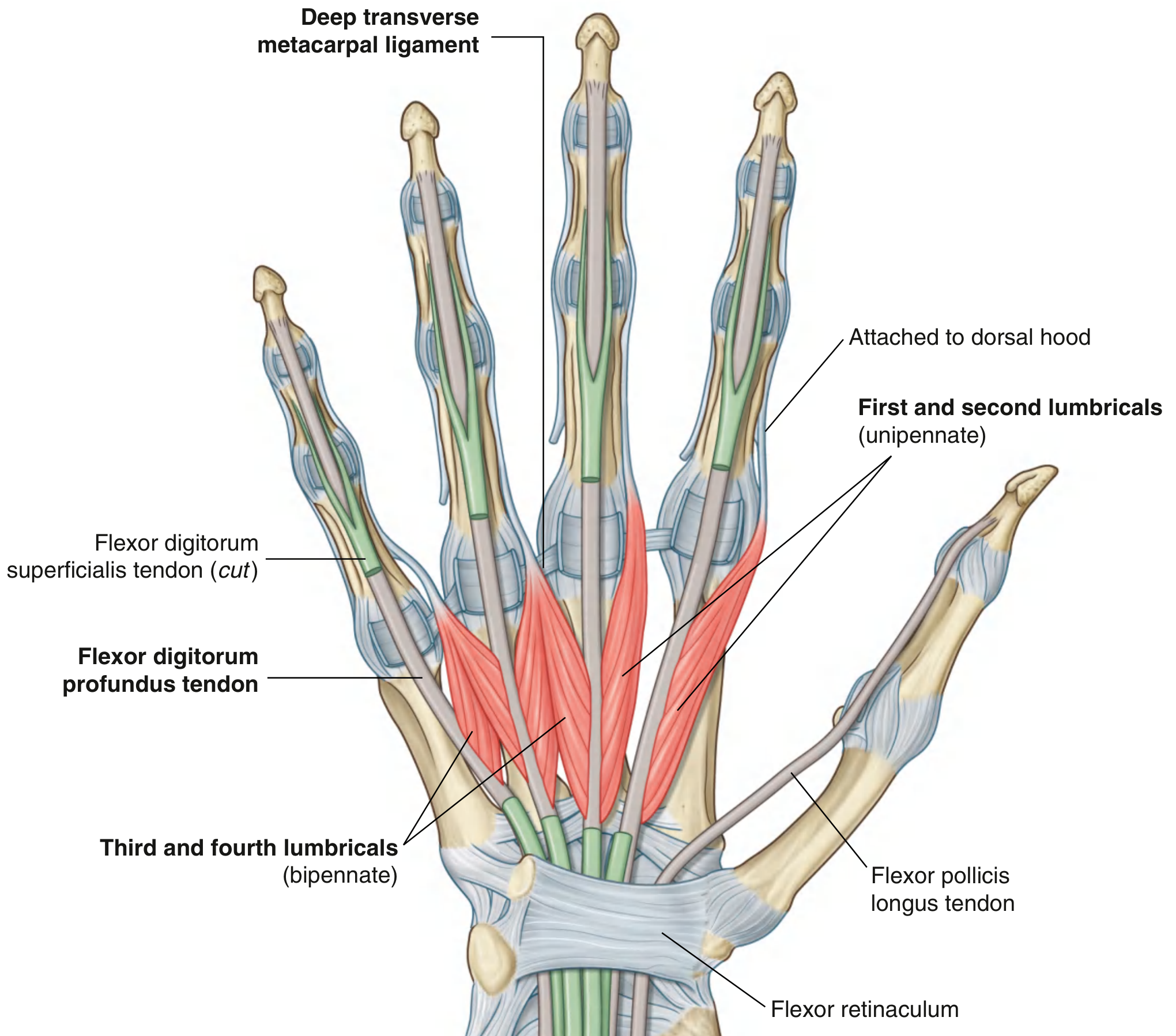
The lumbricals (Latin: lumbricus = earthworm) are four small, worm-shaped intrinsic muscles of the hand. They are unique in the entire body because they originate from a tendon rather than bone - specifically, from the tendons of flexor digitorum profundus (FDP).
Number & Naming
There are four lumbricals, numbered 1st through 4th (radial to ulnar), for the index, middle, ring, and little fingers respectively.
Origin
Each lumbrical arises from the radial side of the FDP tendon of its corresponding digit, in the palm:
| Lumbrical | Arises from FDP tendon of |
|---|---|
| 1st | Index finger (unipennate) |
| 2nd | Middle finger (unipennate) |
| 3rd | Ring finger (bipennate - from adjacent FDP tendons) |
| 4th | Little finger (bipennate - from adjacent FDP tendons) |
- 1st and 2nd lumbricals are unipennate (single head, from one FDP tendon each)
- 3rd and 4th lumbricals are bipennate (two heads, from adjacent FDP tendons)
Insertion
Each lumbrical passes palmar to the deep transverse metacarpal ligament on the radial side of the corresponding digit, then curves dorsally to insert into the dorsal digital expansion (extensor hood) of each finger:
- 1st lumbrical → dorsal digital expansion of index finger (2nd digit)
- 2nd lumbrical → dorsal digital expansion of middle finger (3rd digit)
- 3rd lumbrical → dorsal digital expansion of ring finger (4th digit)
- 4th lumbrical → dorsal digital expansion of little finger (5th digit)
Innervation
This is a high-yield exam point:
| Lumbricals | Nerve | Spinal Level |
|---|---|---|
| 1st & 2nd (lateral/radial two) | Median nerve (digital branches) | C8, T1 |
| 3rd & 4th (medial/ulnar two) | Deep branch of ulnar nerve | C8, T1 |
Mnemonic: "2 Median, 2 Ulnar" - the two radial lumbricals are median, the two ulnar lumbricals are ulnar.
All intrinsic hand muscles are predominantly C8, T1. The three thenar muscles and the two lateral lumbricals are the only intrinsic muscles innervated by the median nerve; the rest are innervated by the ulnar nerve.
Action
The lumbricals perform a seemingly paradoxical action through their unique position:
MCP joints (2nd-5th): Flexion
PIP and DIP joints (2nd-5th): Extension
This simultaneous flexion at the MCP + extension at the IP joints is their defining function - the "intrinsic-plus" position. Because the force of the lumbricals is applied to the extensor hood distal to the fulcrum of the MCP joint, they flex the MCP while the force is simultaneously transmitted dorsally through the hood to extend the IP joints.
This type of precision movement is used, for example, in the upstroke when writing the letter 't', and is fundamental to fine, precision grip.
Relation to the Dorsal Digital Expansion (Extensor Hood)
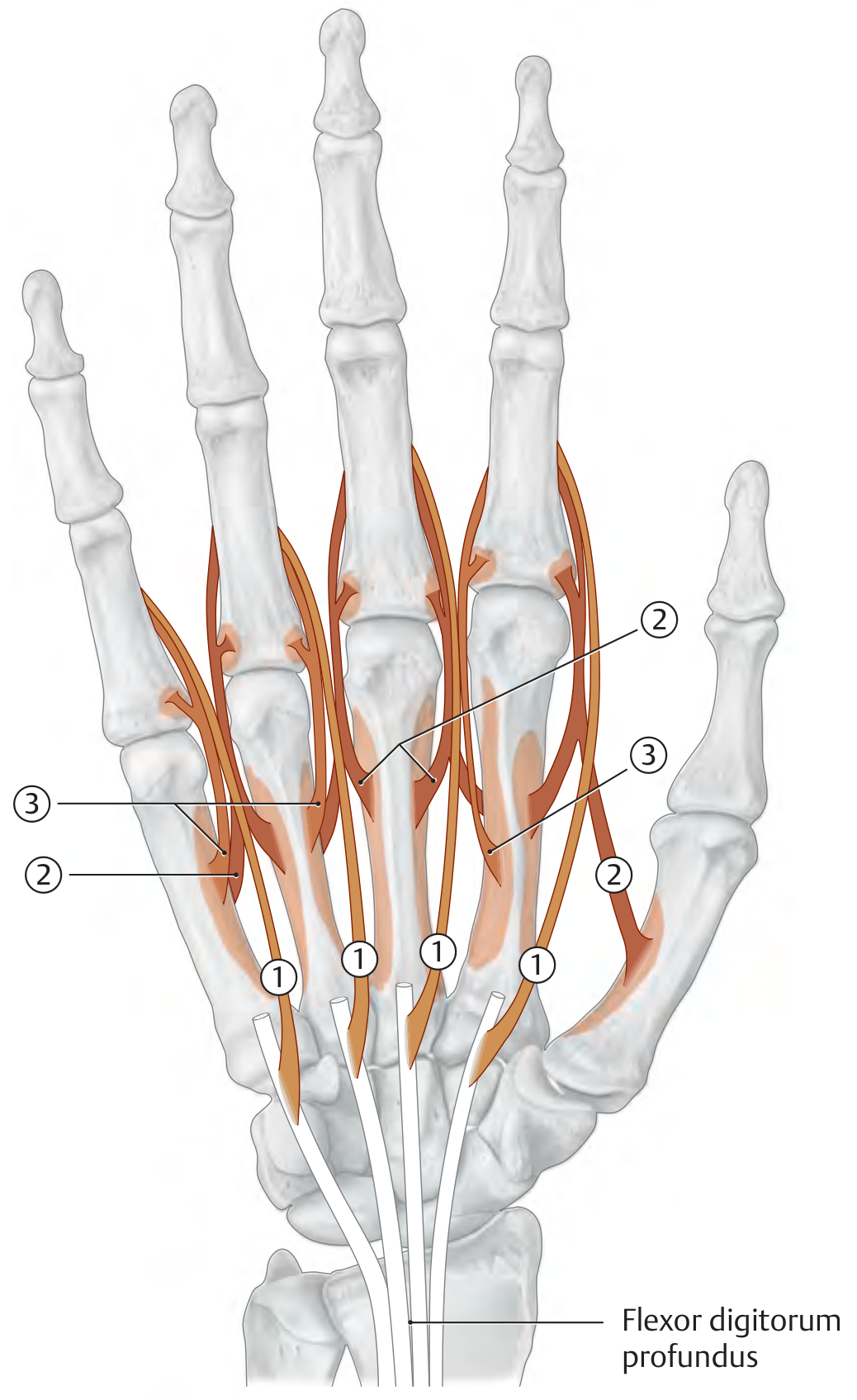
The lumbrical tendons insert into the lateral part of the dorsal digital expansion alongside the interosseous tendons. The dorsal digital expansion is more than a simple aponeurosis - it is a complex system of interwoven fiber bands. This arrangement means the lumbricals (and interossei), acting through the extensor hood, can control all three joints of the finger simultaneously.
Clinical Significance
1. Lumbrical-Plus Finger
When the FDP tendon is cut distal to the lumbrical origin (e.g., in a zone I/II tendon injury) and the FDP retracts into the palm, the lumbrical origin moves proximally with it. When the patient attempts finger flexion (contracting FDP), paradoxically the PIP joint extends because the retracted FDP pulls the lumbrical origin proximally, increasing tension on the extensor hood. This is the lumbrical-plus finger (or paradoxical extension deformity).
- Treated with release of the radial lateral band.
2. Quadrigia Effect
The FDP tendons to the middle, ring, and small fingers share a common muscle belly. Tethering one FDP tendon (e.g., after repair or adhesion) limits excursion of the others, reducing grip strength in adjacent fingers. The 3rd and 4th lumbricals are affected as they arise from these shared tendons.
3. Intrinsic-Plus and Intrinsic-Minus Deformities
- Intrinsic-plus (spastic): MCP flexion + IP extension - seen in spasticity, rheumatoid arthritis (intrinsic tightness)
- Intrinsic-minus (claw hand): MCP hyperextension + IP flexion - seen in ulnar nerve palsy (ring and little fingers), median + ulnar palsy (all four fingers). The lumbricals normally prevent clawing; loss of their function causes it.
4. Ulnar Nerve Palsy - "Ulnar Claw"
In ulnar nerve palsy, the 3rd and 4th lumbricals (and all interossei) are paralyzed. The ring and little fingers develop clawing (MCP hyperextension, IP flexion) because the intact FDP is unopposed at the IP joints and the lost intrinsics no longer flex the MCP joints. The index and middle fingers are spared because their 1st and 2nd lumbricals remain intact (median nerve).
Summary Table
| Feature | 1st & 2nd Lumbricals | 3rd & 4th Lumbricals |
|---|---|---|
| Shape | Unipennate | Bipennate |
| Origin | Single FDP tendon | Two adjacent FDP tendons |
| Innervation | Median nerve | Deep ulnar nerve |
| Spinal level | C8, T1 | C8, T1 |
| Insertion | Dorsal digital expansion (radial side) | Dorsal digital expansion (radial side) |
| Action | MCP flexion + IP extension | MCP flexion + IP extension |
Sources: Gray's Anatomy for Students, p. 925-929 | THIEME Atlas of General Anatomy & Musculoskeletal System, p. 366-367 | Schwartz's Principles of Surgery 11th Ed, p. 1956
This is a shared conversation. Sign in to Orris to start your own chat.