
Appendicitis
appendicitis anatomy appendix inflammation
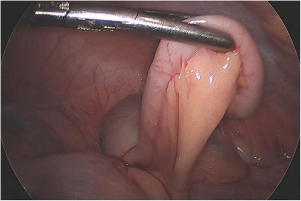
This intraoperative laparoscopic photograph shows a grossly normal vermiform appendix in a pediatric patient. The imaging modality is diagnostic laparoscopy, capturing the right lower quadrant of the abdomen. The appendix is held by a surgical grasper, revealing a smooth, glistening serosal surface with a healthy pinkish-tan hue. There are no visual markers of acute appendicitis, such as luminal distension, erythema, edema, or fibrinopurulent exudate. The mesoappendix and surrounding mesenteric fat appear unremarkable, lacking the hyperemia or 'fat stranding' characteristic of acute inflammation. This image serves as a clinical comparison for medical students to distinguish a healthy appendix from one with inflammatory changes, such as those seen in typical acute appendicitis or cases mimicking it, like Multi-system Inflammatory Syndrome in Children (MIS-C). Educational focus is on the laparoscopic appearance of visceral abdominal anatomy and the visual exclusion of transmural inflammation during a surgical workup for acute abdominal pain.
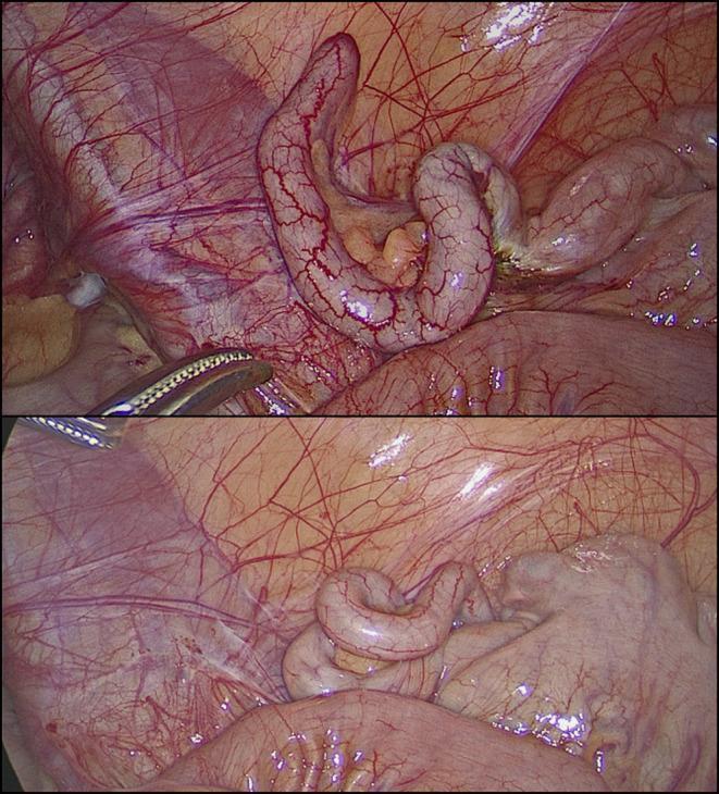
This clinical photograph consists of two laparoscopic views showing a macroscopically normal vermiform appendix during a diagnostic laparoscopy. The appendix appears as an elongated, tubular, and slightly coiled structure with a smooth, pale pink serosal surface. Fine, branching subserosal blood vessels are clearly visible across its length, indicating normal vascularity without the congestion or purulent exudate typical of acute appendicitis. In the upper frame, a laparoscopic grasper is visible, gently manipulating the appendix to facilitate inspection. The surrounding anatomy includes the mesoappendix containing adipose tissue and the glistening peritoneal lining of the right iliac fossa. There are no visual signs of inflammation, perforation, or abscess formation. This image serves as a reference for a 'grossly negative' appendix in the context of clinical suspicion of appendicitis, where symptoms may actually stem from non-inflammatory conditions such as follicular lymphoid hyperplasia or obstructive fecaliths.
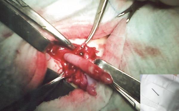
This clinical photograph captures an intraoperative view of an inflamed vermiform appendix during a surgical exploration for an Amyand's hernia. The primary image shows a red, swollen, and hyperemic appendix being retracted with surgical instruments. A visible perforation is noted at the distal apex of the appendix, caused by an ingested foreign body. The surrounding hernia sac contains serous-purulent exudate and hemorrhagic fluid, indicative of acute inflammation and localized infection. An inset photograph in the bottom-right corner displays the extracted metallic domestic pin against a white surgical cloth. The image demonstrates a rare surgical complication where a foreign body causes perforated appendicitis within a right inguinal hernia sac. Educational focus includes surgical anatomy of the inguinal canal, identification of acute appendicitis during hernioplasty, and management of foreign body-induced visceral perforation.
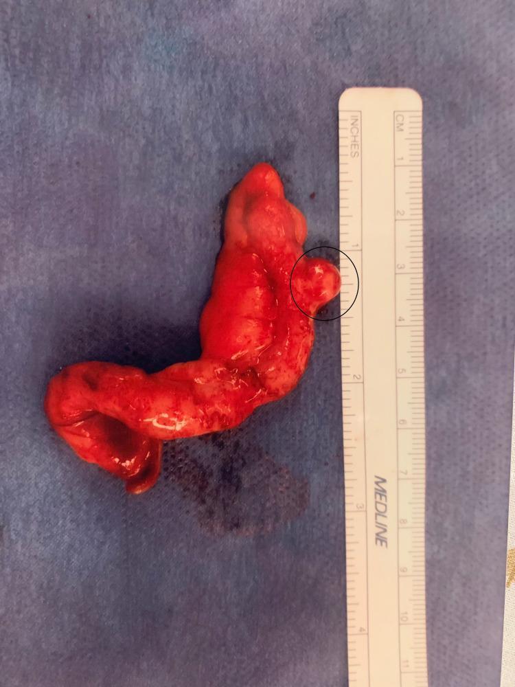
Content Type: Clinical Photograph (Gross Pathology Specimen). Anatomy: Appendix. This image displays a surgically resected vermiform appendix positioned against a sterile blue drape and a metric ruler for scale. The specimen measures approximately 7.5 cm in length and 1.5 cm in width. The serosal surface is diffusely erythematous and hyperemic, with irregular contours consistent with acute inflammation and focal serositis. A black circle highlights a prominent, rounded outpouching measuring roughly 0.5 cm in diameter located along the distal third of the organ. This visual finding is characteristic of an appendiceal diverticulum. The overall presentation demonstrates appendiceal diverticulosis in the setting of acute appendicitis. Clinical Significance: Appendiceal diverticulitis is a rare variant of appendicitis that carries a higher risk of perforation and may be associated with underlying neoplasms, though this specimen primarily illustrates the morphological findings of diverticular inflammation.
"appendicitis"[MeSH Terms] AND ("diagnosis" OR "management")
Appendicitis
Overview
Anatomy
- Retrocecal (most common)
- Subcecal
- Pelvic
- Preileal / postileal
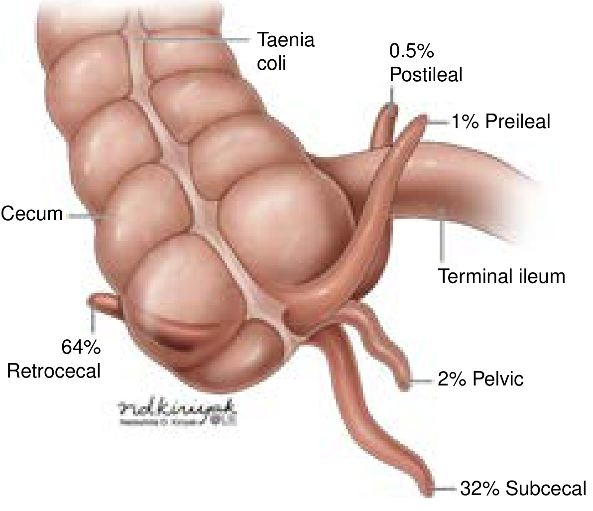
Pathophysiology
- Obstruction - most commonly by a fecalith/appendicolith; also lymphoid hyperplasia, parasites, neoplasm, or foreign body
- Continued mucus secretion → increased intraluminal pressure
- Bacterial overgrowth, venous congestion
- Distension of the narrow appendiceal lumen → stretches visceral afferent nerve fibers → vague periumbilical pain (first symptom)
- Arterial inflow obstruction → ischemia → necrosis → gangrene → perforation ± abscess
Clinical Presentation
| Feature | Detail |
|---|---|
| Initial pain | Vague, periumbilical/epigastric; colicky |
| Migration | Shifts to right lower quadrant (RLQ) over hours |
| Nausea/vomiting | Follows pain onset (if vomiting precedes pain, consider another diagnosis) |
| Anorexia | Very common |
| Fever | Low-grade initially; high fever suggests perforation |
| Tachycardia | Reflects systemic inflammation |
-
McBurney's point tenderness - 1/3 of the way from the right anterior superior iliac spine to the umbilicus
-
Rovsing's sign - RLQ pain on palpation of the LLQ (indicates peritoneal irritation)
-
Psoas sign - pain on extension of the right hip (retrocecal appendix)
-
Obturator sign - pain on internal rotation of the right hip (pelvic appendix)
-
Rebound tenderness, guarding, rigidity (suggests peritonitis/perforation)
-
Current Surgical Therapy 14e; Sabiston Textbook of Surgery
Diagnosis
Laboratory
- WBC: Leukocytosis in ~90% of cases (typically 10,000-18,000/μL with left shift); a normal WBC does not exclude appendicitis (10% have normal count)
- CRP / procalcitonin: Elevated; CRP is most sensitive of the biomarkers
- Urinalysis: Usually normal; trace pyuria may occur due to appendix proximity to bladder
- Urine hCG: Must rule out ectopic pregnancy in women of childbearing age
Scoring Systems
| Score | Notes |
|---|---|
| Alvarado score | Best studied; useful for ruling out appendicitis but not sufficiently specific alone |
| Appendicitis Inflammatory Response (AIR) score | Recommended over Alvarado in current consensus guidelines |
| Adult Appendicitis Score (AAS) | Recommended in current guidelines |
| Pediatric Appendicitis Score (PAS) | For children |
Imaging
| Modality | Sensitivity / Specificity | Notes |
|---|---|---|
| CT (multidetector, low-dose ~2 mSv) | ≥94% / ≥94% | Preferred in adults; detects perforation; non-contrast nearly as good as contrast-enhanced |
| Ultrasound | ~83% / ~93% | First-line in children and pregnant women; if positive, very helpful; negative does not exclude |
| MRI | Comparable to CT | Preferred alternative in pregnancy when CT contraindicated |
| Plain radiograph | Poor sensitivity/specificity | Limited role |
- Goldman-Cecil Medicine; Sabiston Textbook of Surgery
Differential Diagnosis
- Mesenteric adenitis (especially in children)
- Ovarian pathology (torsion, ruptured cyst, ectopic pregnancy)
- Pelvic inflammatory disease
- Meckel's diverticulitis
- Crohn's disease (terminal ileitis)
- Ureteral colic
- Perforated peptic ulcer
- Psoas abscess
Treatment
Uncomplicated Appendicitis
- Laparoscopic appendectomy is preferred over open - lower complication rates, faster return to normal activity
- Preoperative IV antibiotics (e.g., cefotetan 2 g IV or cefoxitin 2 g IV, or ticarcillin-clavulanate) reduce infectious complications
- Timing within 12-24 hours of diagnosis
- Viable alternative in selected patients without fecalith
- The 2024 Cochrane meta-analysis (PMID 38682788) confirms antibiotics can resolve uncomplicated appendicitis but with higher recurrence rates than surgery
- The 2024 SAGES guideline (PMID 38740595) provides evidence-based recommendations for both operative and antibiotic approaches
- ~20-35% of patients managed nonoperatively will require appendectomy within 1 year
- Contraindications to nonoperative approach: fecalith, perforation, abscess, clinical deterioration
Complicated/Perforated Appendicitis
- Perforation increases mortality from 0.0002% to 3% and morbidity from 3% to 47%
- Open appendectomy preferred when perforation is evident
- Perioperative broad-spectrum antibiotics (covering gram-negatives and anaerobes)
- If presenting late (>5 days) with an abscess: interval appendectomy strategy - IR drain placement first, then elective appendectomy in 6-8 weeks
Special Populations
Pregnancy
- Most common surgical emergency in pregnancy
- Same incidence as non-pregnant women but higher perforation rate due to diagnostic delays
- Appendix may displace out of RLQ in late pregnancy (though <25% move beyond RLQ even in 3rd trimester)
- Physiologic leukocytosis of pregnancy confounds lab interpretation
- Imaging: Ultrasound first, then MRI if inconclusive (avoid CT radiation if possible)
- ROSEN's Emergency Medicine advises that laparoscopy is safe in all trimesters
Pediatric
- Most common acute surgical problem in children
- Young children (2-6 years) frequently present already perforated due to diagnostic difficulty
- Ultrasound and MRI preferred over CT to avoid ionizing radiation
- Nonoperative management achieves resolution in 80-90% within 24-48 hours, but recurrence rates can reach 40% at medium-term follow-up
- Laparoscopic appendectomy remains the standard of care
- Current Surgical Therapy 14e, p. 1500
Complications
| Complication | Notes |
|---|---|
| Perforation | 16-40% of cases; greatly increases morbidity/mortality |
| Peritonitis | Local or diffuse |
| Appendiceal abscess/phlegmon | May be managed with IR drainage + interval appendectomy |
| Sepsis / septic shock | Risk increases ~2-fold in pregnancy |
| Bowel obstruction | Post-inflammatory or post-surgical adhesions |
| Negative appendectomy | Rate ~15% without imaging; imaging reduces this significantly |
Key Recent Evidence (2024)
- The Cochrane systematic review (Doleman et al., 2024 - PMID 38682788) confirms antibiotics are effective for uncomplicated appendicitis but with higher recurrence vs surgery
- The SAGES 2024 guideline (PMID 38740595) recommends considering both operative and nonoperative options with shared decision-making
- AIR and AAS scores are now recommended over the Alvarado score for risk stratification