
A 45 year old woman presents to the outpatient clinic with sudden onset of drooping of the left corner of the mouth, inability to close the left eye, and absence of forehead wrinkles on the same side. she also complains of the alterd taste sensation on the anterior part of the tongue and hypertensitivity to sounds on the affected side. a. Which cranial nerve is most likely to be affected in this case? b. What are the functional components of this nerve? c. Mention the different branches of this nerve d. Give a brief anatomical explaination of the features of the above scenario.
Reading File
Finding Sources
Finding Sources
Reading File
Reading File
Finding Sources
Finding Sources
Reading File
Reading File
Finding Sources
Searching Images
Bell's palsy facial nerve paralysis forehead sparing lower motor neuron
Finding Sources
Loading Image
Reading File
I now have comprehensive information from multiple authoritative textbooks. Let me compile the full answer.
Facial Nerve (CN VII) - Comprehensive Analysis
a. Cranial Nerve Affected
Cranial Nerve VII - the Facial Nerve (specifically its left peripheral trunk) is affected.
The combination of:
- Drooping of the left corner of the mouth (orbicularis oris weakness)
- Inability to close the left eye (orbicularis oculi weakness)
- Absent forehead wrinkles on the left (frontalis weakness)
- Altered taste on the anterior tongue
- Hypersensitivity to sounds (hyperacusis)
...all point unequivocally to a lower motor neuron (LMN) lesion of CN VII, consistent with Bell's palsy. The fact that the forehead is involved (no wrinkles) is the key distinguishing sign of a peripheral (LMN) facial nerve lesion, as opposed to an upper motor neuron (UMN) lesion where the forehead is spared due to bilateral cortical representation.
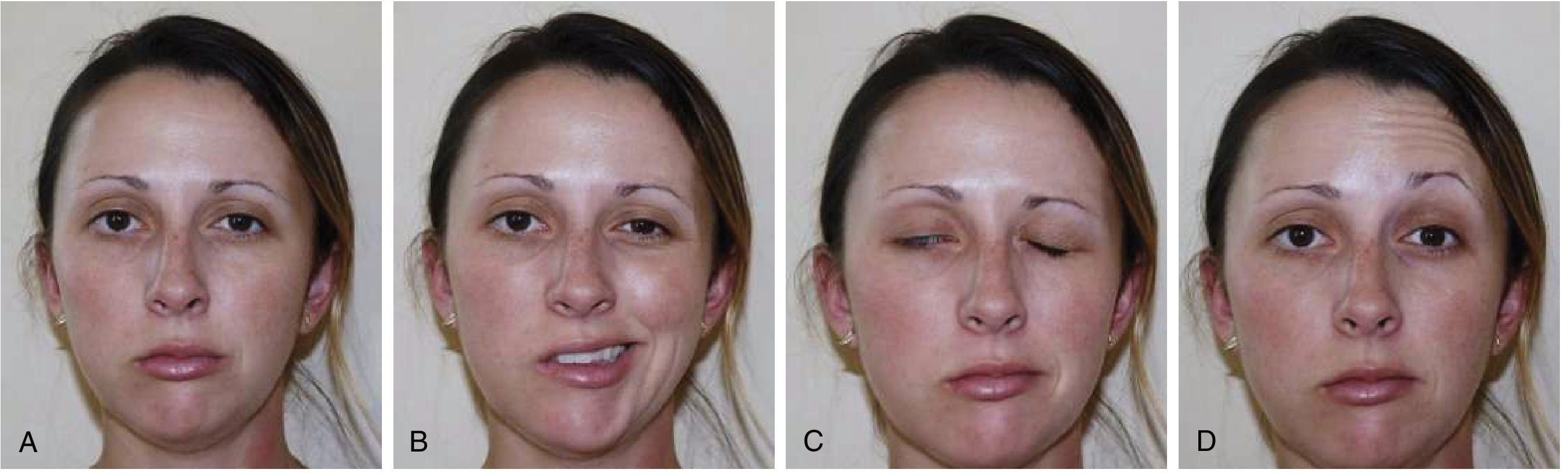
Bell's palsy: (A) rest, (B) attempting to smile - asymmetric, (C) attempting to close eyes - left eye fails to close, (D) attempting to wrinkle forehead - left side absent
b. Functional Components of CN VII
CN VII carries five fiber types (functional components):
| Component | Type | Function |
|---|---|---|
| Special Visceral Efferent (SVE) - Branchial motor | Motor to muscles of facial expression, stapedius, stylohyoid, posterior belly of digastric | Main motor output; muscles derived from the 2nd pharyngeal arch |
| General Visceral Efferent (GVE) - Parasympathetic | Preganglionic secretomotor fibers | Innervates lacrimal gland (via GSPN and pterygopalatine ganglion), submandibular and sublingual glands (via chorda tympani and submandibular ganglion), and nasal cavity seromucinous glands |
| Special Visceral Afferent (SVA) - Taste | Sensory for taste | Anterior 2/3 of tongue (via chorda tympani), tonsillar fossae, and posterior palate (via GSPN); cell bodies in the geniculate ganglion; central projections to the nucleus tractus solitarius |
| General Somatic Afferent (GSA) | Cutaneous sensation | Skin of the external auditory canal (EAC) and concha of the auricle; proprioception from facial muscles |
| General Visceral Afferent (GVA) | Visceral sensation | Mucosa of the nose, pharynx, and palate |
Sources: Shambaugh Surgery of the Ear, p. 68; Cummings Otolaryngology, p. 2451; Localization in Clinical Neurology, p. 779
c. Branches of CN VII
Intrapetrosal (Intratemporal) Branches
-
Greater Superficial Petrosal Nerve (GSPN) - arises from the anterior aspect of the geniculate ganglion; carries preganglionic parasympathetic fibers to the lacrimal gland and nasal/palatine glands via the pterygopalatine ganglion; also carries taste fibers from the palate.
-
Nerve to the Stapedius Muscle - arises from the mastoid (vertical) segment near the pyramidal eminence; controls the stapedius muscle in the middle ear.
-
Chorda Tympani - arises from the lower part of the mastoid segment; joins the lingual nerve (CN V3); carries:
- Preganglionic parasympathetics to submandibular/sublingual glands
- Taste afferents from anterior 2/3 of tongue
Extracranial Branches (after exit through stylomastoid foramen)
-
Posterior Auricular Nerve - to the occipitalis, posterior auricular, and oblique/transverse auricular muscles.
-
Digastric Branch - to the posterior belly of the digastric muscle.
-
Stylohyoid Branch - to the stylohyoid muscle.
Terminal Branches (Five, arising at the pes anserinus within the parotid gland)
| Branch | Target muscles |
|---|---|
| Temporal | Frontalis, orbicularis oculi (upper), corrugator supercilii |
| Zygomatic | Orbicularis oculi (lower), zygomaticus |
| Buccal | Buccinator, orbicularis oris, muscles of the upper lip/nose |
| Marginal Mandibular | Depressor anguli oris, muscles of lower lip and chin |
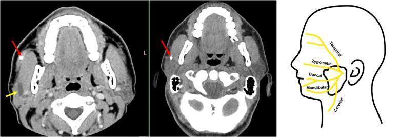
| Cervical | Platysma |
d. Anatomical Explanation of the Clinical Features
Why the entire left face is affected (LMN pattern - including the forehead)
The forehead (frontalis) muscle receives bilateral cortical input from both motor cortices. In an UMN (central) lesion - such as a stroke - the contralateral motor cortex is damaged, but the forehead is still partially supplied by the ipsilateral (intact) cortex, so forehead wrinkling is preserved. In a peripheral (LMN) lesion of CN VII itself, ALL facial muscles on that side lose their only motor nerve supply - including the frontalis - so the forehead is also paralyzed. This patient has absent forehead wrinkles, confirming a peripheral CN VII lesion.
Why she cannot close her left eye
The orbicularis oculi (the muscle that closes the eyelid) is supplied by the zygomatic branch of CN VII. With its paralysis, the eye cannot be closed (lagophthalmos), and the eyeball may roll upward on attempted closure (Bell's phenomenon).
Why the left corner of the mouth droops
The depressor anguli oris and orbicularis oris are supplied by the buccal and marginal mandibular branches of CN VII. Their paralysis causes drooping of the mouth corner and inability to show teeth on that side.
Why taste is altered on the anterior tongue
The chorda tympani - a branch arising from the mastoid segment of CN VII while still within the fallopian canal - carries special visceral afferent (taste) fibers from the anterior 2/3 of the tongue to the geniculate ganglion and then to the nucleus tractus solitarius in the brainstem. In this patient, the lesion is proximal to the point where the chorda tympani joins the main facial nerve trunk (i.e., within the fallopian canal above the chorda tympani origin), so taste is impaired. This is a key localizing sign - if the lesion were distal to the chorda tympani take-off (i.e., at the stylomastoid foramen or below), taste would be intact.
Why there is hypersensitivity to sounds (hyperacusis)
The stapedius muscle in the middle ear is innervated by the nerve to the stapedius - a branch arising from the mastoid segment of CN VII, just above the chorda tympani. The stapedius normally dampens excessive vibration of the ossicular chain, protecting the inner ear from loud sounds. When CN VII is lesioned proximal to this branch, the stapedius is paralyzed, and every sound is perceived as abnormally loud or harsh (hyperacusis). The presence of hyperacusis alongside taste loss confirms the lesion is within the mastoid segment of the fallopian canal, at or above the nerve to stapedius.
Summary of lesion localization
CN VII course (proximal → distal):
Pons → IAC → Geniculate ganglion
  ↓
  [GSPN branches off here - lacrimation]
  ↓
Tympanic segment
  ↓
Mastoid segment
  ├─ Nerve to stapedius  ← lesion is AT or ABOVE this point
  ├─ Chorda tympani      ← lesion is AT or ABOVE this point (taste affected)
  ↓
Stylomastoid foramen
  ↓
Posterior auricular nerve, digastric, stylohyoid
  ↓
Parotid gland → 5 terminal branches → all ipsilateral facial muscles
This patient's lesion involves the mastoid segment of CN VII within the fallopian canal - above the nerve to stapedius and chorda tympani - but lacrimation is not mentioned as affected, suggesting the geniculate ganglion/GSPN may be spared. This pattern is entirely consistent with Bell's palsy, the most common cause of acute peripheral facial nerve palsy, most likely due to viral reactivation (Herpes simplex virus) causing inflammation and edema of the nerve within the narrow bony fallopian canal.
Sources: Localization in Clinical Neurology 8e, p. 779; Cummings Otolaryngology, pp. 2451-52; Neuroanatomy through Clinical Cases 3e, p. 270; Adams and Victor's Principles of Neurology 12e; Shambaugh Surgery of the Ear, p. 68
This is a shared conversation. Sign in to Orris to start your own chat.