
How do tram track capacities in Cartagena syndrome look on the chest X-ray? Describe me a chest X-ray for Cartagena syndrome. And cystic fibrosis.
Kartagener syndrome bronchiectasis tram track chest X-ray
cystic fibrosis chest X-ray bronchiectasis hyperinflation
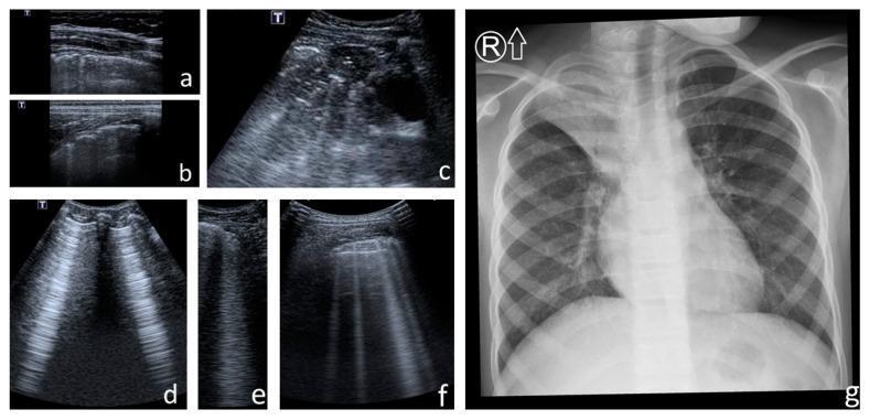
A collation of clinical images comparing lung ultrasound (LUS) and chest X-ray (CXR) findings in the context of cystic fibrosis. Panels (a) and (b) demonstrate pleural line abnormalities, including irregularity and fragmentation. Panel (c) shows a large subpleural consolidation with heterogeneous echogenicity and static air bronchograms. Panels (d), (e), and (f) highlight characteristic LUS artifacts: (d) illustrates Am-lines (broad vertical artifacts containing parallel horizontal lines), (e) shows a single confluent B-line, and (f) displays multiple discrete B-lines (comet-tail artifacts) arising from the pleural line. Panel (g) is a posterior-anterior chest X-ray showing advanced pulmonary changes including a barrel chest deformity, hyperinflation (increased transparency) of the left upper and middle lobes, and right upper lobe opacity consistent with cirrhosis and atelectasis. Evidence of bilateral bronchiectasis is present, characterized by peribronchial thickening and tram-track signs. These images represent a diagnostic comparison between LUS scoring and traditional radiographic assessment (modified Chrispin–Norman score) for monitoring chronic lung disease progression.
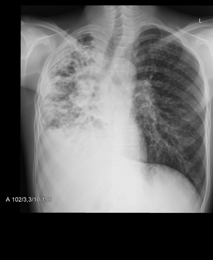
This diagnostic image is a posterior-anterior (PA) chest x-ray demonstrating advanced pulmonary pathology, characteristic of chronic obstructive or infectious lung disease such as cystic fibrosis. The right lung field exhibits significant volume loss (atelectasis) and a large inferior opacification consistent with pleural effusion or dense consolidation. The upper and middle zones of the right lung show a disorganized pattern of cystic and linear opacities, indicating extensive bronchiectasis and honeycombing. A resulting mediastinal shift toward the right is evident. In contrast, the left lung field displays compensatory hyperinflation with flattened diaphragmatic contours and increased radiolucency. Diffuse nodular and linear interstitial opacities are present throughout the left lung, suggesting widespread small-airway involvement. The radiographic findings are quantified by a modified Chrispin-Norman score of 27, reflecting high disease severity. This image serves as a teaching tool for recognizing the complex interplay of parenchymal destruction, airway remodeling, and volume shifts in chronic respiratory conditions.
radiological findings for kartagner on cxr