
Structure present above the thalamus
The structure present above the thalamus is the epithalamus.
Searching sources...
Reading File
Finding Sources
Finding Sources
Reading File
Reading File
Finding Sources
Reading File
Loading Image
Epithalamus - The Structure Above the Thalamus
The structure present above (dorsal to) the thalamus is the Epithalamus.
Position Within the Diencephalon
The diencephalon is divided into four regions based on their position relative to the third ventricle:
| Region | Position relative to thalamus |
|---|---|
| Epithalamus | Dorsal (above) |
| Thalamus | Central / lateral walls |
| Subthalamus | Ventrolateral |
| Hypothalamus | Ventral (below) |
The diagram below from embryology clearly illustrates this arrangement - note panel E (transverse section) showing the epithalamus dorsally, the thalamus laterally, and the hypothalamus ventrally:
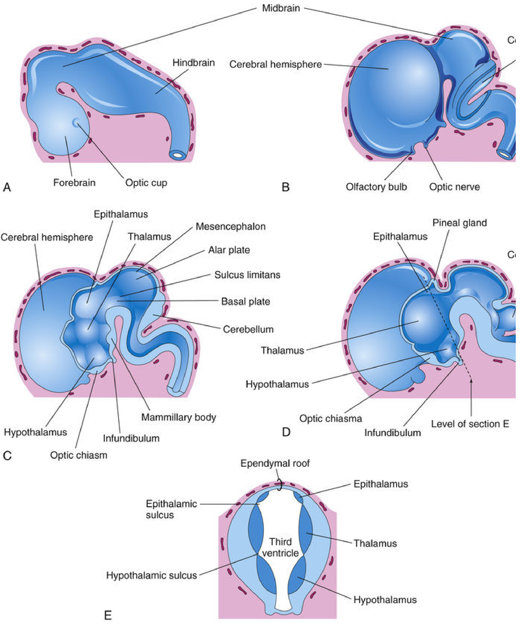
(The Developing Human: Clinically Oriented Embryology, Fig. 17.22E)
Components of the Epithalamus
The epithalamus consists of several small nuclei and structures:
-
Pineal gland (pineal body)
- An endocrine gland that secretes melatonin, which regulates circadian rhythms and sleep-wake cycles
- It projects posteriorly from the roof of the diencephalon
-
Habenula (habenular nuclei)
- A bilateral nuclear complex involved in limbic-motor integration
- Plays a role in regulating reward, aversion, and stress responses
- Connected to other limbic structures via the stria medullaris thalami
-
Parts of the pretectum
- Involved in the pupillary light reflex and other visual reflexes
-
Posterior commissure
- A white matter bundle crossing the midline just above the pineal gland
Development
Embryologically, the epithalamus develops from the roof and dorsal portion of the lateral wall of the diencephalon. The thalamus is separated from the epithalamus by the epithalamic sulcus (visible in panel E of the figure above).
In a nutshell: The structure above the thalamus is the epithalamus, part of the diencephalon, consisting mainly of the pineal gland, habenular nuclei, and parts of the pretectum.
- Neuroanatomy through Clinical Cases, 3rd Ed., p. 306-307
- The Developing Human: Clinically Oriented Embryology, p. 1059-1060
Show the sagital section brain having all the structures abive the mid brain
Let me search the medical library for a sagittal brain section diagram.
Searching sources...
Finding Sources
Finding Sources
Finding Sources
Finding Sources
Finding Sources
Finding Sources
Finding Sources
Finding Sources
Reading File
Reading File
Loading Image
Loading Image
Now let me get a more detailed sagittal/medial surface diagram with all the labelled structures:
Finding Sources
Finding Sources
Here are the best sagittal/schematic brain diagrams from the textbooks showing all structures above and around the midbrain:
Sagittal Brain - Structures Above the Midbrain
Diagram A - Major Brain Subdivisions (sagittal schematic)
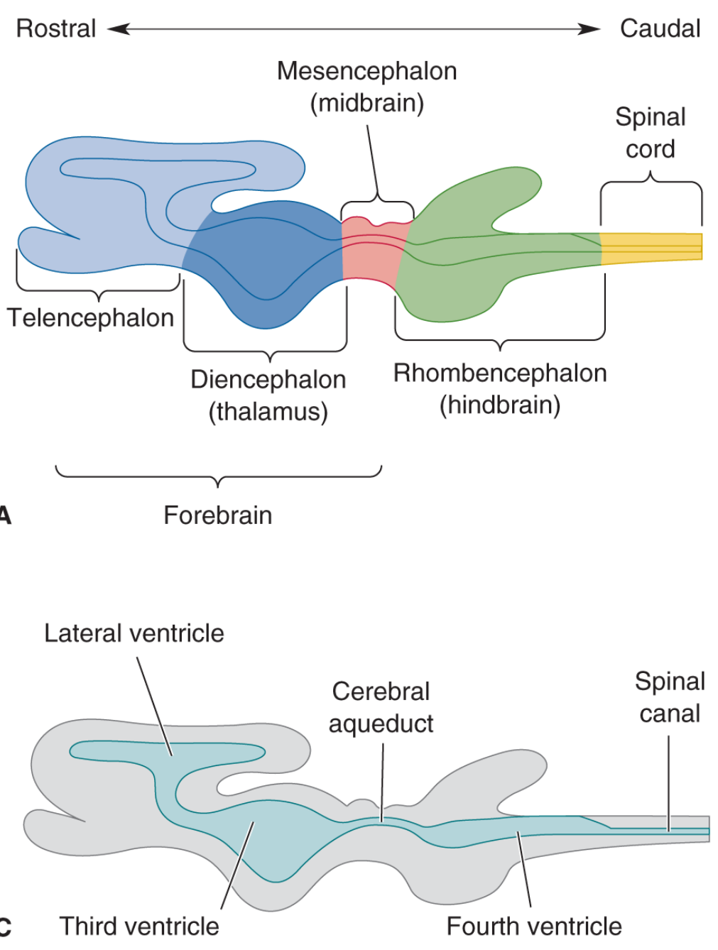
(Neuroscience: Exploring the Brain, 5th Ed. - Fig. 7.23A & C)
This shows the rostral-caudal arrangement:
- Telencephalon (forebrain) - cerebral cortex + basal telencephalon
- Diencephalon - thalamus region (above the midbrain)
- Mesencephalon (midbrain) - the reference landmark (shown in red)
- Below: Rhombencephalon (hindbrain) - pons, medulla, cerebellum
- Ventricular system shown in panel C - lateral ventricles → 3rd ventricle → cerebral aqueduct → 4th ventricle
Diagram B - Detailed Structures in Each Division
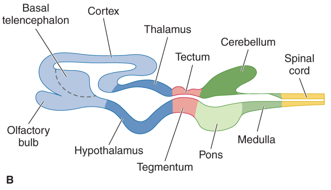
(Neuroscience: Exploring the Brain, 5th Ed. - Fig. 7.23B)
Structures Above the Midbrain (Rostral to it)
| Division | Structures |
|---|---|
| Telencephalon | Cerebral cortex, basal ganglia (caudate, putamen, globus pallidus), olfactory bulb, hippocampus, amygdala |
| Diencephalon | Thalamus, hypothalamus, epithalamus (pineal gland, habenula), subthalamus |
| Midbrain (reference) | Tectum (superior & inferior colliculi), tegmentum, cerebral aqueduct, cerebral peduncles |
Diagram C - Embryological Sagittal View (with epithalamus)
(The Developing Human: Clinically Oriented Embryology - Fig. 17.22)
Panels C and D clearly show the medial/sagittal view with the pineal gland, epithalamus, thalamus, hypothalamus, optic chiasm, and mammillary bodies labeled on the developing brain. Panel E (transverse) confirms the dorsal-ventral stacking: Epithalamus (top) → Thalamus (middle) → Hypothalamus (bottom).
Key takeaway: All structures above the midbrain belong to the diencephalon (thalamus, hypothalamus, epithalamus, subthalamus) and the telencephalon (cerebral cortex, basal ganglia, limbic structures). The midbrain (mesencephalon) sits at the junction, connecting the forebrain to the hindbrain, with the cerebral aqueduct running through it.
- Neuroscience: Exploring the Brain, 5th Ed., pp. 628-630
- The Developing Human: Clinically Oriented Embryology, pp. 1059-1060
This is a shared conversation. Sign in to Orris to start your own chat.