Histology of testis
Reading File
Finding Sources
Finding Sources
Finding Sources
Finding Sources
Finding Sources
Finding Sources
Reading File
Reading File
Loading Image
Loading Image
Reading File
Loading Image
Reading File
Reading File
I now have thorough content from both Junqueira's and Ross Histology. Let me compile the comprehensive answer.
Histology of the Testis
Sources: Histology: A Text and Atlas (Ross & Pawlina, 9e) | Junqueira's Basic Histology (17e)
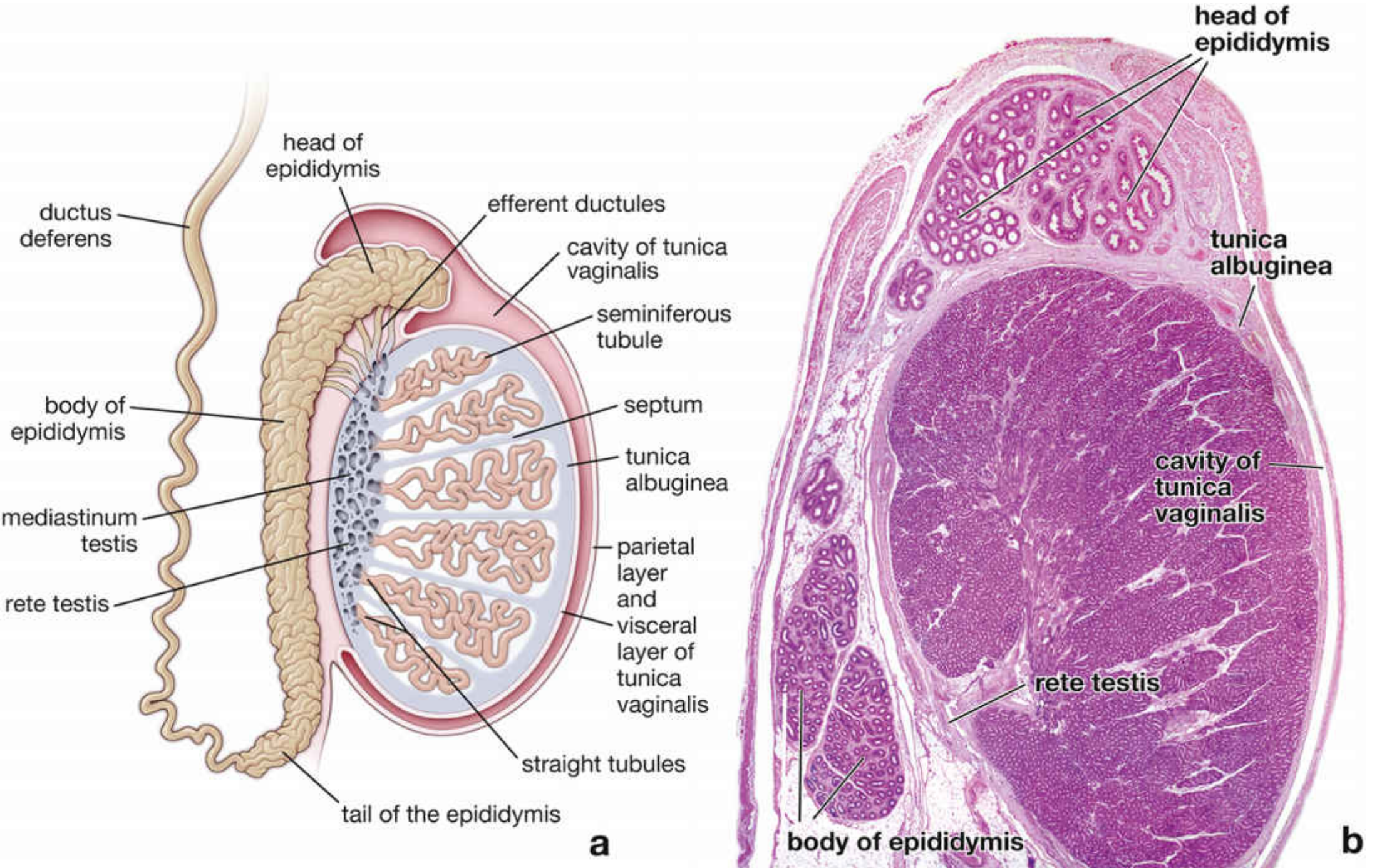
1. General Organization
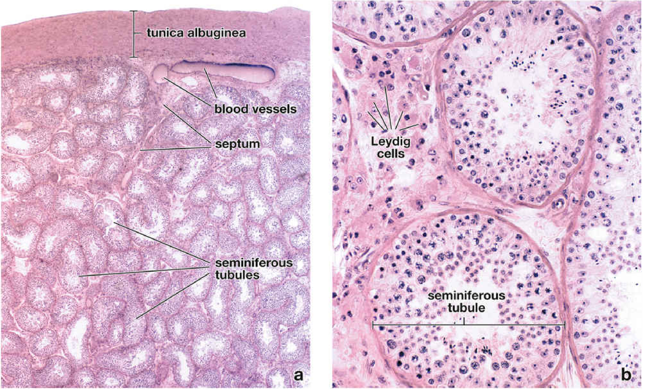
Each testis is covered by a thick, dense irregular connective tissue capsule called the tunica albuginea. Its inner layer, the tunica vasculosa, is a loose connective tissue containing blood vessels and lymphatics. Externally, a mesothelium-lined serous sac - the tunica vaginalis (derived from peritoneum) - encloses the testis.
Along the posterior surface, the tunica albuginea thickens and projects inward as the mediastinum testis, through which blood vessels, lymphatics, and genital ducts pass. Connective tissue septa extend from the capsule inward, dividing the testis into approximately 250 lobules, each containing one to four seminiferous tubules.
2. Seminiferous Tubules
- Number: 250-1000 per testis
- Dimensions: ~150-250 µm in diameter; each tubule ~50 cm long (range 30-80 cm) and highly convoluted
- Shape: Each tubule forms a loop; both ends open near the mediastinum into short straight tubules (tubuli recti), which drain into the rete testis
- Wall: Lined by a complex stratified seminiferous (germinal) epithelium, resting on a basement membrane (basal lamina)
Tunica Propria (Peritubular Tissue)
Surrounds the basal lamina: 3-5 layers of myoid cells (peritubular contractile cells) with smooth-muscle-like actin filaments and rER for collagen synthesis. Their rhythmic peristaltic contractions move spermatozoa and testicular fluid toward the excurrent ducts.
3. Cells of the Seminiferous Epithelium
The seminiferous epithelium contains two fundamental cell populations:
A. Sertoli Cells (Supporting/Sustentacular Cells)
- Tall columnar cells extending full thickness of the epithelium, from basal lamina to lumen
- Nuclei are large, ovoid/triangular, with prominent nucleoli; often indented - a key identifying feature on H&E
- Extensive lateral and apical cytoplasmic processes embrace surrounding spermatogenic cells
- Do not replicate after puberty
- Adjacent Sertoli cells are joined near their bases by tight junctions (occluding junctions) forming the blood-testis barrier (BTB)
Blood-Testis Barrier
The BTB divides the seminiferous epithelium into two compartments:
- Basal compartment (below the junctions): contains spermatogonia and preleptotene spermatocytes; exposed to blood-derived substances
- Adluminal (abluminal) compartment (above the junctions): contains primary/secondary spermatocytes and spermatids; immunologically privileged, protected from autoimmune attack since meiotic/post-meiotic cells express novel antigens
Functions of Sertoli Cells
| Function | Detail |
|---|---|
| Structural support | Scaffold for spermatogenic cells |
| Phagocytosis | Engulf residual bodies shed by spermatids |
| Secretion | Androgen-binding protein (ABP), inhibin, Mullerian inhibiting substance, transferrin, plasminogen activator |
| Blood-testis barrier | Immunological protection of meiotic cells |
| Spermiation | Release of mature spermatids into lumen |
| Endocrine response | FSH receptors; amplify testosterone action |
B. Spermatogenic Cells
Arranged in poorly defined layers from periphery to lumen in order of maturation:
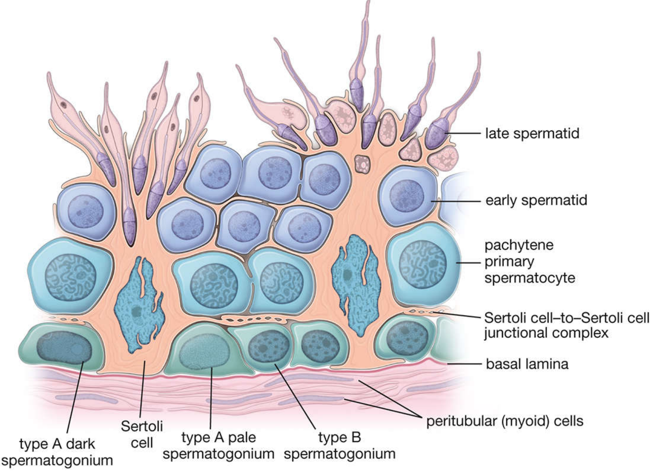
Spermatogonia (basal compartment, resting on basal lamina)
- Type A dark (Ad): Reserve stem cells; serve as renewal stem cells; dark oval nuclei with fine chromatin
- Type A pale (Ap): Committed progenitors; pale oval nuclei; give rise to type B
- Type B: Round nuclei with peripheral heterochromatin; divide to form primary spermatocytes
Primary Spermatocytes
- Largest cells in the epithelium; most commonly seen in prophase I (long duration: ~22 days in humans)
- Recognized by their large size and prominent chromosomes (thread-like in leptotene, paired in zygotene, thick/crossing over in pachytene, short/condensed in diplotene)
- Undergo meiosis I → two secondary spermatocytes
Secondary Spermatocytes
- Smaller than primary; undergo meiosis II rapidly (~few hours)
- Rarely seen in routine sections due to brief existence
Spermatids (early and late)
- Early (round) spermatids: Small, round cells with round nuclei; located near lumen; product of meiosis II; haploid
- Late (elongated) spermatids: Elongated dark nuclei directed toward lumen; product of spermiogenesis
4. Spermatogenesis
Spermatogenesis proceeds through three phases:
- Spermatogonial phase - mitotic renewal and commitment
- Spermatocyte phase (meiosis) - reduction to haploid cells (spermatids)
- Spermatid phase (spermiogenesis) - morphological transformation into spermatozoa
Spermiogenesis (Spermatid → Spermatozoon)
Key events:
- Acrosome formation: Golgi apparatus produces proacrosomal granules that fuse into the acrosomal vesicle, flattening to cap the anterior nucleus
- Nuclear condensation: Chromatin condenses; nucleus elongates
- Flagellum formation: Centriole organizes the axoneme
- Mitochondrial sheath: Mitochondria migrate to surround the midpiece
- Cytoplasmic reduction: Excess cytoplasm shed as residual bodies, phagocytosed by Sertoli cells
The full duration of spermatogenesis in humans is approximately 74 days.
Cycle of the Seminiferous Epithelium
At any given point in a tubule cross-section, spermatogenic cells exist in a specific, reproducible cell association (stage). The series of stages between two successive occurrences of the same pattern at one site constitutes a cycle (~16 days in humans). The sequential displacement of stages along the length of the tubule is called the wave of the seminiferous epithelium.
5. Interstitial Tissue (Leydig Cells)
The connective tissue stroma between tubules contains fibroblasts, lymphatics, blood vessels, and Leydig (interstitial) cells.
Leydig Cell Features
- Large, polygonal eosinophilic cells, occurring singly or in clusters
- Central round nuclei
- Cytoplasm rich in smooth ER (accounts for eosinophilia; site of steroidogenesis enzymes)
- Mitochondria with tubulovesicular cristae - hallmark of steroid-secreting cells
- Lipid droplets (cholesterol substrate)
- Lipofuscin pigment
- Distinctive rod-shaped crystals of Reinke (~3 × 20 µm) - pathognomonic for human Leydig cells; exact function unknown, likely a protein product
Function
- Produce testosterone in response to LH (= ICSH in males) from pituitary
- Also secrete INSL3 (stimulates testicular descent, promotes meiosis) and oxytocin (stimulates myoid cell contraction)
- Active in fetal life (stimulated by hCG), quiescent from ~5 months fetal life, then reactivated at puberty
6. Intratesticular Duct System
| Structure | Epithelium | Notes |
|---|---|---|
| Straight tubules (tubuli recti) | Simple cuboidal to columnar (only Sertoli cells, no spermatogenic cells) | Short transition from seminiferous tubules |
| Rete testis | Simple cuboidal/squamous with microvilli | Anastomosing channels in mediastinum |
| Efferent ductules | Pseudostratified; alternating groups of tall ciliated columnar and short non-ciliated cells ("festooned/scalloped" luminal border) | Absorb ~90% of testicular fluid |
7. Temperature Regulation
Spermatogenesis requires ~34°C (below core body temperature of 37°C), maintained by:
- Pampiniform plexus - venous network around testicular artery in spermatic cord: countercurrent heat exchange
- Dartos muscle (scrotal skin) and cremaster muscle - relax/contract to adjust testicular position
- Scrotal sweat evaporation
Key Clinical Correlations
- Cryptorchidism: Failure of testicular descent; bilateral cases cause infertility if not corrected by 2-3 years (sustained 37°C damages spermatogenic cells). Markedly increases risk of germ cell tumors.
- Hydrocele: Excess fluid in tunica vaginalis cavity.
- Testicular germ cell tumors: 95% of testicular malignancies; arise after puberty; strongly associated with cryptorchidism.
- Leydig cell tumors: Rare, usually benign; contain Reinke crystals; cause precocious puberty in prepubertal boys or gynecomastia/feminization in adults.
- Aging: Progressive thickening of tunica propria, decreased spermatogenesis.
Histology: A Text and Atlas (Ross & Pawlina), pp. 2091-2103 | Junqueira's Basic Histology, 17e, pp. 1058-1065
This is a shared conversation. Sign in to Orris to start your own chat.