
Cor pulmonale
"cor pulmonale" AND management
cor pulmonale right ventricular hypertrophy chest X-ray ECG
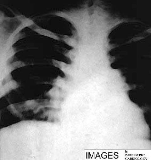
**Imaging Modality:** Frontal Chest X-ray (CXR). **Anatomical Region:** Thoracic cavity, focusing on the cardiomediastinal silhouette and pulmonary vasculature. **Observed Pathology:** Findings are consistent with cor pulmonale secondary to severe pulmonary hypertension. **Characteristic Visual Features:** * **Cardiac Morphology:** Pronounced cardiomegaly with a "boot-shaped" appearance, characterized by an upturned apex and a prominent right heart border, indicating right ventricular and right atrial hypertrophy. * **Vascular Markings:** Significant prominence and dilation of the central pulmonary arteries (hilar engorgement). There is a marked discrepancy between the enlarged central vessels and the "pruning" or attenuation of peripheral pulmonary vascular markings. * **Mediastinum:** Expansion of the transverse cardiac diameter and a bulging pulmonary trunk segment along the left heart border. * **Lung Fields:** Increased lucency in the peripheral lung zones due to decreased perfusion (oligemia). **Diagnostic Cues:** The combination of central pulmonary artery dilation and right-sided heart enlargement without signs of left-sided failure (e.g., absence of Kerley B lines or pleural effusions) is highly suggestive of pulmonary arterial hypertension leading to chronic cor pulmonale.
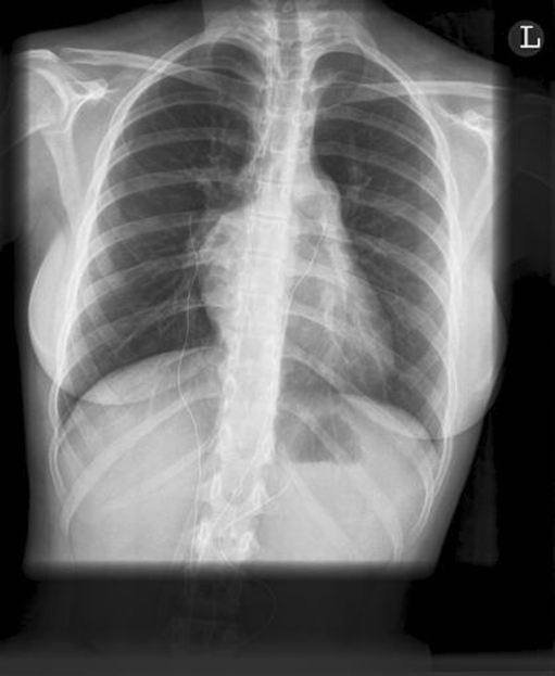
**Modality:** Posteroanterior (PA) Chest Radiograph (X-ray). **Anatomical Region:** Thorax, including the lungs, mediastinum, and cardiac silhouette. **Observed Pathology:** Findings are highly suggestive of pulmonary arterial hypertension (PAH) with associated cor pulmonale. **Characteristic Visual Features:** * **Cardiovascular:** Marked prominence of the main pulmonary artery segment (pulmonary trunk). Significant enlargement of the right heart borders, consistent with right atrial and right ventricular hypertrophy/dilation. The cardiac silhouette is widened with a shifted apex. * **Pulmonary Vasculature:** Demonstrable "pruning" effect, characterized by dilated central hilar pulmonary arteries followed by rapid tapering and rarification (oligaemia) of the peripheral pulmonary vessels. * **Lung Parenchyma:** Clear lung fields without focal consolidation, pleural effusion, or overt pulmonary edema. **Diagnostic Features:** The constellation of central pulmonary artery dilation alongside peripheral vascular pruning and right-sided cardiomegaly serves as a classic radiographic indicator of elevated pulmonary vascular resistance and secondary right heart strain. **Clinical Context:** Consistent with chronic pulmonary arterial hypertension.
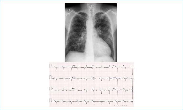
This composite educational image includes a posteroanterior (PA) chest radiograph and a 12-lead electrocardiogram (ECG) demonstrating signs of congenital heart disease and hemodynamic stress. The chest X-ray reveals a moderately enlarged cardiac silhouette with an elongated, rounded apex and an excavated middle arch, suggesting ventricular dominance. A notable anatomical variation is the right-sided aortic arch. The pulmonary vasculature appears prominent, particularly in the hila and lower lung fields, which may indicate systemic-pulmonary collateral circulation. The accompanying ECG shows a sinus rhythm with markers of right-sided heart strain and cavity overload. Key findings include peaked P waves (P-pulmonale) in leads I, II, and left precordial leads, and a significant right-axis deviation of the QRS complex (+180º) with deep S waves from V3 through V6. T-wave inversions in the precordial leads further support right ventricular strain. This combination of findings is characteristic of complex cyanotic congenital heart disease, such as Tetralogy of Fallot or pulmonary atresia with a right-sided aortic arch.
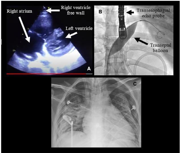
This composite clinical figure illustrates the diagnosis and management of severe pulmonary hypertension and its complications. Panel A shows a transthoracic echocardiogram (TTE) in a patient with acute cor pulmonale. Labeled structures include a dilated right atrium and left ventricle, with an arrow highlighting the right ventricle free wall, demonstrating significant right heart strain. Panel B is a fluoroscopic image capturing an atrioseptostomy procedure. It depicts a transesophageal echo (TEE) probe positioned in the esophagus and a transeptal balloon inflated across the interatrial septum to create a right-to-left shunt. Panel C is an anteroposterior thoracic X-ray demonstrating bilateral pneumothorax with limited lung expansion. Visible medical devices include bilateral thoracic drainage tubes (chest tubes), an endotracheal tube, and ECG leads. The image set provides a comprehensive visual summary of hemodynamic monitoring, interventional rescue maneuvers for right ventricular failure, and subsequent pulmonary complications in a critical care setting.
Cor Pulmonale
Definition
- Robbins & Kumar Basic Pathology, p. 363
- Robbins, Cotran & Kumar Pathologic Basis of Disease, p. 522
Types
| Feature | Acute | Chronic |
|---|---|---|
| Onset | Sudden | Insidious |
| Typical cause | Massive pulmonary embolism | COPD, pulmonary fibrosis, PPH |
| RV morphology | Dilation only (no hypertrophy) | Hypertrophy + dilation |
| RV wall thickness | Normal or mildly increased | Up to 1.0 cm or more |
Causes / Predisposing Disorders
Diseases of the Pulmonary Parenchyma
- Chronic obstructive pulmonary disease (COPD) - most common cause
- Diffuse pulmonary interstitial fibrosis
- Pneumoconioses (silicosis, coal-worker's pneumoconiosis)
- Cystic fibrosis
- Bronchiectasis
Diseases of the Pulmonary Vessels
- Primary (idiopathic) pulmonary arterial hypertension
- Recurrent pulmonary emboli (chronic thromboembolic PH - CTEPH)
- Pulmonary vasculitis
Disorders Affecting Chest Movement or Alveolar Ventilation
- Obesity-hypoventilation syndrome (Pickwickian syndrome)
- Obstructive sleep apnea
- Severe kyphoscoliosis
- Neuromuscular disease affecting respiratory muscles
- High-altitude chronic hypoxia
Pathophysiology
- Lung disease (obstruction or restriction) → loss of pulmonary vascular surface area + impaired gas exchange
- Hypoxemia ± hypercarbia → potent stimuli for pulmonary vasoconstriction and vascular remodeling
- Chronic hypoxia → increased erythropoietin → polycythemia → increased blood viscosity → further elevates PVR
- Endothelial dysfunction → reduced prostacyclin and NO, increased endothelin → exacerbates vasoconstriction
- Elevated pulmonary artery pressure → RV pressure and volume overload
- Compensated phase: RV hypertrophy maintains output
- Decompensated phase: RV dilation, tricuspid regurgitation, right heart failure
- Fuster and Hurst's The Heart, 15th Ed., p. 1774-1775
Morphology (Gross Pathology)
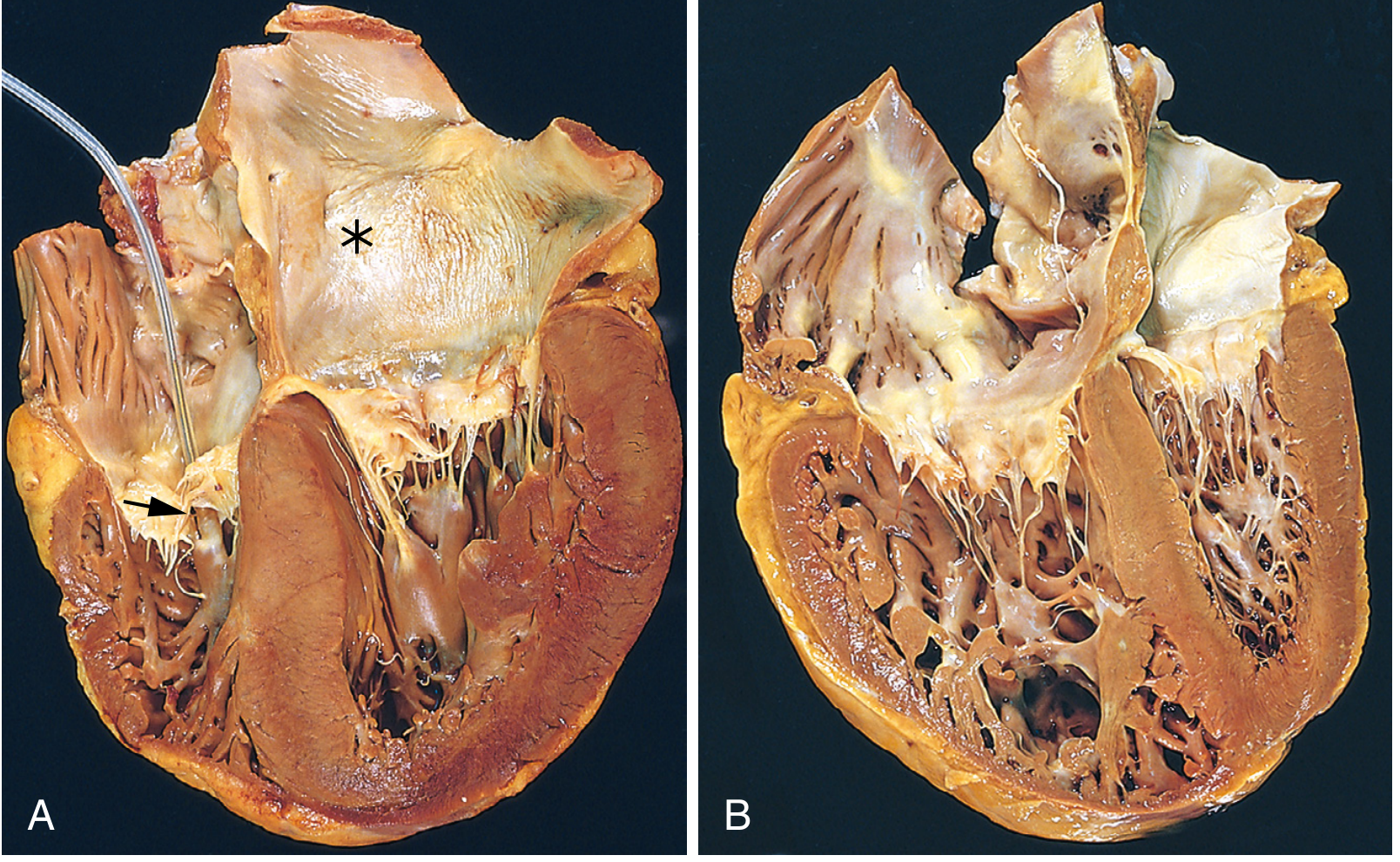
- RV wall thickens - sometimes up to or exceeding LV wall thickness
- The normally crescent-shaped RV chamber becomes ovoid
- Hypertrophy may manifest as thickened muscle bundles in the outflow tract below the pulmonary valve, or thickening of the moderator band
- RV compression of the LV may occur
- Tricuspid regurgitation and valve fibrous thickening may develop
- Pulmonary arteries show abnormal intimal thickening
Clinical Features
- Dyspnea - initially on exertion, progressing to rest
- Fatigue, reduced exercise tolerance
- Cyanosis (central, from hypoxemia)
- Signs of RV failure:
- Elevated JVP with prominent 'a' wave (pre-decompensation) and 'v' wave (TR)
- Right-sided S3 or S4 gallop
- Parasternal heave (RV lift)
- Loud P2 (pulmonic component of S2)
- Tricuspid regurgitation murmur (pan-systolic, louder on inspiration - Carvallo's sign)
- Hepatomegaly, ascites
- Peripheral edema - pitting, bilateral
- Fuster and Hurst's The Heart, 15th Ed., p. 1775
Investigations
ECG
- P pulmonale - tall, peaked P waves (>2.5 mm) in II, III, aVF (right atrial enlargement)
- Right axis deviation (QRS axis >+90°)
- Right bundle branch block (partial or complete)
- Deep S waves in I, aVL; prominent R in aVR
- RV strain pattern: T-wave inversions in V1-V4
- S1Q3T3 pattern in acute cor pulmonale (massive PE)
Echocardiography (non-invasive gold standard for screening)
- RV enlargement and hypertrophy
- Increased RV/LV ratio and eccentricity index ("D-shaped" septum)
- Increased tricuspid regurgitant jet velocity (estimates RVSP/PAP)
- TAPSE (tricuspid annular plane systolic excursion): normal ≥16 mm; TAPSE <10 mm = severe RV dysfunction
- Paradoxical septal motion
- Right heart catheterization remains the gold standard for confirming pulmonary artery pressures
Chest X-Ray
- Cardiomegaly with prominent right heart border
- Enlarged pulmonary trunk (prominent left heart border at level of pulmonary artery)
- Dilated central pulmonary arteries with peripheral vascular pruning ("knuckle sign")
- RPA >16 mm or LPA >18 mm suggests PH
- Increased retrosternal density on lateral view (RV enlargement)
- Underlying lung disease (hyperinflation in COPD, fibrosis pattern, etc.)
CT
- Greater detail for underlying parenchymal disease (COPD, ILD, bronchiectasis)
- CT pulmonary angiography: drug of choice for acute PE and CTEPH
- Can identify intravascular webs/bands in CTEPH; excludes mimics (pulmonary artery sarcoma, mediastinal fibrosis)
MRI
- Assesses pulmonary arterial stiffness and compliance (altered early in PH)
- Provides detailed RV functional assessment
ABG
- Hypoxemia (low PaO2)
- Hypercapnia (elevated PaCO2) in ventilatory failure
- Respiratory acidosis (chronic or acute-on-chronic)
CBC
- Polycythemia (raised hematocrit and Hb from chronic hypoxia)
Management
1. Treat Underlying Cause
- Bronchodilators + corticosteroids for COPD/asthma exacerbations
- Anticoagulation for CTEPH; surgical pulmonary endarterectomy for operable CTEPH
- Treatment of ILD, OSA, obesity-hypoventilation
2. Correct Hypoxia - Long-Term Oxygen Therapy (LTOT)
- The most effective intervention for reducing PVR and slowing progression
- Indicated when PaO2 <55 mmHg (or <60 mmHg with polycythemia or cor pulmonale)
- Target SaO2 >90%; goal: >15 hours/day
- Reverses hypoxic vasoconstriction and reduces polycythemia
3. Treat Hypercapnia / Ventilatory Failure
- Non-invasive positive pressure ventilation (NIV/BiPAP) for hypercapnic respiratory failure
- CPAP for obstructive sleep apnea
4. Diuretics
- For peripheral edema and fluid overload
- Use cautiously to avoid volume depletion and reduced RV preload
5. Pulmonary Vasodilators
- Used primarily in Group 1 PAH (idiopathic/heritable)
- Prostanoids: epoprostenol (IV), treprostinil, iloprost
- Endothelin receptor antagonists: bosentan, ambrisentan, macitentan
- PDE-5 inhibitors: sildenafil, tadalafil
- sGC stimulators: riociguat
- Benefit in COPD-associated PH is less established; use only in "disproportionate" PH
6. Anticoagulation
- Strongly indicated in CTEPH
- May be considered in severe PAH when risk-benefit is favorable
7. Phlebotomy
- For symptomatic polycythemia (Hct >55-60%) when LTOT is insufficient
- Reduces blood viscosity and right heart afterload acutely
8. Heart-Lung or Lung Transplantation
- Last resort in end-stage disease unresponsive to medical therapy
Disproportionate Pulmonary Hypertension
- Fuster and Hurst's The Heart, 15th Ed., p. 1777
Key Points Summary
| Aspect | Key Fact |
|---|---|
| Definition | RV hypertrophy/failure due to pulmonary disease (not LHF or CHD) |
| Most common cause | COPD |
| Acute cause | Massive pulmonary embolism |
| Core mechanism | Hypoxia → pulmonary vasoconstriction → elevated PVR → RV pressure overload |
| Key ECG findings | P pulmonale, RAD, RBBB, RV strain (V1-V4 T inversions) |
| Key echo finding | RV dilation/hypertrophy, TAPSE <10 mm = severe dysfunction |
| Gold standard Dx | Right heart catheterization |
| Most effective Rx | Treat underlying disease + LTOT |
| Edema mechanism | Elevated RA pressure + RAAS activation + renal hypoperfusion |
- Robbins & Kumar Basic Pathology, pp. 326, 363
- Robbins, Cotran & Kumar Pathologic Basis of Disease, pp. 504-505, 522
- Fuster and Hurst's The Heart, 15th Ed., pp. 1773-1778
- Fishman's Pulmonary Diseases and Disorders