
Phagocytosis
Reading File
Phagocytosis is the process by which cells (mainly neutrophils and macrophages) engulf and destroy large particles like bacteria, dead cells, and debris. It involves receptor-mediated recognition, pseudopod extension, phagosome formation, and fusion with lysosomes to digest the target.
Searching sources...
Finding Sources
Finding Sources
Reading File
Reading File
Reading File
Reading File
Loading Image
Loading Image
Here is a thorough breakdown of phagocytosis drawn from multiple authoritative textbooks.
Phagocytosis
Definition
Phagocytosis is the process by which certain cells - primarily neutrophils and macrophages - engulf and destroy large particulate material such as bacteria, fungi, dead cells, and foreign debris. It is a cornerstone of both innate immunity and the inflammatory response.
- Robbins & Kumar Basic Pathology
- Guyton and Hall Textbook of Medical Physiology
Phagocytic Cells
| Feature | Neutrophils | Macrophages |
|---|---|---|
| Origin | HSCs in bone marrow | HSCs (inflammatory) / yolk sac/fetal liver (tissue-resident) |
| Life span in tissues | 1-2 days | Days to weeks (inflammatory); years (tissue-resident) |
| Capacity | ~3-20 bacteria per cell | ~100 bacteria; can ingest whole RBCs, malarial parasites |
| ROS production | Rapid, via respiratory burst | Less prominent |
| Nitric oxide | Low/none | Induced via iNOS (transcriptional) |
| Cytokine production | Low/none | Major functional activity |
| Lipase content | Absent | Present - can digest lipid coats (e.g., M. tuberculosis) |
| Survival after phagocytosis | Typically dies after | Can survive and function for months |
- Robbins & Kumar Basic Pathology, Table 2.4
Steps of Phagocytosis
1. Recognition and Attachment
Phagocytes express surface receptors that recognize targets directly or indirectly:
- Direct recognition: Pattern-recognition receptors such as the mannose receptor (recognizes terminal mannose residues on microbial glycoproteins) and scavenger receptors.
- Indirect recognition (Opsonization): Efficiency is greatly enhanced when the target is coated with opsonins - molecules that bridge the microbe to phagocyte receptors:
- IgG antibodies (via Fc receptors, FcgRs)
- C3b - the cleavage product of complement (via complement receptors, e.g., Mac-1/CR3)
- Plasma lectins (e.g., mannose-binding lectin)
"The C3 molecules attach to receptors on the phagocyte membrane, thus initiating phagocytosis. This process whereby a pathogen is selected for phagocytosis and destruction is called opsonization." - Guyton & Hall
2. Engulfment
After receptor binding, the phagocyte undergoes dramatic cytoskeletal rearrangement:
- Pseudopodia project outward in all directions around the particle.
- The pseudopodia meet on the opposite side and fuse, enclosing the particle.
- The enclosed membrane-bound vesicle invaginates into the cytoplasm and pinches off as a phagosome.
A neutrophil can phagocytize 3 to 20 bacteria before it becomes inactivated and dies; a macrophage can handle up to 100.
3. Phagolysosome Formation
Lysosomes (and other cytoplasmic granules) rapidly fuse with the phagosome, forming the phagolysosome. Lysosomal contents are discharged directly into the vesicle, safely away from the cell's cytoplasm.
4. Intracellular Killing and Digestion
Two main mechanisms destroy the ingested material:
a) Reactive Oxygen Species (Respiratory Burst)
- Enzymes in the phagosome membrane assemble phagocyte oxidase (NADPH oxidase), generating:
- Superoxide (O2-)
- Hydrogen peroxide (H2O2)
- Hydroxyl ions (OH-)
- Myeloperoxidase (MPO), a lysosomal enzyme, then catalyzes: H2O2 + Cl- → hypochlorous acid (HOCl) - an extremely potent bactericidal agent.
- This is especially prominent in neutrophils.
b) Lysosomal Enzymes
- Proteolytic enzymes digest bacterial proteins.
- Lipases (present in macrophage lysosomes) digest thick lipid membranes of bacteria like M. tuberculosis.
- Antimicrobial peptides (defensins, lysozyme) directly disrupt microbial membranes.
c) Reactive Nitrogen Species
- Macrophages produce nitric oxide (NO) via inducible nitric oxide synthase (iNOS), which is toxic to pathogens.
5. Exocytosis
After digestion, the phagosome migrates to and fuses with the plasma membrane, expelling any remaining cellular debris by exocytosis. Macrophages can then reuse their components and continue functioning.
Diagram: The Phagocytosis Process
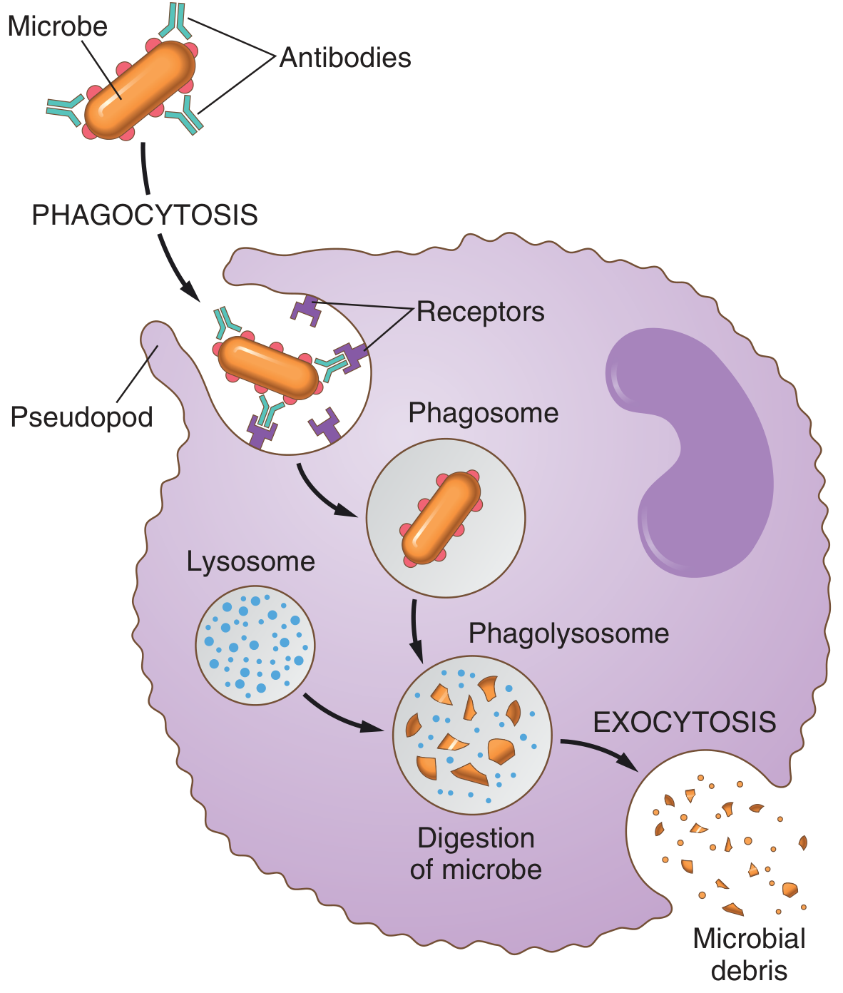
Fig. 34.3 from Guyton & Hall - showing antibody-coated bacteria being engulfed, phagosome formation, phagolysosome digestion, and exocytosis of microbial debris.
Selectivity - Why Phagocytes Don't Eat Normal Cells
Three factors govern selectivity (Guyton & Hall):
- Surface texture: Normal cells have smooth surfaces that resist phagocytosis; rough surfaces (like bacterial cell walls) promote it.
- Protective protein coats: Normal body cells have surface proteins that repel phagocytes; dead cells and foreign particles lack these.
- Opsonization: Antibodies and complement bound to pathogens flag them specifically for destruction.
Evasion of Phagocytosis by Pathogens
Some pathogens have evolved strategies to escape:
| Mechanism | Example |
|---|---|
| Resist lysosomal digestion (lipid coat) | Mycobacterium tuberculosis |
| Escape from phagosome into cytosol | Listeria monocytogenes, Shigella spp. |
| Inhibit phagosome-lysosome fusion | M. tuberculosis, Legionella |
| Anti-phagocytic capsules | Streptococcus pneumoniae, Klebsiella |
- Janeway's Immunobiology 10e
Clinical Relevance
- Chronic Granulomatous Disease (CGD): Defect in NADPH oxidase → impaired respiratory burst → recurrent infections with catalase-positive organisms.
- Chediak-Higashi Syndrome: Defective lysosomal trafficking → phagocytes cannot fuse lysosomes with phagosomes effectively.
- Myeloperoxidase deficiency: Mild susceptibility to Candida infections in immunosuppressed individuals.
- Tuberculosis: M. tuberculosis survives inside macrophage phagosomes by inhibiting phagolysosome fusion, which is the basis of its chronic, intracellular persistence.
Sources:
- Guyton and Hall Textbook of Medical Physiology, Ch. 34
- Robbins & Kumar Basic Pathology, Ch. 2
- Harper's Illustrated Biochemistry, Ch. 54
- Janeway's Immunobiology, 10e
This is a shared conversation. Sign in to Orris to start your own chat.