
Peri orbital edima is present but when lie down it disappeared
Reading File
Periorbital edema that disappears on lying down is a classic clinical finding pointing to dependent edema - fluid shifts with gravity. The most common cause is hypoalbuminemia (nephrotic syndrome, liver disease, malnutrition). Let me pull detailed textbook content on this.
Searching sources...
Finding Sources
Finding Sources
Searching PubMed
periorbital edema hypoalbuminemia nephrotic syndrome
Good start. Let me pull the detailed content on nephrotic syndrome edema and the Tintinalli finding about dependent periorbital edema.
Reading File
Reading File
Finding Sources
Finding Sources
Finding Sources
Excellent - found the exact relevant content in Comprehensive Clinical Nephrology. Let me read it.
Reading File
Reading File
I now have excellent textbook content. Let me fetch the nephrotic edema image which is directly relevant.
Loading Image
I now have everything I need for a comprehensive answer. Here it is:
Periorbital Edema That Disappears on Lying Down
This is a classic, textbook sign of nephrotic syndrome (or other hypoalbuminemic states). The explanation is purely gravitational.
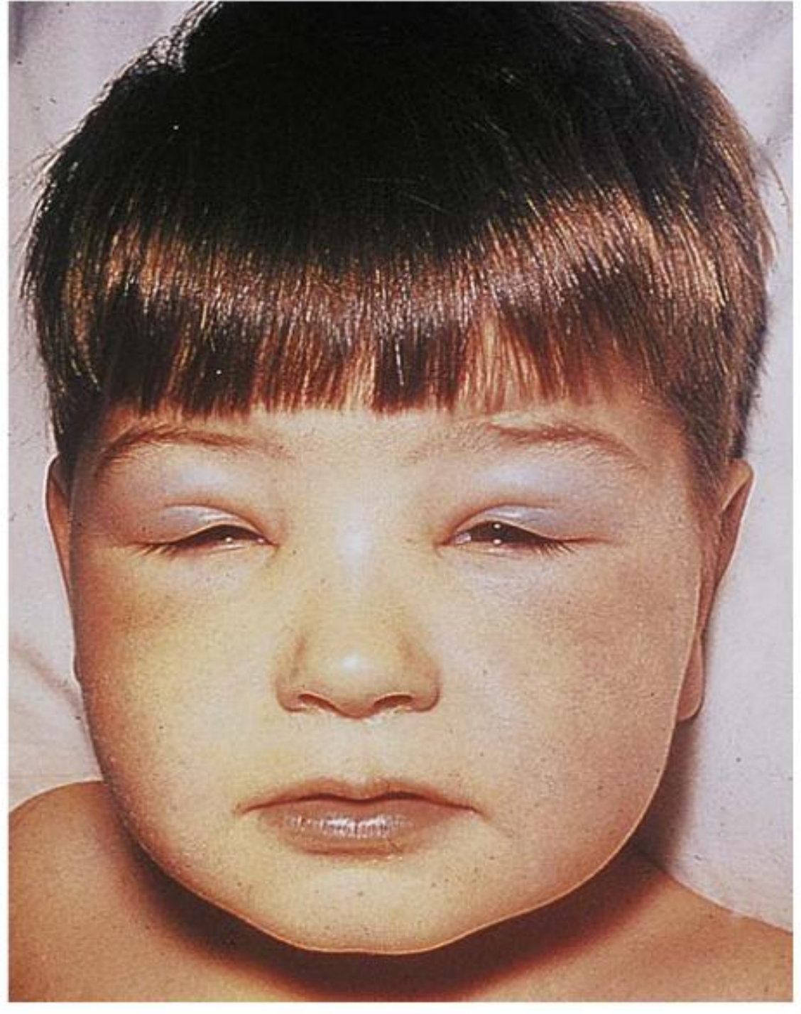
Why Does It Behave This Way?
The loose connective tissue around the orbit has very low tissue pressure, making it the path of least resistance for edema fluid. The behavior depends entirely on posture and gravity:
- When lying flat (sleeping) - fluid shifts from lower extremities up to the face/periorbital region. By morning, the eyelids are prominently swollen.
- When upright during the day - gravity redistributes the fluid downward (to ankles, legs, sacrum in bedridden patients). The periorbital edema resolves.
As Comprehensive Clinical Nephrology (7th Ed.) states:
"In the nephrotic patient, edema is often periorbital in the morning, whereas the face is NOT affected overnight in edema associated with heart failure (edema distributes by gravity, and patients with heart failure often cannot lie flat due to orthopnea)."
This is also confirmed by Harrison's (22nd Ed.):
"The edema [in nephrotic syndrome] is diffuse, symmetric, and most prominent in the dependent areas; periorbital edema is most prominent in the morning."
Key Differential Diagnosis
The behavior (disappears on lying down / worst in morning) points to low oncotic pressure edema, not high hydrostatic pressure edema:
| Feature | Nephrotic Syndrome | Heart Failure | Cirrhosis |
|---|---|---|---|
| Periorbital edema in morning | YES - classic | No (can't lie flat - orthopnea) | No (ascites prevents lying flat) |
| Disappears with upright posture | Yes | Usually persists | Usually persists |
| Proteinuria | Massive (≥3.5 g/day) | Mild if any | Mild |
| Hypoalbuminemia | Yes | Not primarily | Yes |
| JVP | Normal/low | Elevated | Normal/low |
| Orthopnea/PND | No | Yes | No |
Mechanism of Edema in Nephrotic Syndrome
- Massive proteinuria (≥3.5 g/day) → hypoalbuminemia (<3.0 g/dL)
- Reduced colloid oncotic pressure in plasma capillaries
- Starling forces favor fluid movement into interstitium
- Reduced effective circulating volume → activates RAAS → Na+ and water retention
- Retained fluid accumulates in dependent/low-resistance tissues - including the periorbital space
- Harrison's Principles of Internal Medicine, 22nd Ed.
- Comprehensive Clinical Nephrology, 7th Ed.
Important Contrast with Sinusitis-Related Edema
Tintinalli's Emergency Medicine notes one more scenario where periorbital edema disappears during the day:
"Sinusitis can be associated with reactive edema and mild inflammation of the eyelids noted upon awakening that regresses during the day as dependent edema resolves. Unilateral periorbital edema that does NOT regress may indicate [periorbital] cellulitis."
So if edema is unilateral and does not resolve - suspect periorbital cellulitis, not nephrotic syndrome.
Summary
| Symptom | Most Likely Diagnosis |
|---|---|
| Bilateral periorbital edema, worst on waking, disappears when upright | Nephrotic syndrome (or other hypoalbuminemic state) |
| Associated with massive proteinuria + hypoalbuminemia | Confirms nephrotic syndrome |
| Unilateral + does NOT disappear | Periorbital cellulitis |
| Non-pitting, does not shift with posture | Myxedema (hypothyroidism) |
Workup to confirm nephrotic syndrome: urine protein/creatinine ratio or 24-hr urine protein, serum albumin, lipid panel, renal function (creatinine, eGFR), and urinalysis for casts.
What are the causes of nephrotic syndrome?
Nephrotic syndrome causes are divided into primary (intrinsic glomerular disease) and secondary (systemic disease affecting the kidney). Common primary causes include minimal change disease (especially in children), focal segmental glomerulosclerosis (FSGS), membranous nephropathy, and membranoproliferative GN. Secondary causes include diabetes, lupus, amyloidosis, infections, and drugs.
Searching sources...
Finding Sources
Finding Sources
Reading File
Reading File
Reading File
Reading File
Reading File
Reading File
I have comprehensive data from multiple authoritative textbooks. Here is the full answer:
Causes of Nephrotic Syndrome
Nephrotic syndrome is caused by a derangement in the glomerular capillary wall that increases permeability to plasma proteins. Causes are divided into primary (intrinsic glomerular disease) and secondary (systemic diseases affecting the kidney). Primary disease accounts for ~95% in children and ~60% in adults. - Robbins & Cotran Pathologic Basis of Disease
PRIMARY Glomerular Diseases
These are intrinsic to the kidney, often idiopathic or immune-mediated:
| Disease | Children | Adults | Key Notes |
|---|---|---|---|
| Minimal Change Disease (MCD) | 75% | 8% | Most common cause in children; podocyte foot process effacement on EM; steroid-responsive (>80% remission) |
| Focal Segmental Glomerulosclerosis (FSGS) | 10% | 35% | Most common in Black patients (~50% of cases); risk of missing on biopsy due to focal distribution; highest thromboembolism risk |
| Membranous Nephropathy | 3% | 30% | Most common in white adults and elderly; caused by anti-PLA2R antibody in ~60% of primary cases; strong association with malignancy in >60 yr |
| Membranoproliferative GN (MPGN) | 10% | 10% | Mixed nephrotic-nephritic presentation; complement-mediated |
| IgA Nephropathy | 2% | 17% | Primarily nephritic, but can present with nephrotic syndrome |
- Robbins & Cotran Pathologic Basis of Disease; Frameworks for Internal Medicine
SECONDARY Causes
1. Metabolic / Systemic Disease
- Diabetes mellitus - Most common secondary cause overall in adults (Kimmelstiel-Wilson nodular glomerulosclerosis on biopsy). Suspect an alternate diagnosis if: no retinopathy, active urine sediment, rapid renal function decline, or short duration of diabetes.
- Amyloidosis - Diagnosed by Congo red staining + apple-green birefringence under polarized light. AL amyloidosis (plasma cell dyscrasia) and AA amyloidosis (chronic inflammation).
- Systemic Lupus Erythematosus (SLE) - Can cause both nephrotic AND nephritic syndrome. Membranous lupus nephropathy (Class V) in 10-15% of lupus nephritis cases.
- Preeclampsia - Nephrotic-range proteinuria in a pregnant woman; resolves after delivery in most cases, though fetal prognosis is poor.
2. Drugs
| Drug | Notes |
|---|---|
| NSAIDs | Very common; cause membranous nephropathy or MCD |
| Gold / Penicillamine | Classic in rheumatoid arthritis treatment |
| Captopril | ACE inhibitor associated |
| Lithium | Causes MCD or FSGS |
| Tamoxifen | Associated with membranous nephropathy |
| Heroin | Associated with FSGS |
- Frameworks for Internal Medicine; Robbins & Cotran
3. Infections
| Type | Examples |
|---|---|
| Bacterial | Infective endocarditis, syphilis |
| Viral | HIV, Hepatitis B (membranous), Hepatitis C |
| Protozoal | Malaria, toxoplasmosis |
| Helminthic | Schistosomiasis, filariasis |
4. Malignancies
The glomerular disease is often a paraneoplastic phenomenon:
- Hodgkin's lymphoma - associated with Minimal Change Disease
- Non-Hodgkin's lymphoma, Multiple myeloma
- Carcinomas (lung, breast, colon, prostate) - associated with Membranous Nephropathy
- Renal cell carcinoma
Malignancy is found in 5-25% of patients with membranous nephropathy, with the strongest association in patients over 60 years old. - Symptom to Diagnosis, 4th Ed.
5. Other
- Hereditary nephritis (Alport syndrome)
- Bee-sting / allergic reactions
- Other autoimmune diseases: thyroiditis, cryoglobulinemia
Age-Based Summary (Most Likely Cause)
| Age Group | Most Common Cause |
|---|---|
| Children (1-8 yr) | Minimal Change Disease (75%) |
| Adolescents/Young adults | FSGS, MCD |
| Adults (White) | Membranous Nephropathy |
| Adults (Black) | FSGS |
| Elderly (>60 yr) | Membranous Nephropathy, Diabetic nephropathy, Amyloidosis |
| Diabetic patients | Diabetic glomerulosclerosis |
| SLE patients | Lupus membranous nephropathy (Class V) |
Pathophysiology (Why All These Causes Lead to the Same Syndrome)
All causes converge on one final common pathway - injury to the glomerular filtration barrier (endothelium + GBM + podocytes), increasing permeability to plasma proteins:
"The glomerular capillary wall acts as a size and charge barrier. Increased permeability resulting from structural or physicochemical alterations allows proteins to escape into the urinary space, resulting in proteinuria → hypoalbuminemia → reduced oncotic pressure → edema." - Robbins & Cotran, Pathologic Basis of Disease
Treatment
Treatment of nephrotic syndrome involves general/supportive measures (edema, proteinuria, lipids, thrombosis, infection) plus disease-specific immunosuppression depending on the underlying cause. Corticosteroids are first-line for MCD; membranous and FSGS require different approaches.
Searching sources...
Finding Sources
Finding Sources
Reading File
Reading File
Finding Sources
Finding Sources
Reading File
Reading File
Excellent - I now have comprehensive treatment data from multiple nephrology textbooks. Here is the full answer:
Treatment of Nephrotic Syndrome
Treatment has two pillars: (1) General/supportive measures that apply to ALL patients, and (2) Disease-specific immunosuppression tailored to the underlying cause.
PART 1: General / Supportive Treatment (All Patients)
From Tietz Textbook of Laboratory Medicine (Box 49.4 - Management of Nephrotic Syndrome):
| Measure | Details |
|---|---|
| Diet | Low-sodium diet; protein intake 1.0 g/kg/day |
| Edema | Loop diuretics (furosemide 1-2 mg/kg). If profound hypoalbuminemia makes diuretics ineffective, give albumin infusion (0.5-1.0 g/kg) followed by furosemide (ICU monitoring needed) |
| Hypertension | RAAS blockade (ACE inhibitor or ARB) - also reduces proteinuria |
| Hyperlipidemia | Statins (HMG-CoA reductase inhibitors) |
| Thromboembolism | Anticoagulation in high-risk patients: serum albumin <20 g/L, or nephrotic syndrome due to membranous nephropathy or MPGN |
| Infection | Vigilance; pneumococcal (23-valent) and varicella vaccines (especially in children, given in remission, off steroids) |
| Hypovolemic shock | Isotonic IV fluid - even if edema is present |
| Patient support | Education and psychological support |
- Tietz Textbook of Laboratory Medicine, 7th Ed.; Tintinalli's Emergency Medicine
PART 2: Disease-Specific Immunosuppression
A. Minimal Change Disease (MCD)
- First-line: Oral corticosteroids (prednisone 2 mg/kg/day, max 60 mg/day OR 60 mg/m²/day)
- In children aged 2-12 with typical presentation, biopsy is NOT needed before starting steroids
- Remission typically within 2 weeks of steroids
- Taper after remission, total course minimum 12 weeks (longer duration reduces relapse)
- ~60% experience multiple relapses
- Steroid-sparing / refractory disease: cyclophosphamide, azathioprine, tacrolimus, cyclosporine, mycophenolate mofetil (MMF), rituximab
- Cyclophosphamide limited to 8-12 weeks (gonadal toxicity risk)
"Systemic glucocorticoids are used as first-line treatment for minimal change disease in adults, with remission achieved in >80% of cases." - Frameworks for Internal Medicine
B. Focal Segmental Glomerulosclerosis (FSGS)
| Setting | Treatment | Notes |
|---|---|---|
| Nephrotic primary FSGS | Prednisone (daily or alternate day) | First-line |
| Steroid-resistant FSGS | Calcineurin inhibitors (cyclosporine preferred; tacrolimus) | RCT evidence for cyclosporine |
| Refractory FSGS | MMF ± high-dose dexamethasone | |
| Subnephrotic proteinuria (any FSGS) | ACEi or ARB ± aldosterone antagonist + low-sodium diet | No immunosuppression needed |
- Immunosuppression is NOT used in secondary FSGS
- Relapse rate >50%; even partial remission slows progression
- National Kidney Foundation Primer on Kidney Diseases, 8th Ed.
C. Membranous Nephropathy (MN)
- Conservative management first for low/medium-risk patients (ACEi/ARB, statins, anticoagulation)
- High-risk patients (progressive GFR decline, severe/persistent proteinuria):
- Alternating corticosteroid + cyclophosphamide (Ponticelli regimen) - 6-month protocol, months alternating steroids and cyclophosphamide; RCT-proven superior to steroids alone
- Rituximab - emerging as preferred option in many centers; B-cell depletion reduces anti-PLA2R antibodies; MENTOR trial showed rituximab non-inferior to cyclosporine with lower relapse
- Calcineurin inhibitors (cyclosporine or tacrolimus) - effective but high relapse rate (~38-50%) on stopping
- MMF - conflicting results, high relapse
"The STARMEN trial indicates that alternating treatment with corticosteroids and cyclophosphamide is superior to sequential treatment with tacrolimus and rituximab in primary membranous nephropathy." - Comprehensive Clinical Nephrology, 7th Ed.
D. Secondary Causes - Treat the Underlying Disease
| Secondary Cause | Specific Treatment |
|---|---|
| Diabetic nephropathy | Tight glycemic control, ACEi/ARB, SGLT2 inhibitors (reduce proteinuria) |
| SLE (Lupus nephritis Class V) | Hydroxychloroquine; MMF or tacrolimus for active nephrotic syndrome |
| Amyloidosis | Treat underlying cause (chemotherapy for AL; treat infection/inflammation for AA) |
| Drug-induced | Stop the offending drug (NSAIDs, gold, penicillamine, lithium, captopril) |
| Infection-related | Treat the infection (antivirals for HBV/HCV/HIV; antimalarials) |
| Malignancy-related | Treat the malignancy; NS often resolves with cancer remission |
| Preeclampsia | Delivery; NS resolves postpartum in most cases |
Second-Line / Steroid-Sparing Agents (Summary)
| Drug | Mechanism | When Used |
|---|---|---|
| Cyclophosphamide | Alkylating agent | MCD relapse, MN (Ponticelli), FSGS-refractory |
| Cyclosporine | Calcineurin inhibitor | FSGS-steroid resistant, MN, MCD-relapse |
| Tacrolimus | Calcineurin inhibitor | MN, MCD, FSGS; high relapse on stopping |
| MMF | Antimetabolite | SLE, refractory MCD/MN/FSGS |
| Rituximab | Anti-CD20 (B-cell depletion) | MN (anti-PLA2R), FSGS-steroid resistant, MCD-refractory |
| Azathioprine | Antimetabolite | Steroid-sparing in MCD |
- Tintinalli's Emergency Medicine; Comprehensive Clinical Nephrology, 7th Ed.; National Kidney Foundation Primer, 8th Ed.
When to Admit
Admit patients with:
- Severe edema or pulmonary effusions/respiratory symptoms
- Signs of systemic infection or thrombotic complications (e.g., renal vein thrombosis, PE)
- Profound hypoalbuminemia requiring IV albumin + diuresis under monitoring
Children with mild/moderate edema can be managed outpatient with low-salt diet (<2 g/day) and close nephrology follow-up. - Tintinalli's Emergency Medicine
Some time it is not present in morning
This is a very clinically important observation. Periorbital edema that is sometimes absent in the morning tells us the fluid is highly mobile and redistributes freely with posture - this actually refines the diagnosis further.
Searching sources...
Finding Sources
Finding Sources
Finding Sources
Finding Sources
Finding Sources
Finding Sources
Finding Sources
Finding Sources
The textbooks don't cover this specific variation directly, but the answer is well-explained by established physiology from content we've already retrieved. Here is the explanation:
Why Periorbital Edema is Sometimes Absent in the Morning
This is actually not contradictory to nephrotic syndrome - it depends on three factors:
1. Sleeping Position
The periorbital edema in nephrotic syndrome forms because fluid shifts to the face when lying flat overnight. But this only happens when the person sleeps in the supine (flat) position.
- Sleeping on their side or elevated on pillows - fluid distributes to the side of the face or stays in the lower trunk. Periorbital edema may be minimal or absent in the morning.
- Sleeping semi-upright (e.g., with extra pillows) - gravity keeps fluid in the legs/ankles. The face is spared.
The textbook states it clearly: "The edema resolves during the day under the influence of gravity" and "edema distributes by gravity" - this works in both directions, upward when flat, downward when upright. - Comprehensive Clinical Nephrology, 7th Ed.
2. Severity / Stage of Disease
Periorbital edema is most prominent when albumin is very low (severe hypoalbuminemia). If the disease is:
- Mild/early stage - total body fluid excess is small; the periorbital tissues may not fill noticeably every morning
- In partial remission (e.g., responding to steroids) - as albumin rises, the overall fluid load decreases and periorbital puffiness becomes inconsistent
- Fluctuating proteinuria - some days protein leak is less, oncotic pressure recovers slightly, and edema is less visible
3. Fluid Intake and Diuretic Use
- If the patient is on diuretics (furosemide), total body fluid is reduced - periorbital edema may be intermittent
- Low fluid intake the previous evening can reduce overnight facial accumulation
Clinical Significance - What This Pattern Tells You
| Pattern | Interpretation |
|---|---|
| Periorbital edema always present in morning, clears by day | Classic nephrotic - moderate to severe disease |
| Periorbital edema sometimes present in morning | Mild/early nephrotic, partial remission, or positional variation |
| Edema never in face, only ankles/legs | Suggests heart failure (can't lie flat due to orthopnea) or moderate nephrotic with upright sleeping |
| Edema fixed, not shifting with posture | Suggests lymphedema or myxedema (non-pitting, not gravity-dependent) |
Key Clinical Pearl
Even when periorbital edema is absent in the morning, the urine will still show heavy proteinuria - this is the constant finding that does not shift with gravity.
So if you suspect nephrotic syndrome despite intermittent facial edema, check first morning urine for protein (dipstick or spot protein:creatinine ratio). That finding will be present regardless of whether the face is puffy or not. - Harrison's Principles of Internal Medicine, 22nd Ed.
The diagnosis does not depend on seeing periorbital edema - it depends on finding proteinuria ≥3.5 g/day (or urine PCR >300 mg/mmol) with hypoalbuminemia.
analyse_media
Thank you for sharing the photo. Here is the clinical visual assessment:
Visual Assessment of the Photo
Periorbital Region
- Mild bilateral infraorbital fullness (under-eye puffiness) is present, but there is no severe or acute periorbital edema
- The upper eyelids are not swollen and the palpebral fissures are normal and symmetric
- There is bilateral dark infraorbital pigmentation (dark circles under the eyes) which can be due to thin skin, venous congestion, or fatigue - but can also be seen with chronic fluid redistribution in the periorbital tissue
- The skin does not appear tense or stretched
Face Overall
- No moon face (no Cushingoid features)
- No generalized facial swelling or myxedema
- Normal facial contours with a defined jawline
Clinical Interpretation in Context
Given everything discussed - periorbital edema present some mornings but not others, disappears on lying down - this photo likely represents:
| Finding | Significance |
|---|---|
| Mild bilateral infraorbital puffiness | Consistent with early or mild nephrotic syndrome with fluctuating edema |
| Dark circles under eyes | May indicate chronic low-grade venous/fluid congestion in the periorbital tissue |
| No severe swelling | Suggests mild disease, partial remission, or early stage - not severe hypoalbuminemia |
Important Caveat
A photograph cannot replace a clinical examination. The most important next steps are:
- Urine dipstick or spot protein:creatinine ratio - check for proteinuria (this will be positive regardless of whether the face looks puffy)
- Serum albumin - if low (<3.0 g/dL), confirms hypoalbuminemia
- Check ankles/feet - when upright all day, is there pitting ankle edema by evening?
- Serum creatinine and lipid panel
Even if this morning the face looks almost normal, the urine protein will tell the truth. Please get this tested and consult a nephrologist.
No pedal edema
This is a very important finding that significantly narrows the differential diagnosis. Let me think through this carefully.
Reasoning...
Finding Sources
Finding Sources
Finding Sources
Finding Sources
Finding Sources
Finding Sources
Reading File
Finding Sources
Excellent information. Now I have enough to give a complete, clinically reasoned answer.
No Pedal Edema - This Changes Everything
The absence of pedal edema is a critical finding. In nephrotic syndrome, fluid distributes by gravity - when upright all day, you would expect ankle/foot pitting edema to be the dominant finding. Its absence makes nephrotic syndrome much less likely as the primary diagnosis.
Revised Differential Diagnosis
With periorbital edema only (intermittent, mornings) and no pedal edema, the differential shifts significantly:
1. Angioedema (Most Likely)
- Localized subcutaneous swelling from fluid leaking out of venules due to increased vascular permeability
- Classically affects the face, lips, eyelids - NOT dependent areas like legs
- Self-limited - onset in minutes to hours, resolves spontaneously within hours to days - this explains why it is sometimes absent in the morning
- Causes:
- Allergic - food, pollen, insect bite, medication
- ACE inhibitor-induced - bradykinin-mediated (very common)
- Hereditary Angioedema (HAE) - C1 inhibitor deficiency, autosomal dominant; onset often at puberty
- Acquired - lymphoproliferative disease, autoimmune disease
- No pitting, no gravity-dependence, no pedal edema
- Swanson's Family Medicine Review
2. Allergic Reaction / Atopic Disease
- Contact allergy (face wash, cosmetics, eye drops)
- Seasonal allergies - bilateral periorbital puffiness, often with itching, watering eyes
- Intermittent - explains why not present every morning
3. Hypothyroidism (Myxedema)
- Causes non-pitting periorbital edema and facial puffiness due to glycosaminoglycan deposition
- Does not shift with gravity - so pedal edema is not the dominant feature early on
- Usually accompanied by fatigue, cold intolerance, constipation, dry skin, coarse hair, loss of lateral eyebrows
- Important to check TSH
4. Early / Very Mild Nephrotic Syndrome
- Still possible but now less likely given no pedal edema
- If present, disease is very early and total fluid excess is minimal
- Requires urine protein check to include or exclude
5. Sleep-Related / Positional
- Sleeping face-down causes fluid to pool in periorbital tissue overnight
- Completely benign - resolves quickly on standing
6. Chronic Venous Congestion Around the Eyes
- Thin skin + prominent subcutaneous venous plexus
- Worse with poor sleep, high salt intake, alcohol
Key Distinguishing Features
| Feature | Nephrotic | Angioedema | Hypothyroid | Allergy |
|---|---|---|---|---|
| Pedal edema | Yes (when upright) | No | No (early) | No |
| Periorbital only | Early/mild | Yes - classic | Yes | Yes |
| Disappears lying down | Yes | No (not gravity-dependent) | No | No |
| Disappears in morning | Goes to legs | Resolves spontaneously | Persistent | Intermittent |
| Pitting | Yes | No | No (non-pitting) | No |
| Proteinuria | Heavy | None | None | None |
| Itching/urticaria | No | Sometimes | No | Yes |
Wait - An Important Reconsideration
You said the edema:
- Present some mornings ✓
- Disappears on lying down ✓
- No pedal edema ✓
If it disappears when lying down (not just in the daytime), that is unusual for angioedema (which does not redistribute with gravity). This could still fit mild early nephrotic where total fluid load is small - the little fluid present shifts to whichever area is most dependent.
What to Do Next
| Test | Looking For |
|---|---|
| Urine dipstick + spot PCR | Proteinuria (nephrotic) |
| Serum albumin | Hypoalbuminemia |
| TSH | Hypothyroidism |
| Serum C3, C4, C1q | Low C4 suggests angioedema/HAE |
| CBC, renal function, LFTs | General screen |
| Allergy history | Foods, drugs, ACE inhibitors? |
| Full blood count | Eosinophilia (allergy) |
Please see a physician for a proper clinical examination - particularly to assess whether the periorbital swelling is pitting or non-pitting, and whether there is any itching, urticaria, or lip swelling associated with episodes.
When getting up during mid night it is more
This is a highly significant clinical clue. Let me reason through this carefully.
Finding Sources
Finding Sources
This finding is explainable purely from established physiology already in context. Here is the full clinical reasoning:
"Worse at Midnight When Getting Up" - The Clinical Logic
This single observation strongly points back to nephrotic syndrome (or another hypoalbuminemic state) and here is exactly why:
The Gravity-Time Sequence Explained
Think of the fluid as water that constantly moves to wherever gravity pulls it:
| Time | Body Position | Where is the Fluid? | What You See |
|---|---|---|---|
| Daytime (upright) | Standing/sitting | Fluid pools in legs/ankles | No periorbital edema - BUT also no pedal edema (if total fluid is small) |
| Early night (lying flat) | Supine | Fluid slowly redistributes from legs → face | Edema begins building around eyes |
| Midnight (2-4 hrs lying) | Still supine | Fluid has now fully accumulated in periorbital tissue | Maximum periorbital edema - most prominent |
| Morning (6-8 hrs lying) | Still supine | Fluid has been lying there long enough... may have partly reabsorbed OR the person may have turned | Edema may be less than midnight, or variable |
| After getting up | Upright again | Fluid drains back down by gravity | Edema disappears |
Why Midnight > Morning?
When you lie flat, gravity-dependent fluid redistribution to the face happens progressively over hours. The edema:
- Peaks after ~3-5 hours of lying flat (around midnight if sleeping since 9-10 PM)
- By morning (after 7-8 hours), some fluid may have been partially reabsorbed by lymphatics, or the person may have shifted position/rolled sideways
This is why midnight > morning - it is at the peak of supine redistribution before any reabsorption occurs.
This pattern is described in Comprehensive Clinical Nephrology (7th Ed.):
"Periorbital edema in the early morning in a nephrotic child. The edema resolves during the day under the influence of gravity."
The midnight peak is simply the earlier, maximum point of that same overnight gravity-driven redistribution.
Why No Pedal Edema?
This now makes more sense too. If total body excess fluid is mild/small (early disease or partial remission):
- When upright during the day, the small amount of excess fluid distributes across the entire lower extremity - spread thin across both legs and feet - not enough to be clinically visible as pedal edema
- But when lying flat overnight, that same small amount of fluid concentrates entirely into the loose periorbital tissue - which has the lowest tissue resistance in the body - making it visible there even when it was invisible in the legs
This explains the apparent paradox perfectly.
Clinical Conclusion - This Pattern is Almost Pathognomonic
The complete picture now:
| Feature | This Patient | Classic Nephrotic |
|---|---|---|
| Periorbital edema | Yes, bilateral | Yes |
| Disappears on standing | Yes | Yes |
| Worst at midnight (after hours supine) | Yes | Yes - peak redistribution |
| Sometimes absent in morning | Yes (reabsorption/position) | Can occur |
| No pedal edema | Yes | Mild/early disease |
| Gravity-dependent shifting | Yes | Yes - defining feature |
This is NOT angioedema (angioedema does not redistribute with gravity or posture - it would be the same at midnight and morning).
This is NOT myxedema (non-pitting, non-shifting, would be persistent).
This is gravity-dependent pitting edema - and the most likely cause, especially in a young person, is:
- Nephrotic syndrome (most likely) - especially Minimal Change Disease in young adults
- Hypoalbuminemia from any cause (malnutrition, liver disease - less likely given age/appearance)
Urgent Next Steps
Please get these tests as soon as possible:
| Test | Why |
|---|---|
| First morning urine - dipstick protein | Heavy proteinuria = nephrotic |
| Spot urine protein:creatinine ratio | >300 mg/mmol = nephrotic range |
| Serum albumin | Will be low if nephrotic |
| Serum creatinine + eGFR | Kidney function |
| Lipid panel (cholesterol, triglycerides) | Hyperlipidemia = part of nephrotic syndrome |
| TSH | Rule out hypothyroidism |
| Blood pressure | Often elevated in nephrotic |
The midnight-worse, morning-variable, disappears-upright pattern with no other cause found strongly warrants a nephrology referral and kidney biopsy if proteinuria is confirmed.
Alcohol consumption present
Excellent addition to the history. Alcohol consumption is a very important etiological clue that shifts the differential significantly.
Searching sources...
Finding Sources
Finding Sources
Finding Sources
Finding Sources
Finding Sources
Finding Sources
Finding Sources
Finding Sources
Now I have enough to synthesize a comprehensive, clinically focused answer. Here it is:
Alcohol Consumption - How It Explains Everything
Alcohol is actually the single most elegant explanation for this entire clinical picture. It works through multiple simultaneous mechanisms, all of which converge to produce exactly what this patient has:
Mechanism 1 - Direct Vascular Effect (Explains the Pattern Perfectly)
Alcohol causes direct vasodilation and increased capillary permeability via:
- Acetaldehyde (alcohol metabolite) triggers histamine release
- Histamine causes postcapillary venule leakage - fluid leaks into interstitium
- This is essentially a mild, transient angioedema-like response triggered by alcohol
Timeline fits exactly:
| Time | Event |
|---|---|
| Evening | Alcohol consumed |
| Night (lying flat) | Vasodilation + fluid leaking out, gravity pools it to the face |
| Midnight | Peak effect - most fluid has leaked + gravity has redistributed it to periorbital tissue |
| Morning | Alcohol metabolized, vasodilation waning, fluid being reabsorbed - edema less |
| Days without drinking | No periorbital edema - explains why "sometimes not present in morning" |
This explains all three observations:
- Worst at midnight ✓
- Sometimes absent in morning ✓
- Disappears on lying down... wait - re-read: disappears when getting up ✓ (gravity pulls it away from face)
Mechanism 2 - High Sodium in Alcohol + Mixers
Alcoholic drinks (beer especially) contain significant sodium. Alcohol also:
- Suppresses ADH initially → fluid loss → rebound water retention later
- Stimulates aldosterone → sodium and water retention
- Net effect: fluid retention overnight, preferentially in loose dependent tissues
Mechanism 3 - Chronic Liver Disease (If Long-term)
If alcohol consumption is chronic and heavy, the liver gets damaged:
| Stage | Effect on Edema |
|---|---|
| Fatty liver | Minimal edema, may have mild hypoalbuminemia |
| Alcoholic hepatitis | Decreased albumin synthesis → low oncotic pressure → periorbital + ascites |
| Cirrhosis | Portal hypertension + hypoalbuminemia → ascites first, then pedal edema |
- Cirrhosis classically causes ascites BEFORE pedal edema (unlike cardiac failure) - so absence of pedal edema does NOT rule out early alcoholic liver disease
- However, cirrhosis would eventually produce ascites and pedal edema
Revised Complete Picture
Putting everything together now:
| Feature | Fits Alcohol-Related? |
|---|---|
| Young male | Yes - common demographic |
| Periorbital edema only | Yes - loose tissue, direct vascular effect |
| Worst at midnight | Yes - peak of alcohol's vascular effect + gravity |
| Sometimes absent in morning | Yes - only on nights of drinking |
| Disappears on getting up | Yes - gravity-dependent |
| No pedal edema | Yes - mild/early, not yet cirrhotic |
| Intermittent pattern | Yes - correlates with drinking episodes |
Most Likely Diagnosis Now
In order of probability:
- Alcohol-induced periorbital edema (direct vascular/histamine effect) - most likely, benign but a warning sign
- Early alcoholic liver disease with mild hypoalbuminemia - check LFTs and albumin
- Nephrotic syndrome - still possible (ask: is there foamy urine?)
- Alcohol + high salt intake causing fluid retention
One Critical Question to Ask
"Does the periorbital edema appear on mornings after drinking the night before, and is it absent on mornings after not drinking?"
If the answer is yes - alcohol is almost certainly the cause.
Investigations Now Prioritized
| Test | Why |
|---|---|
| LFTs (ALT, AST, GGT, ALP) | GGT especially elevated in alcohol use; liver damage |
| Serum albumin | Low = liver not making enough protein |
| AST:ALT ratio | >2:1 strongly suggests alcoholic liver disease |
| Urine protein (dipstick) | Rule out nephrotic syndrome |
| Full blood count | Macrocytosis (MCV elevated) = chronic alcohol use |
| Serum bilirubin | Liver function |
| Abdominal ultrasound | Fatty liver, early cirrhosis, ascites |
| TSH | Rule out hypothyroidism |
Important Advice
Regardless of the exact cause, alcohol is directly contributing to this edema and causing liver stress. The edema is the body's early warning signal. Reducing or stopping alcohol is both diagnostic (if edema resolves, alcohol was the cause) and therapeutic.
No
Could you clarify what "No" refers to? I want to make sure I understand correctly.
Asking for Preferences
What does 'No' refer to?
Important - so alcohol does not trigger or worsen the edema. This rules out alcohol as a direct cause. Let me now ask one more key question to narrow this down further.
Asking for Preferences
Is there foamy or frothy urine (especially in the morning)? · How long has this periorbital edema been happening?
Finding Sources
Finding Sources
Finding Sources
Finding Sources
Reading File
Now let me synthesize the complete clinical picture with all the information gathered.
Updated Clinical Summary - All Clues Together
Let me now consolidate everything we know:
| Feature | Present / Absent |
|---|---|
| Periorbital edema | ✅ Present |
| Bilateral | ✅ Yes |
| Worst at midnight (after hours supine) | ✅ Yes |
| Disappears on getting up (gravity-dependent) | ✅ Yes |
| Sometimes absent in morning | ✅ Yes |
| No pedal edema | ✅ Absent |
| No foamy urine | ✅ Absent |
| Alcohol consumption present | ✅ Yes |
| Edema does NOT correlate with drinking | ✅ Confirmed |
| Duration: few months | ✅ Chronic |
No Foamy Urine + No Pedal Edema = Nephrotic Syndrome Now Unlikely
Foamy urine is caused by heavy proteinuria - protein in urine acts like soap and creates persistent froth. Its absence over several months makes significant proteinuria unlikely, and nephrotic syndrome moves down the differential considerably.
Revised Top Diagnoses
1. Hypothyroidism (NOW Top Suspect)
This fits remarkably well:
- Periorbital puffiness - one of the most classic early signs; caused by glycosaminoglycan (GAG) deposition in the dermis, not fluid per se
- Non-pitting - but in early/mild disease, can have some positional variation
- No pedal edema early on - myxedema affects the face and periorbital area first
- Chronic, months-long course - hypothyroidism is insidious and slowly progressive
- No foamy urine - kidney is not involved
- Young male - can occur at any age
- Alcohol use - does not explain edema but does not exclude hypothyroidism
Other symptoms to ask about: fatigue, weight gain, cold intolerance, constipation, dry skin, coarse hair, loss of outer third of eyebrows, slow speech, bradycardia, constipation, low mood.
Harrison's (22nd Ed.) lists the signs and symptoms of hypothyroidism in descending frequency - periorbital edema/puffiness and dry coarse skin are among the most prominent.
2. Idiopathic / Positional Periorbital Edema
- Very common, especially in young adults with:
- High salt diet (sodium retention → fluid retention → pools in face overnight)
- Sleep position (sleeping face-down or flat causes gravity-dependent facial fluid)
- Alcohol (even if not the trigger every time, can contribute to sodium/water retention on some nights)
- Allergies - subclinical/mild, not always obvious
- Explains: intermittent, gravity-dependent, no systemic disease markers
- Duration of months fits a dietary/lifestyle pattern
3. Subclinical / Mild Nephrotic Syndrome
- Still cannot be ruled out without a urine test
- Foamy urine can be absent or not noticed if proteinuria is mild
- A spot urine protein:creatinine ratio is needed to definitively exclude this
4. Early Alcoholic Liver Disease
- Alcohol consumption present
- Liver produces albumin - early damage = mildly reduced albumin
- Mild hypoalbuminemia → periorbital edema (face first, before legs)
- No foamy urine, no pedal edema - fits early disease
- GGT is the most sensitive liver enzyme for alcohol damage
Most Important Next Step
A simple TSH blood test will confirm or rule out hypothyroidism - the now #1 suspect.
| Priority | Test | Rules Out / Confirms |
|---|---|---|
| 1st | TSH | Hypothyroidism |
| 2nd | Urine dipstick + spot PCR | Nephrotic syndrome |
| 3rd | Serum albumin | Hypoalbuminemia |
| 4th | LFTs + GGT | Liver disease (alcohol) |
| 5th | FBC (MCV) | Alcohol effect (macrocytosis) |
One More Question
Do you have any of these symptoms?
- Feeling tired/sluggish more than usual?
- Weight gain recently?
- Feeling cold when others are not?
- Dry skin or hair becoming coarser?
- Constipation?
- Feeling low in mood or slowed down mentally?
If yes to 2 or more - hypothyroidism is very likely the answer, and a simple TSH test will confirm it.
1
Only 1 symptom from the hypothyroid list - which one is present?
Asking for Preferences
Which symptom is present?
Finding Sources
Finding Sources
Finding Sources
Reading File
This is very helpful. Here is the focused clinical synthesis:
Clinical Picture Now
Fatigue/tiredness is the #1 most common symptom of hypothyroidism - listed first in Harrison's table of symptoms in descending order of frequency:
Symptoms: Tiredness, weakness | Signs: Dry coarse skin; cool peripheral extremities... Puffy face, hands, and feet (myxedema) - Harrison's Principles of Internal Medicine, 22nd Ed.
Putting the Whole Case Together
| Clue | Points To |
|---|---|
| Periorbital puffiness, bilateral | Hypothyroidism (myxedema) ✅ |
| Worse at midnight (after hours lying) | Myxedema + gravity pooling ✅ |
| Sometimes absent in morning | Mild/subclinical - not severe enough to be constant ✅ |
| Disappears on getting up | GAG-related puffiness can have some positional component in mild disease ✅ |
| No pedal edema | Early/subclinical hypothyroidism - legs not yet involved ✅ |
| No foamy urine | Kidney not involved - not nephrotic ✅ |
| Fatigue (the #1 symptom of hypothyroidism) | Hypothyroidism ✅ |
| Duration: few months | Insidious onset typical of Hashimoto's ✅ |
| Young male | Hashimoto's can occur in males, though less common |
| Alcohol use | Unrelated to edema - independent finding |
Most Likely Diagnosis
Subclinical or Early Overt Hypothyroidism (Hashimoto's Thyroiditis)
Why subclinical? Because:
- Only 1 symptom (fatigue)
- Edema is mild and intermittent, not severe
- No cold intolerance, no weight gain, no constipation yet
- Subclinical hypothyroidism = TSH elevated, T4 still normal - a very early stage where the body is compensating
Harrison's states: "There is a phase of compensation when normal thyroid hormone levels are maintained by a rise in TSH... some patients may have minor symptoms - this is subclinical hypothyroidism."
As it progresses untreated → TSH rises further → T4 falls → more symptoms appear
One Single Test Will Confirm This
TSH (Thyroid Stimulating Hormone)
| TSH Result | Interpretation |
|---|---|
| Normal (0.4 - 4.0 mIU/L) | Hypothyroidism ruled out |
| Mildly elevated (4 - 10 mIU/L) | Subclinical hypothyroidism - fits this picture |
| >10 mIU/L | Overt hypothyroidism |
If TSH is elevated, add:
- Free T4 - to stage severity
- Anti-TPO antibodies - confirms Hashimoto's (autoimmune cause)
Action Plan
- Get a TSH test - this is urgent, simple, and cheap
- If TSH elevated → see an endocrinologist
- Treatment: Levothyroxine (T4 replacement) - once daily tablet
- With treatment, both the fatigue and periorbital puffiness should resolve within weeks
- Stop or reduce alcohol - even if not the direct cause, alcohol stresses the liver which is involved in thyroid hormone metabolism
Bottom Line
A young male with months of fatigue + intermittent bilateral periorbital puffiness worse at midnight + no foamy urine + no pedal edema = Subclinical/early hypothyroidism until proven otherwise.
Get a TSH. Do not delay.
This is a shared conversation. Sign in to Orris to start your own chat.