
Acute Abdomen in Gynecology
Reading File
Reading File
Reading File
“Acute abdomen in gynecology” covers a broad emergency topic. Main areas are:
- Initial assessment and resuscitation
- Pregnancy-related emergencies, especially ectopic pregnancy
- Adnexal causes, such as ovarian torsion and ruptured/hemorrhagic cyst
- Pelvic infection, including PID and tubo-ovarian abscess
- Uterine causes, including fibroid degeneration/torsion
- Non-gynecologic mimics and differential diagnosis
- Investigations, ultrasound findings, and emergency management
The immediate priority is to recognize hemorrhage, sepsis, peritonitis, or torsion and arrange urgent gynecologic/surgical review. In anyone of reproductive potential, pregnancy testing is an early essential step, because ectopic pregnancy must be excluded.
Asking for Preferences
Which section should I cover in detail?
“Acute abdomen in gynecology” covers a broad emergency topic. Main areas are:
- Initial assessment and resuscitation
- Pregnancy-related emergencies, especially ectopic pregnancy
- Adnexal causes, such as ovarian torsion and ruptured/hemorrhagic cyst
- Pelvic infection, including PID and tubo-ovarian abscess
- Uterine causes, including fibroid degeneration/torsion
- Non-gynecologic mimics and differential diagnosis
- Investigations, ultrasound findings, and emergency management
The immediate priority is to recognize hemorrhage, sepsis, peritonitis, or torsion and arrange urgent gynecologic/surgical review. In anyone of reproductive potential, pregnancy testing is an early essential step, because ectopic pregnancy must be excluded.
Explain everything in detail based on gynaecology and obstetrics
Acute Abdomen in Gynecology and Obstetrics: Scope
“Acute abdomen” means sudden abdominal or pelvic pain that may require urgent diagnosis and treatment. In gynecology and obstetrics, the priority is identifying conditions that threaten life, fertility, or pregnancy.
Main topics
-
General emergency approach
- Triage, resuscitation, focused history and examination
- Recognition of shock, internal hemorrhage, peritonitis, and sepsis
- Role of pregnancy testing, blood tests, pelvic ultrasound, and laparoscopy
-
Obstetric causes
- Ectopic pregnancy, including ruptured ectopic pregnancy
- Miscarriage and septic miscarriage
- Acute complications of early pregnancy
- Placental abruption and uterine rupture in later pregnancy
- Acute fatty liver, HELLP syndrome, and other non-pelvic obstetric causes when relevant
-
Adnexal and ovarian causes
- Adnexal torsion
- Ruptured ovarian cyst
- Hemorrhagic corpus luteum
- Endometrioma rupture
- Ovarian hyperstimulation syndrome
-
Pelvic infection
- Acute pelvic inflammatory disease
- Pyosalpinx and tubo-ovarian abscess
- Pelvic peritonitis
- Septic abortion
-
Uterine and cervical causes
- Degeneration of leiomyoma, especially red degeneration in pregnancy
- Torsion of a pedunculated subserosal fibroid
- Fibroid-related bleeding or pressure complications
-
Non-gynecologic conditions that mimic gynecologic emergencies
- Acute appendicitis
- Urinary calculi and pyelonephritis
- Intestinal obstruction, perforation, gastroenteritis
- Cholecystitis and pancreatitis
- Surgical emergencies should never be excluded merely because pelvic pathology is suspected.
Core rule
For any patient of reproductive potential with acute lower abdominal pain, perform a pregnancy test early and consider ectopic pregnancy until it has been reasonably excluded. Hemodynamic instability, shoulder-tip pain, syncope, guarding, rigidity, falling hemoglobin, or free intraperitoneal fluid indicate possible major internal bleeding and demand immediate senior gynecologic/surgical involvement.
I can provide a detailed, exam-oriented note on one section at a time. Which should come first?
- General approach, differential diagnosis, investigations, and management algorithm
- Ectopic pregnancy and other early-pregnancy emergencies
- Ovarian torsion, cyst accidents, and fibroid emergencies
- PID, tubo-ovarian abscess, and septic abortion
- A sequential full set of notes, beginning with the general approach
Rupture and torsion in ovaries
Reading File
Finding Sources
Finding Sources
Finding Sources
Searching PubMed
"Adnexal Torsion" AND management
Searching the Web
ACOG adnexal torsion ovarian cyst rupture guidance
Searching Images
ultrasound image ovarian torsion enlarged ovary peripheral follicles whirlpool sign
Reading File
Reading File
Finding Sources
Finding Sources
Finding Sources
query_images_db
Reading File
Reading File
Reading File
Reading File
Reading Web Page
https://www.acog.org/clinical/clinical-guidance/committee-op…
Ovarian Cyst Rupture and Adnexal (Ovarian) Torsion
Both can cause sudden acute lower abdominal pain and are important gynecologic emergencies. The first task is to assess hemodynamic stability and exclude ruptured ectopic pregnancy in every patient who could be pregnant.
| Feature | Ovarian cyst rupture | Adnexal torsion |
|---|---|---|
| Main event | Cyst wall leaks or ruptures, sometimes bleeding into peritoneum | Ovary, often with tube, twists on its vascular pedicle |
| Main hazard | Hemoperitoneum and hemorrhagic shock | Ischemia, infarction, loss of ovarian function |
| Typical pain | Sudden, often after ovulation, intercourse, or exertion | Sudden severe unilateral pain, may be intermittent |
| Nausea/vomiting | May occur | Very common and strongly suggestive |
| Ultrasound | Cyst, complex/hemorrhagic cyst, pelvic free fluid | Enlarged ovary, edema, peripheral follicles, mass, free fluid, possible twisted pedicle |
| Definitive management | Observation if stable; surgery for significant bleeding or acute abdomen | Urgent laparoscopy and detorsion if suspected |
1. Rupture of an ovarian cyst
Definition and pathology
An ovarian cyst rupture means leakage or tearing of a functional or pathological cyst into the peritoneal cavity. The commonest functional cysts are:
- Follicular cyst
- Corpus luteum cyst
- Hemorrhagic cyst
A normal follicle may also rupture at ovulation, producing mittelschmerz, a brief midcycle pain caused partly by a small amount of peritoneal blood and prostaglandin-rich follicular fluid. This is generally self-limiting.
A corpus luteum cyst is the most frequent cyst to rupture with clinically important bleeding and hemoperitoneum. It commonly occurs in the luteal phase, and rupture is more frequent on the right side. It may follow intercourse. Anticoagulant use or a bleeding disorder increases the risk of substantial hemorrhage. Berek & Novak’s Gynecology, pp. 452, 559-560.
Other cysts that may rupture
- Endometrioma: rupture may release old blood into the peritoneum, causing marked pain and chemical peritonitis.
- Dermoid cyst: rupture can produce chemical peritonitis due to leakage of sebaceous material.
- Tubo-ovarian abscess: rupture causes pelvic/generalized peritonitis and sepsis.
- Ovarian neoplasm: may occasionally rupture and bleed.
Clinical features
The presentation depends mainly on the amount and rate of bleeding.
Uncomplicated rupture
- Sudden unilateral lower abdominal or pelvic pain
- Often around midcycle or in the luteal phase
- Pain after exercise, lifting, or sexual intercourse may be reported
- Mild abdominal tenderness
- Stable pulse and blood pressure
- Little or no intraperitoneal blood
Hemorrhagic rupture with hemoperitoneum
- Sudden severe pain, sometimes diffuse abdominal pain
- Dizziness, weakness, presyncope, or syncope
- Shoulder-tip pain from diaphragmatic irritation
- Tachycardia, orthostatic hypotension, pallor
- Abdominal distension, guarding, rebound tenderness
- Falling hemoglobin or hematocrit
- Peritoneal free fluid on ultrasound
The major diagnostic concern is that a ruptured corpus luteum can closely resemble a ruptured ectopic pregnancy. Berek & Novak’s Gynecology, p. 560.
Investigations
-
Immediate pregnancy test
- Urine or serum beta-hCG.
- A positive result requires urgent assessment for ectopic pregnancy.
-
Transvaginal pelvic ultrasound
- Identifies ovarian cyst morphology.
- May show a hemorrhagic cyst.
- Detects and estimates pelvic free fluid or hemoperitoneum.
- Assesses the uterus and adnexa for ectopic pregnancy.
-
Blood tests
- Complete blood count, especially hemoglobin.
- Blood group, Rh status, and cross-match if bleeding is suspected.
- Coagulation studies if anticoagulant use, bleeding disorder, or major hemorrhage is suspected.
-
Serial observations
- Pulse, blood pressure, postural vital signs, abdominal examination, and serial hemoglobin where indicated.
Management
Stable patient, minimal bleeding
Conservative treatment is appropriate when the patient has:
- Stable vital signs
- No significant anemia
- No evidence of increasing hemoperitoneum
- Mild or improving pain
- Reliable observation and follow-up
Management includes analgesia, observation, repeat clinical assessment, and sometimes repeat ultrasound or hemoglobin testing. Uncomplicated functional cyst rupture often resolves without surgery. Berek & Novak’s Gynecology, p. 559.
Indications for surgery
Urgent laparoscopy, or laparotomy if unstable, is indicated with:
- Hemodynamic instability or orthostasis
- Significant anemia or continued fall in hemoglobin
- Large or increasing hemoperitoneum
- Persistent severe pain or peritonitis
- Diagnostic uncertainty, especially inability to exclude ectopic pregnancy
- Ruptured endometrioma or dermoid with chemical peritonitis
- Ruptured tubo-ovarian abscess causing acute abdomen/sepsis
The usual fertility-preserving operation is evacuation of blood, control of bleeding, cystectomy where suitable, and ovarian preservation whenever possible. Berek & Novak’s Gynecology, p. 560.
2. Adnexal torsion
Definition
Adnexal torsion is twisting of the ovary alone or, more often, the ovary and fallopian tube around the vascular pedicle. It is a time-critical surgical emergency because untreated torsion can progress from venous congestion to arterial compromise, ischemia, hemorrhagic infarction, necrosis, infection, and loss of ovarian function.
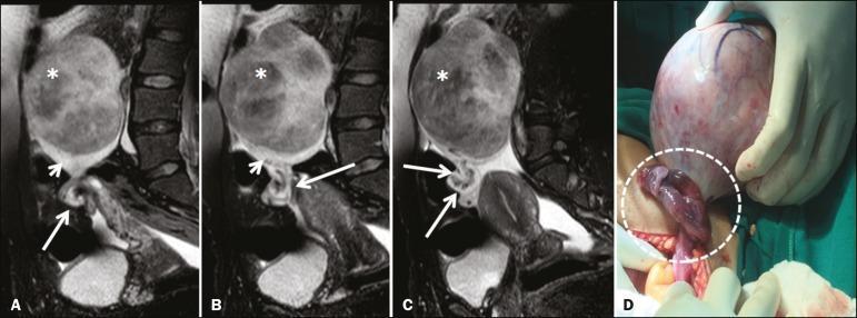
Pathophysiology
- The adnexa twists around its pedicle.
- Lymphatic and venous drainage are obstructed first.
- Ovarian congestion and edema develop, enlarging the ovary.
- Continued twisting impairs arterial inflow.
- Ischemia and hemorrhagic infarction occur.
- Delayed treatment can result in necrosis and loss of the adnexa.
The initial venous obstruction explains why arterial Doppler flow may still be detectable early in torsion. Berek & Novak’s Gynecology, p. 561; Tintinalli’s Emergency Medicine, p. 668.
Predisposing factors
Torsion is usually associated with an enlarged or unusually mobile ovary. Important risk factors include:
- Ovarian cyst or mass, especially a mass about 5 cm or larger
- Benign mature cystic teratoma, also called dermoid cyst
- Functional ovarian cyst or enlarged corpus luteum
- Pregnancy, especially early pregnancy
- Ovulation induction, ovarian hyperstimulation, or assisted reproduction
- Polycystic ovaries
- Paraovarian/paratubal cyst
- Previous tubal ligation
- Pediatric or adolescent age, where torsion can occur despite a normal ovary
Malignant ovarian tumors and inflammatory masses such as endometriomas or abscesses torsion less often, probably because adhesions reduce ovarian mobility. Berek & Novak’s Gynecology, p. 561.
Clinical features
Symptoms
The classical presentation is:
- Sudden-onset severe unilateral lower abdominal or pelvic pain
- Pain may be sharp, colicky, constant, or intermittent
- Pain may wax and wane because intermittent torsion and spontaneous detorsion can occur
- Nausea and vomiting
- Pain following exercise, lifting, or intercourse in some cases
Nausea and vomiting are particularly helpful clinical clues. However, pain is not always abrupt: some patients have gradual or intermittent pain, which contributes to missed diagnosis. Tintinalli’s Emergency Medicine, p. 668.
Signs
- Unilateral lower abdominal tenderness
- Guarding or rebound tenderness
- Unilateral adnexal tenderness on bimanual examination
- Palpable adnexal mass in some patients
- Tachycardia
- Mild fever and leukocytosis may occur later due to ischemic necrosis
A negative pregnancy test is usual, but torsion can coexist with an intrauterine pregnancy, especially with a corpus luteum cyst or ovarian hyperstimulation.
Diagnosis
Torsion is fundamentally a clinical and surgical diagnosis. No symptom, laboratory test, or imaging finding can confidently exclude it when clinical suspicion remains high.
Initial work-up
- Urgent assessment of vital signs and abdominal findings
- Urine or serum beta-hCG
- Complete blood count, electrolytes, urinalysis
- Blood group and cross-match if surgery/bleeding is possible
- Transvaginal ultrasound with Doppler
- Urgent gynecologic review
Ultrasound findings
Typical ultrasound findings include:
- Enlarged ovary, often more than 4 cm
- Ovarian stromal edema
- Peripherally displaced follicles, sometimes described as the “string of pearls” appearance
- Ovarian or adnexal mass/cyst
- Free pelvic fluid
- Thickened fallopian tube
- Twisted pedicle or whirlpool sign, if visualized
- Reduced or absent venous flow, followed later by impaired arterial flow
Important Doppler point
Normal arterial Doppler flow does not exclude torsion.
This is because:
- The ovary has a dual blood supply.
- Venous obstruction occurs before arterial obstruction.
- Torsion may be intermittent or partial.
- The ultrasound study is operator-dependent.
The gynecology text specifically states that Doppler flow to the ovary does not definitively rule out torsion. Berek & Novak’s Gynecology, p. 561. The ACOG torsion guidance likewise states that Doppler flow alone should not determine clinical decision-making.
Role of CT and MRI
- Ultrasound is the first-line imaging test.
- CT is not preferred when torsion is specifically suspected, but may reveal an enlarged or displaced ovary when performed for non-specific abdominal pain.
- MRI can be useful in selected equivocal cases, especially in pregnancy, but should not delay needed surgery.
Management of ovarian torsion
Immediate management
- Nil by mouth
- IV access and fluids
- Analgesia and antiemetics
- Pregnancy test and preoperative blood tests
- Urgent gynecology consultation
- Prompt operative assessment if suspicion is significant
Definitive treatment: urgent laparoscopy
Laparoscopy with detorsion is both diagnostic and therapeutic. The goal is to untwist the adnexa as early as possible and preserve ovarian tissue and future fertility.
Key operative principles:
- Inspect the ovary and tube.
- Detorse the adnexa.
- Preserve the ovary, even if it appears blue-black, enlarged, or necrotic-looking.
- Treat an associated cyst or mass appropriately.
- Perform cystectomy selectively when indicated.
- Consider oophoropexy only in selected recurrent torsion or special high-risk circumstances.
A dark or cyanotic appearance at surgery does not reliably prove irreversible necrosis. Ovarian function can recover after detorsion.
For adolescents, ACOG recommends minimally invasive surgery, detorsion, and preservation of adnexal structures regardless of ovarian appearance; oophorectomy is generally reserved for the rare situation in which the ovary is non-salvageable and falls apart. ACOG’s recommendation supports this fertility-preserving approach.
Oophorectomy or salpingo-oophorectomy
Removal is considered only when:
- The ovary is truly non-viable and disintegrates during surgery
- There is strong concern for malignancy
- Bleeding cannot be controlled with ovarian preservation
- The patient is postmenopausal with a suspicious adnexal mass, depending on operative findings and oncologic assessment
Distinguishing rupture from torsion
| Point | Ruptured cyst | Torsion |
|---|---|---|
| Pain pattern | Sudden pain, may improve as fluid/blood settles | Sudden severe pain, persistent or intermittent/waxing and waning |
| Vomiting | Less prominent | Common |
| Hemodynamic instability | Suggests significant hemorrhagic rupture | Usually late or uncommon unless another complication exists |
| Free fluid | Often present, may be large if hemorrhagic | Can be present but is usually not the main finding |
| Ovary on ultrasound | Hemorrhagic cyst, collapsed cyst, free fluid | Enlarged edematous ovary, peripheral follicles, possible mass and reduced venous flow |
| Main emergency concern | Hemoperitoneum, especially ectopic pregnancy mimic | Ovarian ischemia and loss of fertility |
| Treatment | Observe if stable; surgery for bleeding/peritonitis | Urgent laparoscopy and detorsion |
Exam-oriented emergency algorithm
For a reproductive-age patient with acute unilateral pelvic pain:
-
Assess stability
- Pulse, BP, pallor, syncope, abdominal rigidity, shock.
- Resuscitate and arrange urgent surgery if unstable.
-
Do beta-hCG immediately
- If positive, exclude ectopic pregnancy urgently.
-
Perform transvaginal ultrasound with Doppler
- Free fluid plus hemorrhagic cyst suggests rupture.
- Enlarged ovary, edema, peripheral follicles, mass, or whirlpool sign suggests torsion.
-
Do not dismiss torsion because Doppler arterial flow is present.
-
If torsion remains clinically likely, proceed to urgent laparoscopy.
- Imaging should not delay surgery in a convincing clinical presentation.
-
If cyst rupture is suspected
- Observe if stable with small fluid volume and no anemia.
- Operate if instability, significant hemoperitoneum, worsening anemia, peritonitis, or diagnostic uncertainty.
Red flags requiring emergency care
- Fainting, dizziness, collapse, or shoulder-tip pain
- Severe sudden pelvic pain with vomiting
- Heavy vaginal bleeding or a positive pregnancy test
- Tachycardia, low blood pressure, pallor
- Rigid abdomen, rebound tenderness, fever, or rapidly worsening pain
These findings require emergency in-person assessment, because ruptured ectopic pregnancy, major intra-abdominal bleeding, ovarian torsion, and pelvic sepsis cannot be safely distinguished at home.
Ovarian apoplexy
Reading File
Finding Sources
Finding Sources
Searching PubMed
"Hemorrhagic Ovarian Cyst" AND management
Searching the Web
hemorrhagic ovarian cyst rupture hemoperitoneum management guideline
Reading File
Reading File
query_images_db
Searching Images
transvaginal ultrasound ruptured hemorrhagic corpus luteum cyst hemoperitoneum
Ovarian Apoplexy
Ovarian apoplexy is sudden bleeding into the ovary, with or without rupture of the ovarian surface and bleeding into the peritoneal cavity. In modern English-language gynecology, it is usually described as a hemorrhagic ovarian cyst, ruptured corpus luteum cyst, or ruptured ovarian cyst with hemoperitoneum.
It is an important cause of acute pelvic pain and may mimic a ruptured ectopic pregnancy, appendicitis, or adnexal torsion.
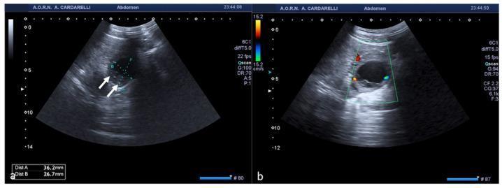
Definition
Apoplexy arises when bleeding occurs into a functional ovarian cyst, most commonly a corpus luteum cyst, and may then rupture through the ovarian capsule. The consequences range from a small, self-limited pelvic bleed to massive hemoperitoneum, hypovolemic shock, and the need for emergency surgery.
The corpus luteum is highly vascular after ovulation. If bleeding occurs within it, pressure can expand the cyst and cause pain; if it ruptures, blood enters the peritoneal cavity. Berek & Novak’s Gynecology, pp. 452, 559-560.
Etiology and risk factors
Common setting
- Reproductive-age patient
- Luteal phase of the menstrual cycle, often around days 20-26
- Corpus luteum cyst, the commonest cyst responsible for significant rupture and hemoperitoneum
- Right ovary more often affected
Precipitating events
Bleeding or rupture may be spontaneous, but pain can begin after:
- Sexual intercourse
- Strenuous exercise
- Lifting heavy objects
- Pelvic examination
- Trauma
Risk factors for severe bleeding
- Anticoagulants
- Inherited or acquired bleeding disorder
- Thrombocytopenia
- Ovulation induction or assisted reproduction
- Enlarged functional ovarian cyst
- Pregnancy, because a corpus luteum may persist and be vascular
Patients taking anticoagulants or with bleeding diatheses are at particular risk of hemorrhage from corpus luteum cyst rupture. Berek & Novak’s Gynecology, p. 452.
Pathogenesis
- Ovulation occurs and the follicle becomes the corpus luteum.
- Bleeding develops in the corpus luteum or another functional cyst.
- The ovarian capsule becomes stretched, causing acute pain.
- The cyst may remain intact, forming a hemorrhagic cyst.
- If it ruptures, blood escapes into the pelvis and abdomen.
- Large blood loss produces hemoperitoneum, tachycardia, hypotension, syncope, and shock.
The degree of bleeding determines the severity. A minor rupture may cause only localized pain, whereas frank hemorrhage may cause major blood loss. Berek & Novak’s Gynecology, p. 559.
Clinical presentation
Symptoms
- Sudden-onset unilateral lower abdominal or pelvic pain
- Pain may start in the midcycle or luteal phase
- Pain may begin after intercourse or exertion
- Increasing pelvic pain progressing to generalized abdominal pain
- Nausea or vomiting
- Dizziness, weakness, or fainting if blood loss is substantial
- Shoulder-tip pain in major hemoperitoneum due to diaphragmatic irritation
Signs
- Lower abdominal tenderness, often unilateral initially
- Guarding or rebound tenderness if peritoneal irritation is present
- Abdominal distension
- Decreased bowel sounds in significant hemoperitoneum
- Cervical-motion or adnexal tenderness on pelvic examination
- Palpable adnexal mass if the cyst has not fully ruptured
- Tachycardia and postural hypotension
- Pallor, cold extremities, and shock in severe internal bleeding
Fever is uncommon. If fever is prominent, consider pelvic inflammatory disease, tubo-ovarian abscess, appendicitis, or another infective/surgical diagnosis. Berek & Novak’s Gynecology, p. 560.
Clinical forms
A practical classification is:
| Form | Main feature | Usual presentation |
|---|---|---|
| Painful form | Bleeding is mainly within the ovary or small-volume peritoneal leak | Sudden pelvic pain, stable vital signs |
| Hemorrhagic/anemic form | Significant intraperitoneal bleeding | Pain, dizziness, syncope, tachycardia, hypotension, falling hemoglobin |
| Mixed form | Both marked pain and clinically important blood loss | Acute abdomen with signs of hypovolemia |
Differential diagnosis
The most important diagnosis to exclude is ectopic pregnancy.
| Condition | Helpful differentiating points |
|---|---|
| Ruptured ectopic pregnancy | Positive beta-hCG, absent intrauterine pregnancy when expected, adnexal ectopic mass, hemoperitoneum |
| Adnexal torsion | Severe unilateral pain with prominent nausea/vomiting; enlarged edematous ovary; Doppler may be abnormal but normal arterial flow does not exclude torsion |
| Acute appendicitis | Migratory right lower abdominal pain, fever, gastrointestinal symptoms, raised inflammatory markers |
| PID or tubo-ovarian abscess | Fever, vaginal discharge, cervical-motion tenderness, raised inflammatory markers |
| Endometrioma rupture | Acute pain and chemical peritonitis, usually less hypovolemia because bleeding is often limited |
| Renal colic | Colicky flank pain radiating to groin, urinary symptoms or hematuria |
| Degenerating fibroid | Known fibroid, pregnancy possible, uterine rather than adnexal mass |
A ruptured corpus luteum can look very similar clinically to a ruptured ectopic pregnancy. Berek & Novak’s Gynecology, p. 560.
Investigations
1. Immediate pregnancy testing
Do urine or serum beta-hCG in every patient of reproductive potential.
- A positive test does not prove ectopic pregnancy, but it makes exclusion of ectopic pregnancy urgent.
- Ovarian apoplexy can occur during an intrauterine pregnancy because of a corpus luteum cyst.
2. Blood tests
- Complete blood count: hemoglobin/hematocrit
- Blood group and Rh typing
- Cross-match if substantial hemorrhage is suspected
- Coagulation profile, especially if anticoagulants or a bleeding disorder are possible
- Serial hemoglobin where ongoing bleeding is suspected
3. Pelvic ultrasound
Transvaginal ultrasonography is first-line. It may show:
- Hemorrhagic corpus luteum or complex ovarian cyst
- Internal echoes, clot, or reticular/lacy cyst contents
- Free pelvic fluid in the pouch of Douglas
- Larger-volume hemoperitoneum
- An intrauterine pregnancy or features concerning for ectopic pregnancy
Ultrasound should assess the cyst, both adnexa, uterus, and the amount of free intraperitoneal fluid. Berek & Novak’s Gynecology, p. 560.
4. Serial clinical assessment
A single normal hemoglobin early after acute bleeding does not exclude substantial hemorrhage. Repeat:
- Pulse and blood pressure, including postural observations
- Abdominal signs
- Hemoglobin/hematocrit
- Ultrasound when the diagnosis or bleeding trend is uncertain
Management
Management depends on hemodynamic stability, volume of hemoperitoneum, severity of pain, trend in hemoglobin, and certainty that ectopic pregnancy has been excluded.
A. Conservative management
Appropriate if the patient is:
- Hemodynamically stable
- Not orthostatic
- Not significantly anemic
- Has no evidence of ongoing major bleeding
- Has only a small amount of pelvic fluid
- Has manageable or improving pain
- Has reliable observation and follow-up
Treatment commonly includes:
- Observation, often in hospital initially
- IV fluids if required
- Analgesia and antiemetics
- Serial vital signs and abdominal examination
- Repeat hemoglobin and ultrasound as clinically indicated
Stable uncomplicated ruptured functional cysts may be managed by observation, hospitalization, or repeat imaging. Berek & Novak’s Gynecology, p. 559.
B. Indications for urgent surgery
Urgent laparoscopy is generally indicated when there is:
- Hemodynamic instability or shock
- Persistent tachycardia or hypotension
- Orthostatic hypotension
- Significant or falling hemoglobin
- Large or increasing hemoperitoneum
- Ongoing active bleeding
- Severe persistent pain despite analgesia
- Peritonitis
- Failure to exclude ectopic pregnancy
- A ruptured endometrioma/dermoid causing chemical peritonitis
- Suspicion of torsion or another surgical emergency
Berek & Novak’s Gynecology notes that orthostasis, significant anemia, or a large amount of intraperitoneal free fluid usually indicates significant hemoperitoneum and need for operative management. Berek & Novak’s Gynecology, p. 560.
C. Operative treatment
The preferred approach, when feasible, is laparoscopy. It allows:
- Confirmation of diagnosis
- Suction and evacuation of hemoperitoneum
- Identification and control of ovarian bleeding
- Cystectomy or suturing/coagulation of the bleeding site when needed
- Preservation of ovarian tissue
Laparotomy may be necessary in profound shock, uncontrolled bleeding, or when laparoscopy is not suitable.
The objective in a young patient is hemostasis with maximum ovarian preservation.
Important distinction: ovarian apoplexy vs ovarian torsion
| Feature | Ovarian apoplexy | Ovarian torsion |
|---|---|---|
| Mechanism | Hemorrhage into or from a cyst, often corpus luteum | Twisting of ovary and often tube on vascular pedicle |
| Main immediate risk | Hemoperitoneum and hemorrhagic shock | Ischemia, infarction, and loss of ovary |
| Pregnancy test | Must exclude ectopic pregnancy | Usually negative, but pregnancy can coexist |
| Ultrasound | Hemorrhagic cyst and free fluid | Enlarged edematous ovary, peripheral follicles, possible twisted pedicle |
| Treatment when stable | Observation and monitoring may be appropriate | Clinical suspicion usually requires urgent laparoscopy |
| Surgical priority | Stop bleeding and preserve ovary | Detorse and preserve adnexa |
Exam summary
Ovarian apoplexy is hemorrhage into the ovary, most often a corpus luteum cyst, with or without rupture into the peritoneal cavity. It presents with acute unilateral pelvic pain and may cause hemoperitoneum, anemia, shock, and an acute abdomen. Ectopic pregnancy must be excluded with beta-hCG and transvaginal ultrasound. Stable patients with minimal bleeding may be observed; hemodynamic instability, falling hemoglobin, substantial hemoperitoneum, or diagnostic uncertainty require urgent operative management, usually laparoscopy with ovarian conservation.
Emergency warning
Sudden severe pelvic pain, fainting, shoulder pain, marked dizziness, rapid pulse, low blood pressure, a positive pregnancy test, or heavy bleeding requires emergency in-person assessment now. These symptoms can indicate ruptured ectopic pregnancy or significant intra-abdominal hemorrhage, which cannot be distinguished safely without examination, blood tests, and ultrasound.
This is a shared conversation. Sign in to Orris to start your own chat.