Scalenus anterior muscle and its relation in thoracic inlet, show me in a diagram.
Detailed anatomical diagram of the scalenus anterior (anterior scalene) muscle showing all its relations at the thoracic inlet. The diagram should be a clear labeled anterior view showing: the scalenus anterior muscle in the center, with the phrenic nerve running on its anterior surface, the subclavian artery passing posterior to the muscle (in the scalene triangle between scalenus anterior and scalenus medius), the subclavian vein passing anterior to the muscle, the brachial plexus roots emerging laterally between scalenus anterior and scalenus medius, the thoracic duct on the left side arching over, the first rib below, clavicle anteriorly, cervical vertebrae origin (C3-C6 transverse processes), and the scalenovertebral triangle medially. Include labels for: Scalenus Anterior, Scalenus Medius, Phrenic Nerve (anterior surface), Subclavian Artery (posterior), Subclavian Vein (anterior), Brachial Plexus (scalene gap), First Rib (insertion), Cervical Vertebrae C3-C6, Scalene Triangle, Thoracic Duct (left side). Clean medical illustration style with color coding for arteries (red), veins (blue), nerves (yellow).
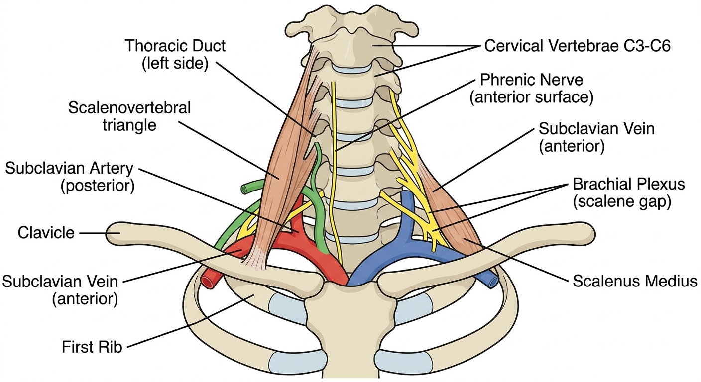
Scalenus Anterior: Anatomy and Relations at the Thoracic Inlet
The Muscle Itself
Key Relations - The "Busy" Muscle
ANTERIOR Surface (structures in front of the muscle)
| Structure | Note |
|---|---|
| Phrenic nerve (C3,C4,C5) | Crosses upper lateral border, then runs obliquely down the anterior surface within prevertebral fascia, exits the lower edge to pass between subclavian vein and artery into the thorax |
| Transverse cervical artery | Branch of thyrocervical trunk, crosses anterior |
| Suprascapular artery | Crosses anterior |
| Ascending cervical artery | Crosses anterior |
| Subclavian vein | Passes anterior to the muscle (to join the internal jugular vein and form the brachiocephalic vein at its medial border) |
| Thoracic duct (left side) | Arches laterally over the muscle before draining into the junction of the left subclavian and internal jugular veins |
POSTERIOR Surface (behind the muscle)
| Structure | Note |
|---|---|
| Subclavian artery (2nd part) | Passes posterior to scalenus anterior; gives off costocervical trunk here |
| Cervical pleura / dome of pleura | Lies just posterior and inferior |
LATERAL Border (scalene gap/triangle)
| Structure | Note |
|---|---|
| Brachial plexus roots | Emerge between scalenus anterior and scalenus medius (the "scalene triangle"); carry a sleeve of prevertebral fascia that becomes the axillary sheath |
| Subclavian artery | Also exits through the scalene triangle alongside the brachial plexus |
MEDIAL Border
| Structure | Note |
|---|---|
| Scalenovertebral triangle | Formed by scalenus anterior laterally and longus colli medially |
| Contents: vertebral artery, vertebral vein, inferior thyroid artery, sympathetic trunk, thoracic duct (left) |
The Thoracic Outlet / Scalene Triangle
- Medially - 1st rib
- Anteriorly - scalenus anterior muscle and clavicle
- Posteriorly - scalenus medius muscle
- A hypertrophied scalenus anterior, cervical rib, or fibrous band can compress the brachial plexus and/or subclavian artery in the scalene triangle
- Adson's test (turn chin to ipsilateral side + deep breath) tenses scalenus anterior and may obliterate the radial pulse
- Surgical decompression involves scalenectomy + 1st rib resection
The textbook diagram above (Gray's Anatomy for Students, Fig. 8.195) shows the root of the neck on the left side with:
- The phrenic nerve (yellow) running on the anterior surface of the anterior scalene (red muscle bulk on the right of the image)
- The subclavian artery (red vessel) posterolateral to the muscle
- The subclavian vein (blue) passing anterior to it, joining the brachiocephalic vein
- The trachea, thyroid, recurrent laryngeal nerve, and vagus nerve on the medial side
Sources: Gray's Anatomy for Students, Fig. 8.195, p. 1173 | Scott-Brown's Otorhinolaryngology, p. 598 | Color Atlas of Human Anatomy, p. 125-126 | Sabiston Textbook of Surgery, Thoracic Outlet Compression section