
Exercise for knee stability
quadriceps strengthening exercise for knee stability
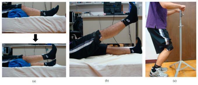
This set of three clinical photographs demonstrates common physical therapy exercises used for quadriceps strengthening and knee osteoarthritis rehabilitation. (a) Short-Arc Exercise (SAE): A supine subject performs knee extension from a slightly flexed position, supported by a bolster under the knee, moving from rest to full extension. (b) Straight Leg Raise (SLR): The subject is supine, lifting one leg while keeping the knee locked in extension, reaching an approximate 45-degree angle of hip flexion. (c) Quadriceps Strengthening Mini-squat (QSM): The subject is in a standing position, performing a partial squat (limited knee flexion) while using a vertical support stand for balance. Across all images, wearable motion sensors are visible, strapped to the distal thigh and proximal shank to track joint range of motion and exercise compliance. These exercises focus on improving joint stability and muscle strength to mitigate impact forces on the knee joint.
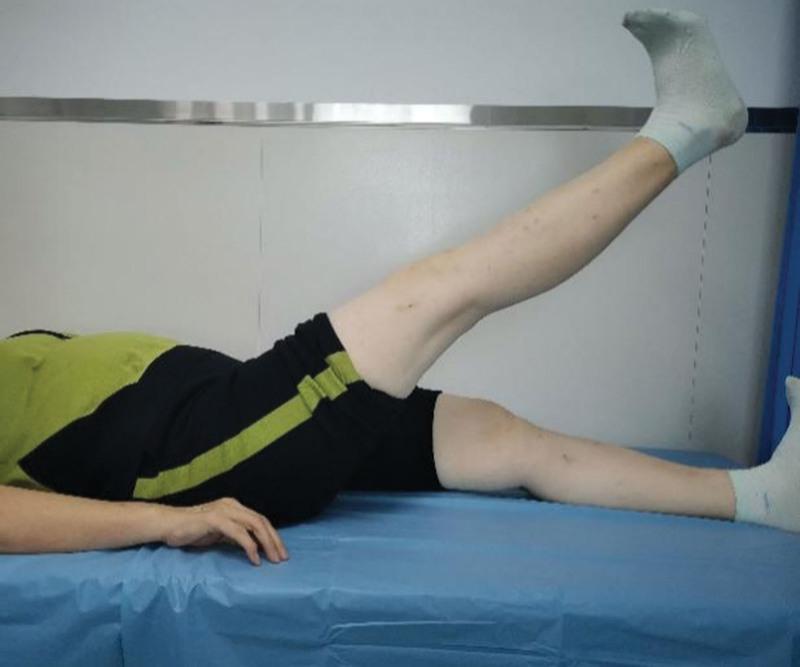
A clinical photograph illustrating a straight leg raise (SLR) exercise for quadriceps strengthening, commonly prescribed for knee osteoarthritis (KOA) rehabilitation. The patient is shown in a supine position on a medical examination table. The 'affected' leg is elevated to an angle of approximately 30 to 45 degrees, maintaining full knee extension (0°). The ankle is held in a neutral position with the heel positioned approximately 25 to 30 cm above the surface of the bed. The contralateral leg remains resting on the table for stability. This procedural image demonstrates a non-weight-bearing isometric and isotonic exercise designed to improve muscle recruitment and joint stability. The visual is intended for orthopedic and physical therapy education, focusing on conservative management strategies for knee joint health and post-treatment recovery.
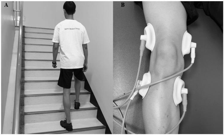
This composite clinical figure demonstrates therapeutic interventions for knee osteoarthritis rehabilitation. Panel A is a clinical photograph of a patient performing stair-climbing training in a controlled indoor environment. The patient is viewed from the posterior aspect, ascending a staircase equipped with a handrail. This exercise focuses on functional mobility and strengthening the quadriceps and hamstring muscles. Panel B is a procedural image showing the application of Interferential Electrotherapy (IFE) to the knee joint. Four circular suction-type electrodes are arranged symmetrically around the patella—two placed proximally on the distal thigh and two placed distally on the proximal calf. The electrodes are connected via cables to an external interferential current (IFC) device. The setup illustrates a multimodal physiotherapy approach combining active exercise with electrotherapy to manage pain and improve postural stability in geriatric patients with knee osteoarthritis.
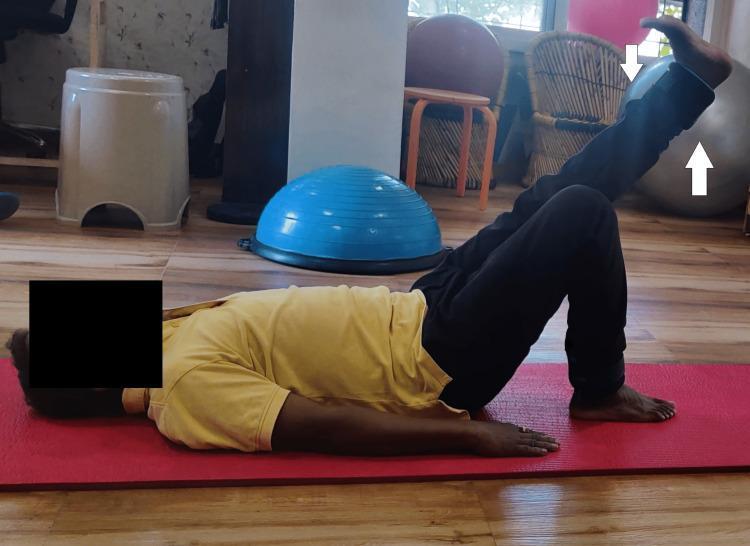
A clinical photograph depicting a male patient in a physiotherapy setting, performing a therapeutic exercise. The patient is positioned supine on a red yoga mat on a wood-laminate floor. The patient is executing a straight leg raise (SLR), with the right lower extremity actively elevated while maintaining knee extension. The contralateral (left) leg is positioned in a crook-lying posture with the knee flexed and the foot flat on the mat to stabilize the lumbar spine and pelvis. The background contains common rehabilitation equipment, including a blue Bosu balance trainer, a gray stability ball, and various stools. This visual demonstrates a post-operative or conservative management exercise focused on strengthening the hip flexors and quadriceps, frequently utilized in rehabilitation protocols for conditions such as avascular necrosis of the femoral head or following total hip replacement.
"knee" AND "exercise therapy" AND stability
best exercises for knee stability strengthening
Exercises for Knee Stability
Core strengthening exercises
- Straight leg raise (SLR): Lying supine, keep the knee locked in full extension and lift the leg to about 30-45 degrees, holding briefly before lowering. This is a non-weight-bearing isometric/isotonic exercise that builds quad activation without stressing the joint - commonly used after ligament injury or surgery - Textbook of Family Medicine 9e.
- Short-arc extension (quad sets): With a pillow or bolster under the knee, actively extend the knee to full extension from a slightly flexed position. Used early in rehab because it loads the quadriceps without full range stress - Tintinalli's Emergency Medicine.
- Quadriceps strengthening is a recognized therapy for knee osteoarthritis that provides pain relief and is one of the best-studied interventions - Rheumatology, 2-Volume Set, 2022.
- Hamstrings work with the quadriceps to protect the knee in the sagittal plane. Hamstring bridges and hamstring curls are standard additions, especially important after PCL or ACL injury where the balance between quad and hamstring pull matters (open-chain quad extension can stress an ACL graft, so many protocols pair it carefully with hamstring work) - Miller's Review of Orthopaedics 9th Edition.
- Hip abductors and rotators control frontal- and transverse-plane stability (dynamic valgus/varus control), which is critical for preventing patellofemoral pain and ACL re-injury. Lateral band walks and clamshells target this.
- Mini-squats: Standing partial squats with support, progressing depth as tolerated - improves joint stability and muscle strength while limiting excessive shear/compressive load.
- Step-ups: Build strength plus balance and coordination; adjustable difficulty via step height.
- Lunges (forward, backward, lateral): Comprehensive strengthening across multiple directions.
- Stair climbing training: Functional strengthening of quads and hamstrings used in structured rehab programs.
- Single-leg stance progressions, use of a wobble board or Bosu, and controlled sit-to-stand transitions retrain the neuromuscular reflexes that stabilize the knee dynamically, not just muscle strength alone.
Clinical context (post-injury)
- ACL/PCL injury or reconstruction: Rehab typically emphasizes quadriceps strengthening and controlled open-kinetic-chain extension, with graded return to closed-chain functional work; PCL injuries (grades I-III) are often managed nonsurgically with bracing plus quad strengthening since the PCL tends to heal - Textbook of Family Medicine, Miller's Review of Orthopaedics.
- Knee osteoarthritis: Quadriceps strengthening plus general exercise therapy is consistently supported for pain relief and function, and is considered a first-line conservative treatment - Rheumatology, 2-Volume Set.
Practical program tips
- Perform strengthening work 2-3 times per week, progressing load/resistance gradually.
- Work within mild discomfort, not sharp pain.
- Pair strengthening with mobility work (e.g., hamstring/calf stretching) since restricted motion alters joint mechanics and increases stress.
- Progress from non-weight-bearing/isometric (SLR, quad sets) to closed-chain functional movements (squats, step-ups, lunges) as tolerated.