
Support of uterus
supports of uterus ligaments pelvic floor anatomy diagram
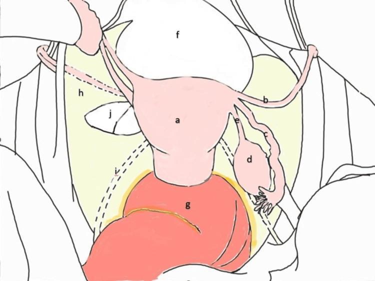
This medical anatomical diagram illustrates a superior view of the female pelvis and internal reproductive organs. Centrally, the uterus (a) is positioned posterior to the urinary bladder (f) and anterior to the rectum (g). Extending laterally from the uterine horns are the round ligaments (b), fallopian tubes (c), and the ovaries (d), which are anchored by the ovarian ligaments (e). The broad ligament (h) is depicted as a wide peritoneal fold draping over these structures and extending to the pelvic side walls. A dashed line indicates the path of the ureteric fold (i) beneath the peritoneum. Notably, the diagram highlights a broad ligament defect (j) on the left side, located caudal to the round ligament. This visual representation serves as an educational guide for identifying Type 1 defects according to the Cilley classification, which are clinically significant as potential sites for internal hernias of the bowel. The diagram uses a combination of solid lines for superficial structures and dashed lines for retroperitoneal or obscured landmarks, providing a clear topographic map of pelvic surgical anatomy.
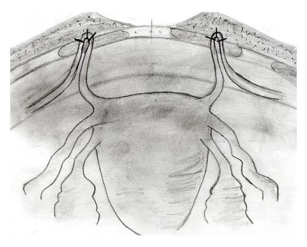
Anatomical Diagram: A black-and-white schematic illustration demonstrating the surgical fixation of the round ligaments to the anterior rectus fascia, likely for the management of pelvic organ prolapse. The diagram provides an anterior view of the uterus and its supporting structures. The uterine fundus is shown in the center, with the round ligaments extending bilaterally. These ligaments are depicted being pulled cranially and looped through small fenestrations in the rectus abdominis muscle layer. The apical 'ans' or loop of each round ligament is secured to the overlying fascia using three distinct sutures, represented by 'x' markers. Additional adnexal structures, including the fallopian tubes and utero-ovarian ligaments, are visible extending from the uterine cornua. The illustration emphasizes the anatomical relationship between the pelvic viscera and the abdominal wall fascia during a suspension procedure, highlighting the point of mechanical support and the method of non-absorbable suture fixation used to stabilize the uterine position.
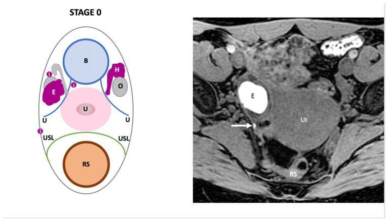
This dual-panel image provides a comparison between a schematic diagram and an axial T1-weighted fat-saturated MR image illustrating 'Stage 0' pelvic endometriosis. The left panel is a labeled diagram identifying key pelvic anatomy including the bladder (B), uterus (U), rectosigmoid (RS), ovaries (O), and uterosacral ligaments (USL). It highlights an endometrial cyst (E), hematosalpinx (H), and superficial peritoneal implants (i). The right panel shows a corresponding diagnostic MR image of the female pelvis. A prominent hyperintense (bright) cystic lesion labeled 'E' represents an ovarian endometrioma, characteristic of blood-filled contents on T1-weighted fat-saturated sequences. Adjacent to this, a white arrow indicates a small focus of intermediate-to-high signal intensity corresponding to a superficial peritoneal implant located near the uterosacral ligament and rectosigmoid (RS). Other visible landmarks include the uterus (Ut) and pelvic musculature. The image demonstrates the diagnostic criteria for early-stage endometriosis (Endo-Stage MRI classification), focusing on the identification of superficial lesions and ovarian involvement without deep pelvic infiltration.
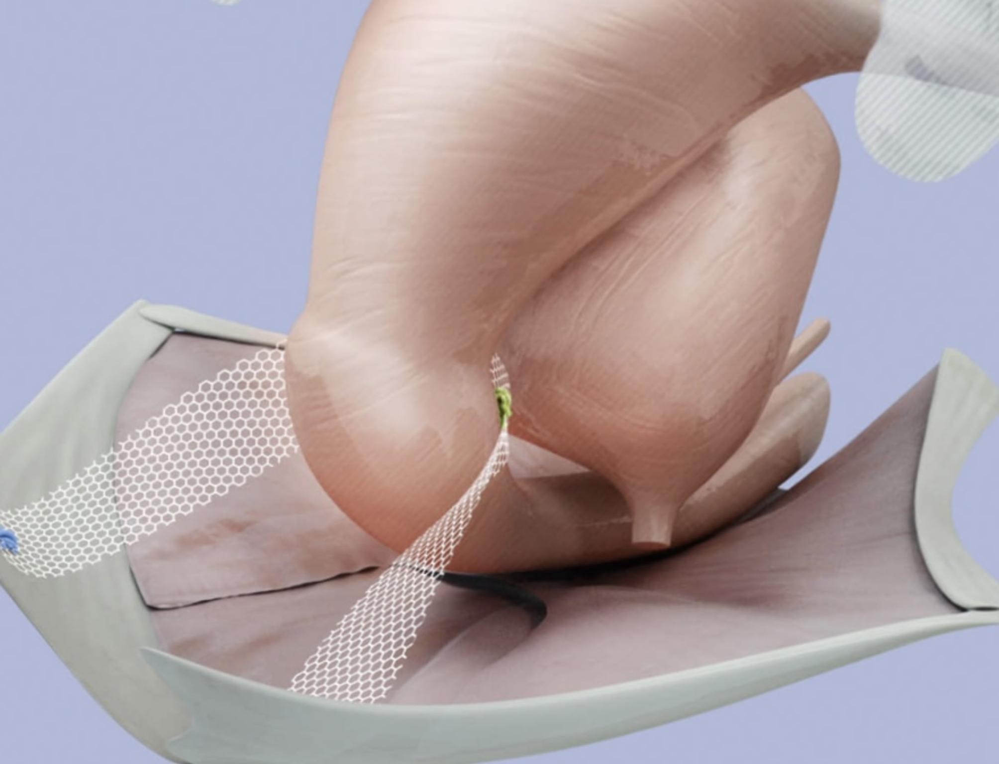
This medical illustration depicts a bilateral sacrospinous colposuspension (BSC) procedure for pelvic organ prolapse (POP) repair. The diagram features a translucent anatomical model of the uterus and cervix in a vertical orientation. A synthetic, white, macroporous mesh with a distinct hexagonal pattern is shown providing apical support. The mesh is anchored bilaterally, extending from the cervical region to simulate the function of natural pelvic ligaments (uterosacral and cardinal ligaments). This surgical setup illustrates the symmetrical suspension of the uterus to the sacrospinous ligaments, intended to recreate a stable pelvic floor and correct apical descent. The illustration is designed for surgical education, highlighting the interaction between prosthetic mesh material and gynecological structures in minimally invasive urogynecological reconstruction.
Support of the Uterus
1. Pelvic Floor Muscles (Most Important)
"The uterus is mainly supported by the pelvic floor muscles, not by the abovementioned ligaments."
- Color Atlas of Human Anatomy, Vol. 2
2. Ligamentous and Fascial Supports
A. Cardinal Ligament (Mackenrodt's / Transverse Cervical Ligament)
- Condensation of endopelvic connective tissue running from the cervix/upper vagina to the lateral pelvic wall.
- The most important ligamentous support against uterine prolapse.
- Together with the uterosacral ligaments, forms the cardinal-uterosacral complex (apical Level I support).
- Attenuation or breaks in this complex are the main cause of uterine prolapse.
B. Uterosacral Ligament (Rectouterine Ligament / Sacrouterine Ligament)
- Runs from the cervix posterolaterally to the sacrum (S2-S4 region).
- Keeps the cervix pulled posteriorly and superiorly, maintaining the normal anteverted-anteflexed position.
- Contains dense subperitoneal connective tissue and autonomic nerve fibers (inferior hypogastric plexus).
- Part of the Level I apical support with the cardinal ligament.
- The McCall culdoplasty technique uses these ligaments to restore apical support after hysterectomy.
C. Round Ligament of the Uterus
- Arises near the uterine horns, runs through the inguinal canal, and ends in the subcutaneous fat of the labia majora.
- Contains smooth muscle cells.
- Derived from the gonadal fold; it is a continuation of the suspensory ligament of the ovary.
- Maintains the anteverted position of the uterus but provides little structural support against prolapse.
D. Broad Ligament of the Uterus
- A peritoneal fold between the lateral margin of the uterus and the lateral pelvic wall.
- Contains connective tissue, vessels, and nerves; it also envelops the fallopian tube, round ligament, and ovarian ligament.
- Does not provide significant mechanical support to the uterus.
E. Pubocervical Fascia
- Fascial condensation anteriorly between the cervix/upper vagina and the pubic bone.
- Supports the bladder base anteriorly.
3. Three-Level Support System (DeLancey Classification)
| Level | Structures | Function |
|---|---|---|
| Level I (Apical) | Cardinal ligament + uterosacral ligament complex | Maintains vaginal/uterine length and axis; prevents apical prolapse (uterine or vault) |
| Level II (Midvaginal) | Paravaginal attachments to arcus tendineus fasciae pelvis (ATFP) and levator fascia | Maintains midline position of vagina; defects cause cystocele/rectocele |
| Level III (Distal) | Muscles and connective tissue around distal vagina and perineum | Supports distal vagina; prevents gaping introitus |
4. Normal Position of the Uterus
Clinical Relevance
- Uterine prolapse results from attenuation of the cardinal-uterosacral complex (Level I failure), often compounded by levator ani injury from vaginal delivery.
- Risk factors: vaginal delivery (55% of women with prolapse have levator ani injury), increasing age, obesity, chronic constipation, and prior pelvic surgery.
- Surgical repair targets Level I (uterosacral/sacrospinous fixation, McCall culdoplasty), Level II (paravaginal repair), or all three.
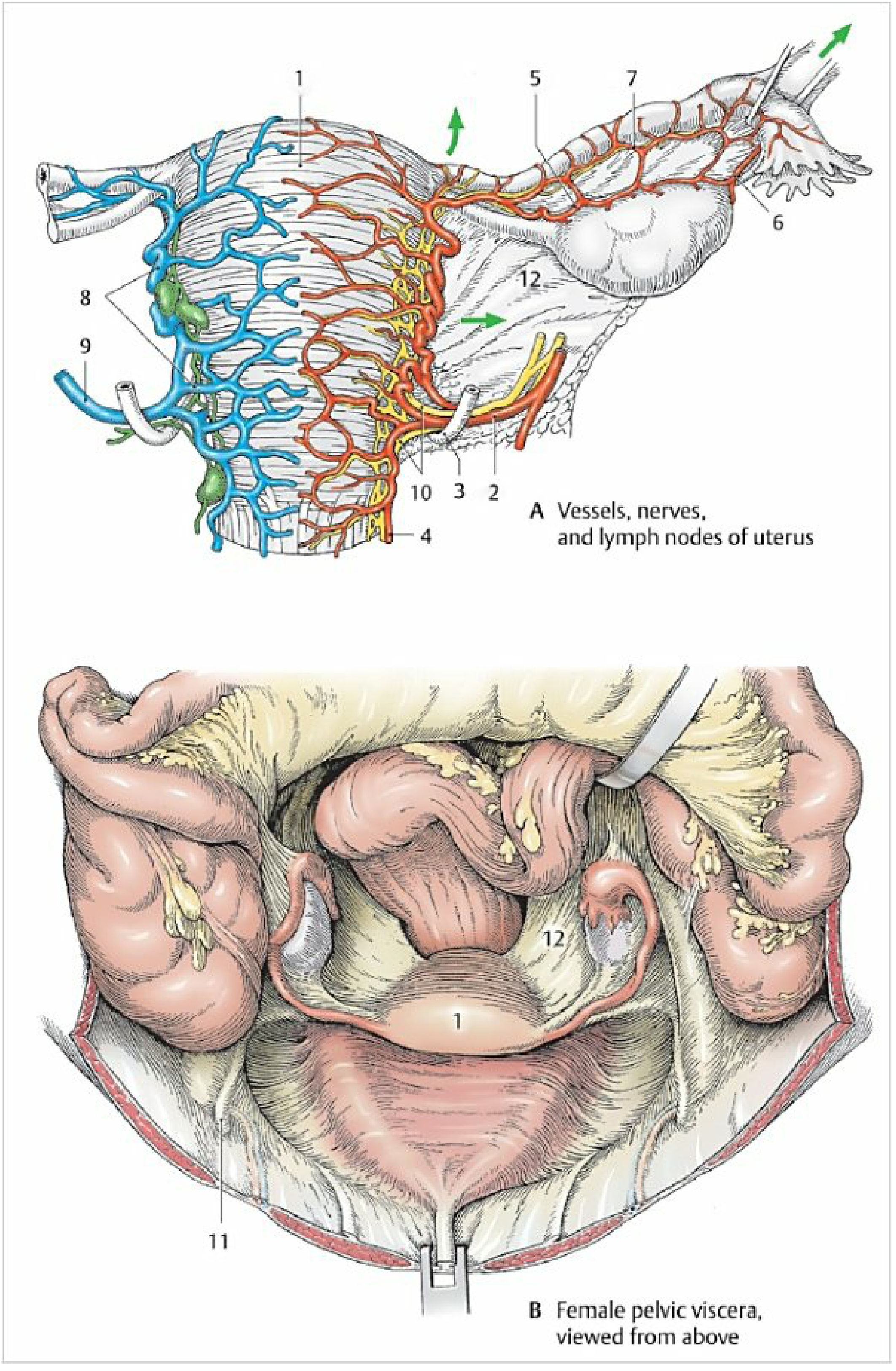
- [Color Atlas of Human Anatomy, Vol. 2](Internal organs, p. 461-462) - primary anatomy reference for uterine ligaments
- [Berek & Novak's Gynecology](p. 1595-1596) - DeLancey three-level support system and pathophysiology of prolapse
- [Campbell-Walsh-Wein Urology](p. uterine prolapse section) - cardinal-uterosacral complex and broad ligament mechanics