
Acro dermatits enterpathyica
acrodermatitis enteropathica skin rash zinc deficiency infant
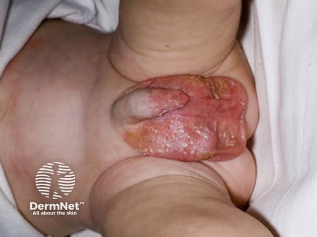
Clinical photograph of an infant displaying periorificial dermatitis associated with acrodermatitis enteropathica (zinc deficiency). This frontal close‑up captures the perioral and periorificial skin with erythematous, scaly plaques, crusting, and fissuring at the oral commissures and surrounding lips. The involved skin is inflamed with a rough, desquamative surface and mild edema; epidermal scaling may be greasy or flaky. Distribution is predominantly periorificial, a pattern typical of zinc‑deficiency dermatitis, though acral involvement may occur in some cases. The image illustrates a classic mucocutaneous eruption seen in congenital/genetic AAC (SLC39A4) or acquired zinc deficiency, and may accompany accompanying symptoms such as diarrhea, irritability, and alopecia in certain patients. Pathophysiology centers on impaired zinc transport and keratinocyte dysfunction, leading to compromised barrier integrity and inflammatory ceruminous changes at skin junctions. Clinically, the finding supports a diagnosis of zinc deficiency dermatitis; zinc level and alkaline phosphatase testing can confirm, and rapid improvement after zinc supplementation is characteristic. Differential diagnoses include seborrheic dermatitis, atopic dermatitis, contact dermatitis, and candidiasis. This image is valuable for education, aiding recognition, differential diagnosis, and monitoring therapeutic response in pediatric dermatology and nutrition clinics. This resource supports clinical documentation and searchable educational annotation.
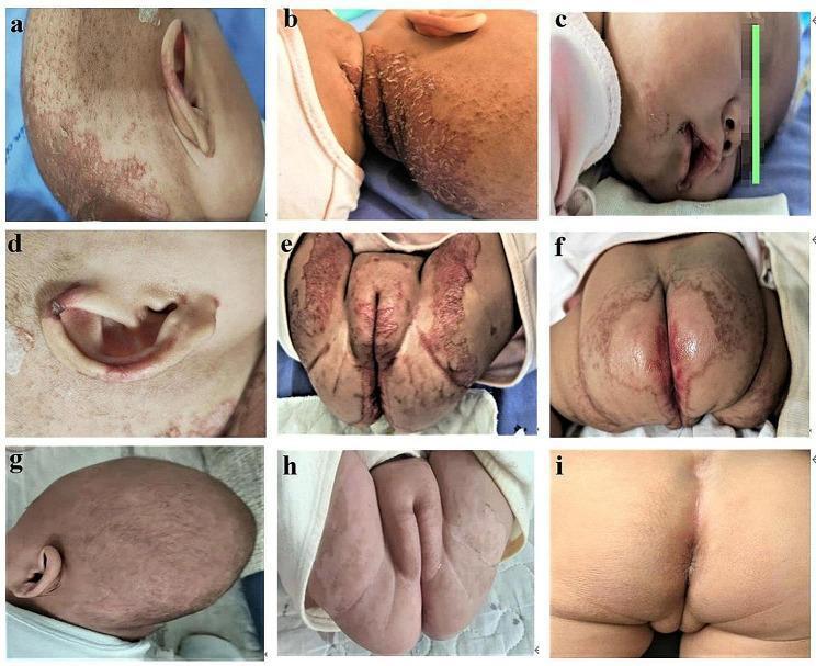
A series of nine clinical photographs (a-i) illustrating the cutaneous manifestations and therapeutic response of acrodermatitis enteropathica-like lesions in an infant. Images a-f depict the pre-treatment state, characterized by well-demarcated, erythematous, and erosive plaques with overlying scaly crusts and scabs. The distribution is classic for zinc deficiency, primarily affecting the acral and periorificial regions: occipital scalp (a, b), perioral area (c), auricle (d), and the perineal and perianal zones (e, f). Diffuse alopecia and sparse hair growth are also visible on the scalp. Images g-i demonstrate the clinical status after 6 days of oral zinc supplementation, showing rapid and significant improvement. The inflammatory erythema and erosions on the scalp (g) and anogenital region (h, i) have largely resolved, leaving behind smooth skin with minimal residual post-inflammatory changes. This comparison serves as a pedagogical tool for recognizing the diagnostic distribution of zinc deficiency-related dermatitis and its dramatic response to targeted replacement therapy.
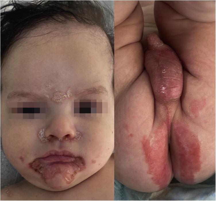
Clinical photograph of an infant presenting with classic dermatological manifestations of zinc deficiency, such as acrodermatitis enteropathica or transient neonatal zinc deficiency. The image is split into two panels. The left panel shows the patient's face with characteristic erythematous, desquamative, and crusted lesions in a symetrical distribution. Primary areas of involvement include the perioral region (surrounding the lips), the perinasal area (alar base and nasolabial folds), and the glabella (interbrow region). The right panel displays the diaper area, showing well-demarcated, bright red erythematous patches and desquamation across the scrotum, penis, and perineal/perianal regions. The overall clinical presentation highlights the classic 'periorificial' distribution of skin lesions typical of nutritional or genetic zinc metabolism disorders. These findings are pedagogically significant for distinguishing nutritional deficiencies from common diaper dermatitis or atopic eczema in pediatric patients.
acrodermatitis enteropathica zinc SLC39A4
Acrodermatitis Enteropathica (AE)
Definition and Genetics
- Emery's Elements of Medical Genetics and Genomics
- Yamada's Textbook of Gastroenterology, 7th ed.
- Schwartz's Principles of Surgery, 11th ed.
Pathophysiology
- DNA polymerase and reverse transcriptase - so deficiency impairs cell proliferation and wound healing
- Alkaline phosphatase - a zinc-dependent enzyme whose serum levels fall in AE (used as a diagnostic marker)
- Enzymes involved in keratinocyte integrity and immune function
Clinical Presentation
| Feature | Details |
|---|---|
| Dermatitis | Eczematous, erythematous, scaly pink plaques in acral (hands, feet) and periorificial regions (perioral, perianal, perinasal, auricular) |
| Diarrhea | Primary GI complication; precedes or accompanies skin findings |
| Alopecia | Scalp, eyebrows, and eyelashes |
- Bottle-fed infants: First few days of life (formula lacks protective zinc-binding ligands in breast milk)
- Breast-fed infants: Days to weeks after weaning (breast milk contains a zinc-binding ligand that permits absorption despite the transport defect)
- Early: erythematous scaly plaques, angular stomatitis (common first sign)
- Later: vesiculation, bullae, erosions
- Chronic: psoriasiform plaques, complete alopecia
Clinical Images
Diagnosis
| Investigation | Finding |
|---|---|
| Serum zinc | Low (<70 mcg/dL; Schwartz's notes <100 mg/dL as abnormal) |
| Serum alkaline phosphatase | Low (zinc-dependent enzyme) |
| Skin biopsy | Psoriasiform epidermal hyperplasia, confluent parakeratosis, pallor/ballooning of upper epidermis |
| Genetic testing | SLC39A4 mutations confirm hereditary form |
| Clinical response to zinc | Rapid improvement is itself diagnostic |
Differential Diagnoses
- Atopic/seborrheic/contact dermatitis
- Candidiasis (diaper area) | Nutritional zinc deficiency (premature infants, severely malnourished patients - produces clinically identical phenotype)
- Necrolytic migratory erythema (glucagonoma)
- Biotin deficiency (multiple carboxylase deficiency)
Treatment
- Zinc sulfate 100-400 mg/day orally (Schwartz's Principles of Surgery)
- Lifelong supplementation is required in the genetic form
- Response is typically dramatic within days of starting therapy (as seen in clinical images above)
Key Points for Exams
- Gene: SLC39A4 → ZIP4 transporter
- Inheritance: Autosomal recessive
- Triad: Dermatitis + Diarrhea + Alopecia
- Distribution: Acral + periorificial (hallmark)
- First sign: Angular stomatitis
- Marker enzymes: Low serum zinc + low alkaline phosphatase
- Treatment: Lifelong oral zinc; rapid response confirms diagnosis
- Breast-fed infants: Delayed onset vs. bottle-fed (protective zinc ligand in breast milk)