
Give me a detailed 10 mark answer for blood retinal barrier
Reading File
Now let me search the medical library and web in parallel for comprehensive content on the blood-retinal barrier.
Finding Sources
Finding Sources
Searching the Web
blood retinal barrier anatomy physiology breakdown clinical significance ophthalmology
Good results. Now let me read the detailed textbook content from the identified blocks.
Reading File
Reading File
Now let me read the BRB-specific section including inner/outer barrier details and the FFA/breakdown sections:
Reading File
Reading File
I now have comprehensive textbook content. Let me also fetch the key diagrams:
Loading Image
Loading Image
I now have all the content and diagrams needed for a thorough 10-mark answer. Here is the complete, exam-ready response:
Blood-Retinal Barrier (BRB)
Definition
The blood-retinal barrier (BRB) is a selective physiological barrier that separates the neural retina from the systemic circulation. It is analogous to the blood-brain barrier and maintains the specialized ionic and molecular microenvironment required for normal visual function. The BRB has two anatomically and functionally distinct components: the inner BRB (iBRB) and the outer BRB (oBRB).
1. Inner Blood-Retinal Barrier (iBRB)
Anatomical basis: The iBRB is formed by the tight junctions (zonula occludentes) between the non-fenestrated endothelial cells of the retinal capillaries.
Structure of retinal capillary walls (from inside outward):
- Endothelial cells - form a single layer on the basement membrane, linked by tight junctions that constitute the actual barrier
- Basement membrane - lies beneath the endothelial cells, with an outer basal lamina enclosing pericytes
- Pericytes - lie external to endothelial cells, have contractile pseudopodal processes that envelop capillaries, and participate in autoregulation of microvascular circulation
Key property: Neither protein-bound nor free fluorescein can cross an intact iBRB. The basement membrane and pericytes play only a minor supporting role.
Distribution: The iBRB covers the inner two-thirds of the retina, supplied by the retinal circulation. Capillary-free zones are present around arterioles and at the foveal avascular zone (FAZ).
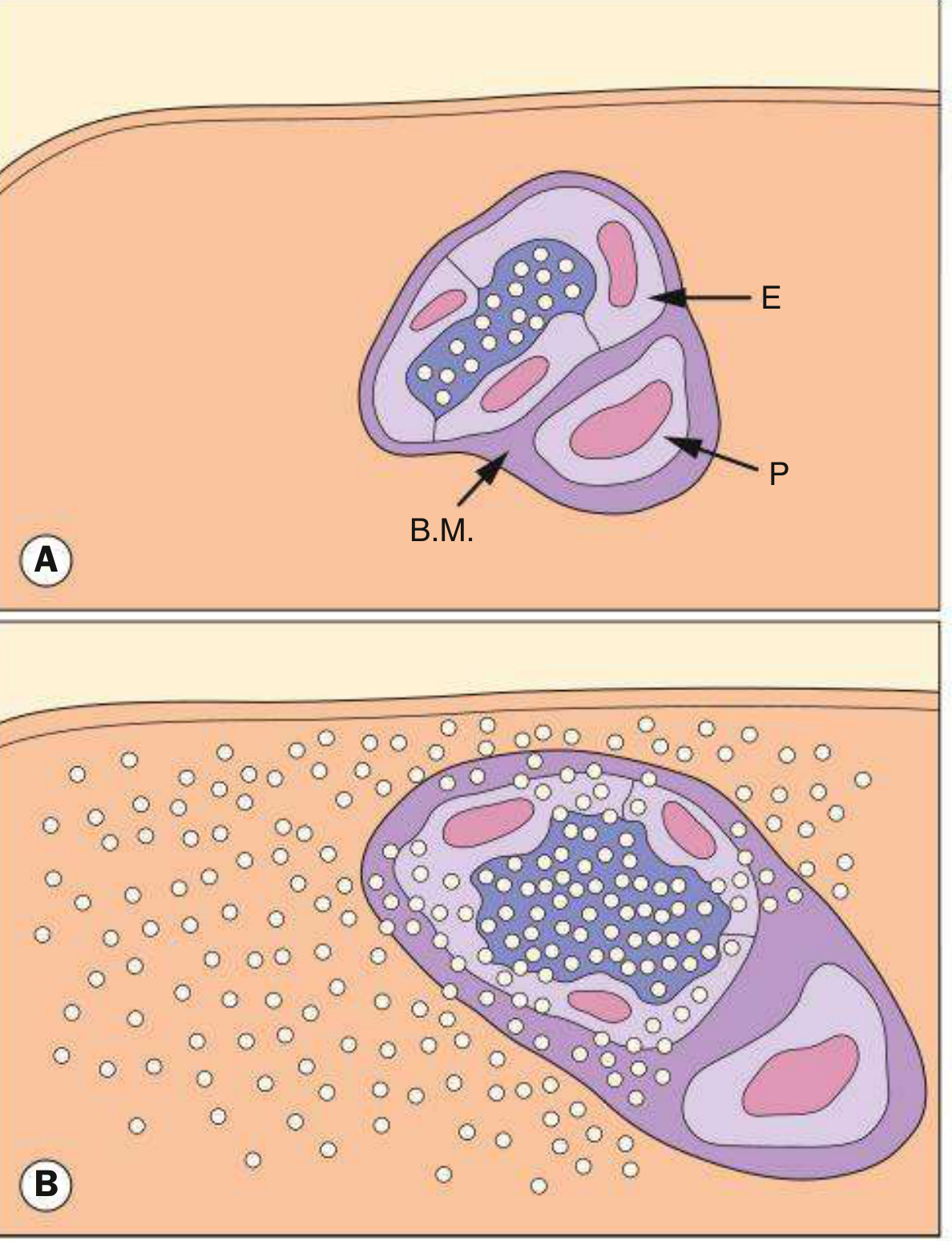
Fig. 14.5 Inner BRB. (A) Intact - E = endothelial cell, P = pericyte, B.M. = basement membrane. (B) Disrupted - fluorescein molecules leak into surrounding retinal tissue. (Kanski's Clinical Ophthalmology, 10th ed.)
2. Outer Blood-Retinal Barrier (oBRB)
Anatomical basis: The oBRB is formed by the RPE cells and their intercellular tight junctions (zonula occludentes), which lie on Bruch membrane.
Structure and mechanism:
- The choriocapillaris is a fenestrated capillary network - its walls allow free and protein-bound fluorescein to escape into the extravascular space
- Fluorescein crosses Bruch membrane freely (Bruch membrane is permeable)
- On reaching the RPE, it is blocked by the tight junctions between adjacent RPE cells
- The RPE also actively pumps ions and water out of the subretinal space, maintaining the dry subretinal microenvironment
Bruch membrane (5-layered structure) separates RPE from choriocapillaris:
- Basal lamina of RPE
- Inner collagenous layer
- Elastic fibre band
- Outer collagenous layer
- Basal lamina of choriocapillaris inner layer
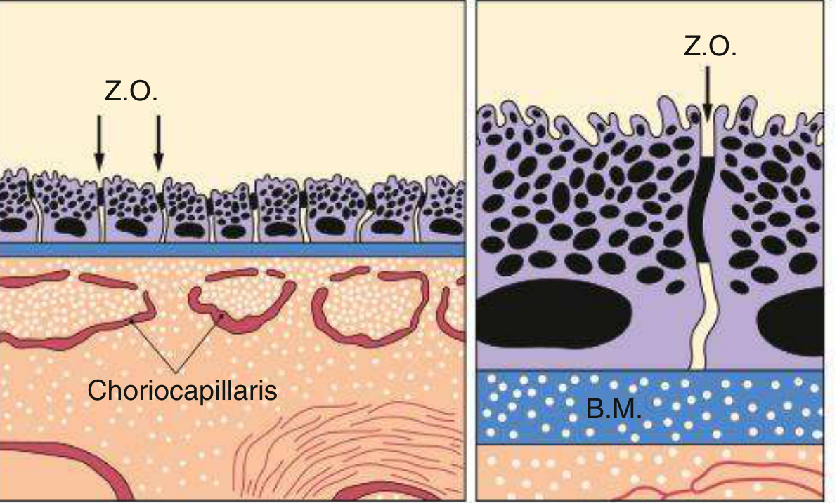
Fig. 14.4 Outer BRB - Z.O. = zonula occludentes (RPE tight junctions); B.M. = Bruch membrane; choriocapillaris below. (Kanski's Clinical Ophthalmology, 10th ed.)
3. Functions of the BRB
| Function | Component |
|---|---|
| Prevents extracellular fluid leaking into subretinal space | oBRB (RPE tight junctions) |
| Maintains ionic homeostasis of the retinal microenvironment | Both iBRB and oBRB |
| Inward transport of metabolites (glucose, amino acids) | RPE (oBRB) |
| Outward transport of metabolic waste products | RPE via Bruch membrane |
| Active pumping of ions/water from subretinal space | RPE |
| Storage, metabolism and transport of Vitamin A (visual cycle) | RPE |
| Absorb stray light (via melanosomes) | RPE |
| Phagocytosis of shed photoreceptor outer segments | RPE |
| Immune privilege of the retina | Both barriers |
4. Breakdown of the BRB - Clinical Significance
A. Breakdown of the Inner BRB
Caused by dysfunction or absence of endothelial tight junctions. On fluorescein angiography (FFA), this causes leakage - early hyperfluorescence that increases in both area and intensity over time.
Causes:
- Dysfunction/loss of existing tight junctions: Background diabetic retinopathy (DR), retinal vein occlusion (RVO), cystoid macular oedema, disc swelling
- Primary absence of tight junctions: Macular neovascularization (MNV), proliferative diabetic retinopathy, Coats disease, tumours
B. Breakdown of the Outer BRB
Caused by RPE tight junction disruption. On FFA, this causes pooling in anatomical spaces.
Causes and FFA patterns:
- Subretinal pooling: Central serous chorioretinopathy (CSC) - small leak that slowly increases in intensity and area
- Sub-RPE pooling: Pigment epithelial detachment (PED) - early hyperfluorescence that increases in intensity but not in size
5. Key Diseases Linked to BRB Disruption
Diabetic Retinopathy (DR)
The most common cause of iBRB breakdown. In DR:
- Pericyte loss is the earliest histological change (before clinically apparent retinopathy)
- The endothelial-to-pericyte ratio normally is ~1:1 in retinal capillaries; selective pericyte dropout occurs in diabetes
- Loss of pericytes leads to loss of retinal blood flow autoregulation
- Subsequent endothelial dysfunction and tight junction loss cause capillary leakage
- Results in retinal oedema, hard exudates, and ultimately macular oedema (most common cause of visual loss in DR)
- Microaneurysms - the earliest clinical sign - may leak plasma constituents into the retina through BRB breakdown
Other Conditions
| Disease | BRB Component Affected |
|---|---|
| Central serous chorioretinopathy | Outer BRB (RPE leak) |
| Age-related macular degeneration | Outer BRB (RPE/Bruch membrane) |
| Retinal vein occlusion | Inner BRB (endothelial damage) |
| Irvine-Gass syndrome (post-cataract surgery CMO) | Inner BRB (inflammatory) |
| Coats disease | Inner BRB (absent endothelial tight junctions) |
| Hypertensive retinopathy | Inner BRB (endothelial damage) |
| Uveitis | Both (inflammatory mediators) |
6. BRB and Fluorescein Angiography (FFA)
The integrity of the BRB is the basis for interpretation of FFA:
- Intact iBRB: Neither bound nor free fluorescein crosses - no leakage seen
- Intact oBRB (RPE): Fluorescein that escapes from fenestrated choriocapillaris is stopped at RPE tight junctions - no pooling in subretinal space
- Bruch membrane is permeable to fluorescein - it does not form part of the barrier
- Window defect (RPE atrophy): Gives hyperfluorescence due to unmasking of choroidal fluorescence (not true leakage) - early, doesn't enlarge
7. Molecular Basis of BRB Tight Junctions
Tight junctions of both the endothelium and RPE are composed of:
- Occludin and claudins (transmembrane proteins forming the actual seal)
- ZO-1, ZO-2, ZO-3 (scaffolding/zonula occludens proteins linking to the actin cytoskeleton)
- JAM (junctional adhesion molecules)
In disease states (especially DR), VEGF (vascular endothelial growth factor) is a major mediator of BRB breakdown - it phosphorylates occludin, leading to tight junction disassembly and increased permeability. This is the rationale for anti-VEGF therapy in diabetic macular oedema.
Summary Table
| Feature | Inner BRB | Outer BRB |
|---|---|---|
| Location | Retinal capillary endothelium | Retinal pigment epithelium (RPE) |
| Tight junction type | Zonula occludentes between endothelial cells | Zonula occludentes between RPE cells |
| Supporting cells | Pericytes, basement membrane | Bruch membrane |
| Blood supply | Retinal circulation (inner 2/3 retina) | Choriocapillaris (outer 1/3 retina) |
| FFA: if intact | No leakage | No pooling in subretinal space |
| FFA: if broken | Leakage (area + intensity increase) | Pooling in anatomical space |
| Key disease | Diabetic retinopathy | Central serous chorioretinopathy, AMD |
References: Kanski's Clinical Ophthalmology: A Systematic Approach, 10th edition - Chapters on Retinal Anatomy, Macular Disease, and Fluorescein Angiography; The Wills Eye Manual, 8th edition.
This is a shared conversation. Sign in to Orris to start your own chat.