
Mrs Vijayabai Prakash Somawanshi, 60F — c/o swelling over (L) thumb, h/o prick by something, 3 days, with fever and pain
Reading File
Now I'll search the medical library for relevant content in parallel.
Finding Sources
Finding Sources
Finding Sources
Searching the Web
felon finger infection management clinical presentation diagnosis treatment
Finding Sources
Finding Sources
Finding Sources
Reading Web Page
https://www.ncbi.nlm.nih.gov/books/NBK430933
Good - I found relevant textbook sections. Let me now read the key textbook blocks for detailed content.
Reading File
Reading File
Reading File
I now have rich textbook content. Let me fetch the clinical images to include in the answer.
Loading Image
Loading Image
Loading Image
I now have all the information needed for a complete, well-illustrated clinical answer.
Clinical Assessment: Mrs Vijayabai Prakash Somawanshi, 60F
Swelling (L) thumb + H/O prick + 3 days + fever + pain
Most Likely Diagnosis: FELON (Pulp Space Abscess)
A felon is a subcutaneous abscess of the distal finger/thumb pulp, arising within the fibrous septae that run from the distal phalangeal periosteum to the epidermis. It is one of the most common hand infections and classically follows a penetrating injury - a prick, splinter, fingerstick, or foreign body.
- Current Surgical Therapy 14e, p. 975
- Bailey and Love's Short Practice of Surgery 28th Ed, Ch. 38
Anatomy - Why Felons Are Painful and Dangerous
The fingertip pulp is divided into multiple closed compartments by vertical fibrous septa. When bacteria are introduced by a prick, infection within these sealed compartments builds pressure rapidly, compressing small blood vessels. This causes:
- Intense throbbing pain (the hallmark)
- Risk of ischemia and tissue necrosis
- Potential spread to the periosteum → osteomyelitis of the distal phalanx
- Proximal spread through the fibrous curtain into the flexor sheath → flexor tenosynovitis
- Extension to the DIP joint → septic arthritis
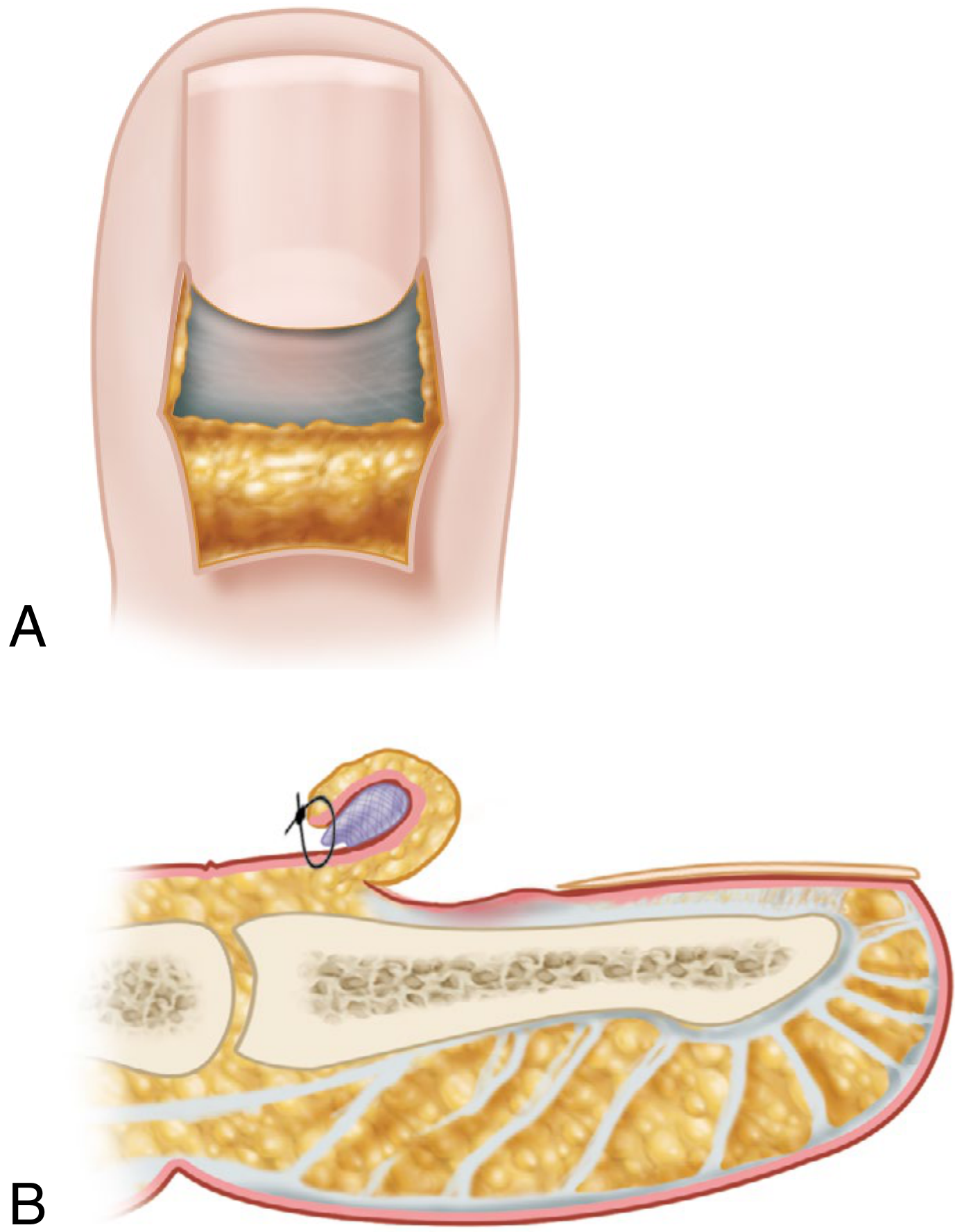
Cross-section of the fingertip showing vertical septa dividing the pulp into compartments - Campbell's Operative Orthopaedics 15th Ed 2026
Clinical Features - This Patient's Presentation Fits Perfectly
| Feature | Felon |
|---|---|
| H/O prick (penetrating trauma) | Classic precipitant |
| 3-day duration | Typical - rapid onset |
| Swelling of the thumb | Entire pulp swollen, tense |
| Pain | Intense, throbbing, worsening |
| Fever | Suggests established infection / systemic response |
"Patients often present with rapid onset of pain, erythema, and significant swelling of the entire pulp of a fingertip. They may endorse a history of a localized injury or retained foreign body." - Current Surgical Therapy 14e
At 60 years, also consider whether she is diabetic (fingerstick users are at elevated risk; glucose monitoring pricks are a recognized cause) and check for immunocompromise.
Differential Diagnoses to Consider
| Diagnosis | How to distinguish |
|---|---|
| Felon (most likely) | Tense, diffuse pulp swelling; no vesicles; post-prick |
| Paronychia | Infection of the nail fold (lateral or dorsal), not the pulp; fluctuance at nail fold margin |
| Herpetic Whitlow | Grouped vesicles, burning/tingling - NOT a prick injury; do NOT incise |
| Cellulitis without abscess | Diffuse erythema, no fluctuance |
| Septic arthritis of DIP | Swelling at the joint, painful on passive motion |
| Flexor tenosynovitis | Kanavel's signs: finger held in flexion, uniform fusiform swelling, pain on passive extension |
Key: DO NOT mistake herpetic whitlow for felon - incision and drainage of whitlow causes added morbidity.
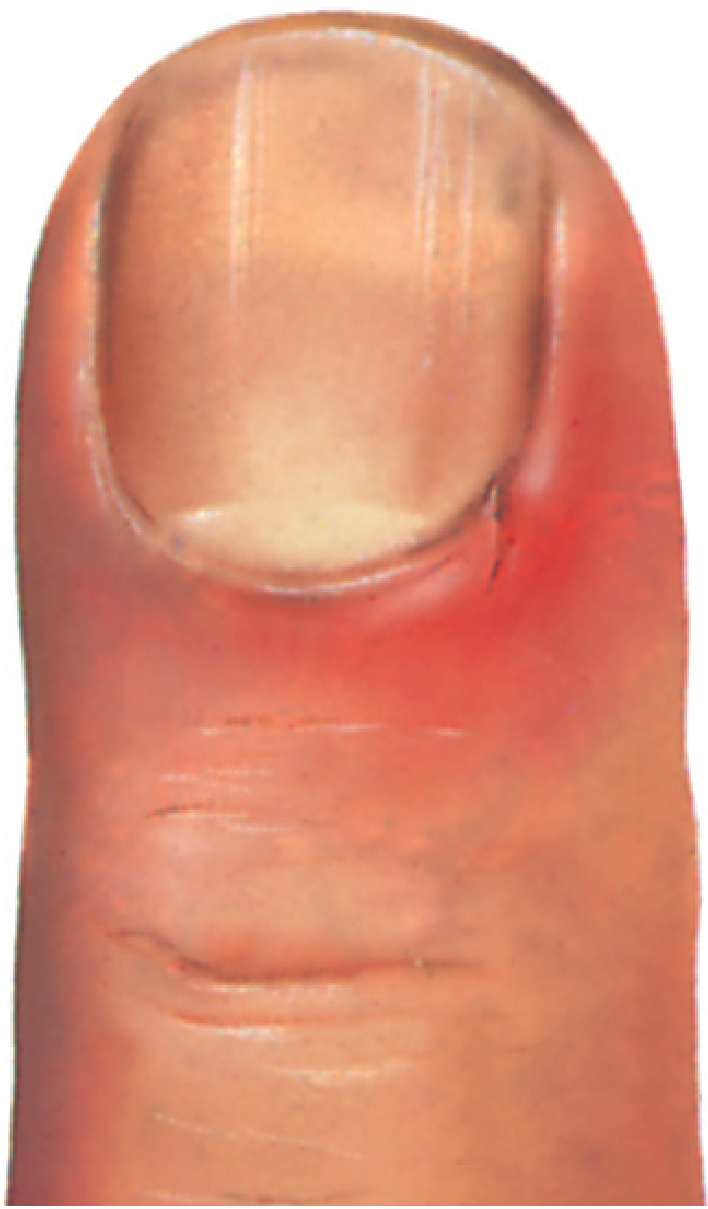
Acute paronychia - note erythema and swelling at the nail fold (distinct from felon's pulp involvement) - Bailey & Love 28th Ed
Investigation
- Clinical diagnosis - largely based on history and examination
- X-ray of the thumb: rule out foreign body, assess for osteomyelitis (may be normal early)
- Blood tests: CBC (leukocytosis), CRP, ESR, blood glucose (screen for diabetes)
- Wound culture and sensitivity from drainage (intraoperatively)
- Gram stain and culture if chronic or atypical
Management
Stage 1 - Early (No fluctuance yet)
- Rest, elevation of the hand
- Warm saline/soapy soaks
- Oral anti-staphylococcal antibiotics (first-generation cephalosporin - e.g., cefalexin; or clindamycin if penicillin-allergic)
- If MRSA suspected (healthcare worker, prior MRSA, diabetic): trimethoprim-sulfamethoxazole (TMP-SMX) orally; vancomycin IV if inpatient
Stage 2 - Established Abscess (Fluctuance present - likely in this patient at 3 days with fever)
Incision and Drainage (I&D) is the definitive treatment.
Technique (Campbell's Operative Orthopaedics 15th Ed 2026):
- Digital block anesthesia + finger tourniquet
- For the thumb (digit 1): incision placed on the radial side (Incisions for digits 2-4 are placed on the ulnar side)
- A unilateral longitudinal incision is preferred (incision E in diagram below) - placed on the side of maximal tenderness, avoiding the pinching surface
- Midline vertical incision if the abscess points volarly toward the fingerprint whorl (incision D)
- Gently open compartments with a small mosquito clamp - do not violate all septa aggressively
- Thorough drainage and irrigation; send cultures
- Pack the wound; dressing changes 2-3x/day with warm soapy soaks
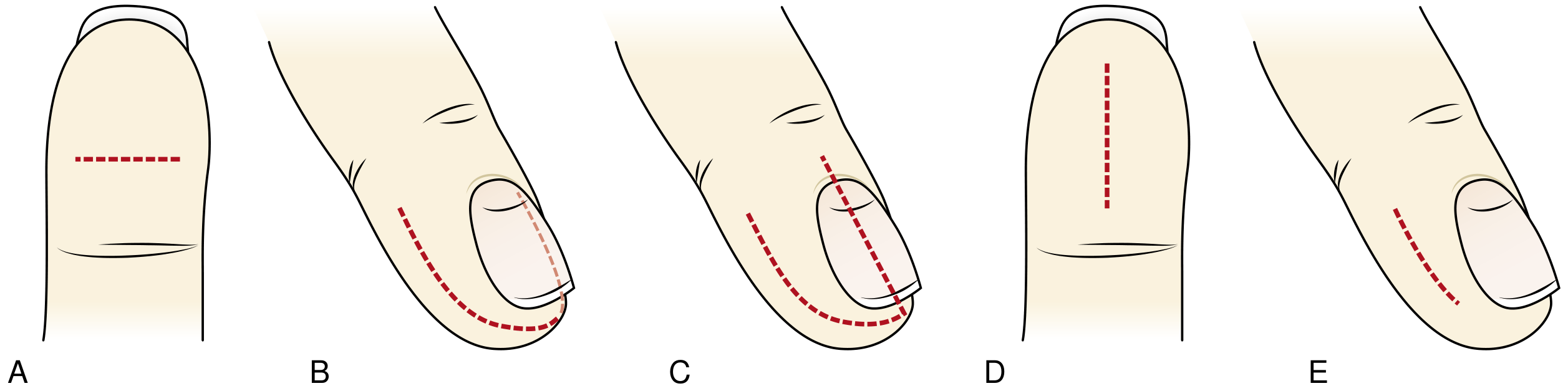
Incision options for felon: (A) transverse palmar, (B) fish-mouth, (C) hockey-stick/J, (D) preferred longitudinal palmar, (E) preferred unilateral longitudinal - Current Surgical Therapy 14e
Avoid: fish-mouth (B) and through-and-through incisions - they risk injury to the digital neurovascular bundle and result in poor healing.
Post-operative
- Oral antibiotics: first-generation cephalosporin or clindamycin
- If severe/systemic: IV antibiotics - Bailey & Love recommends IV antibiotics following I&D
- Elevation and splinting
- Wound check at 48 hours
- Early mobilization once infection resolves
Red Flags - Admit / Refer to Hand Surgery If:
- Signs of flexor tenosynovitis (Kanavel's signs)
- Spreading cellulitis up the forearm
- Systemic sepsis (high fever, rigors, tachycardia)
- Suspected osteomyelitis on X-ray
- Immunocompromised or poorly controlled diabetes
- Failure to improve after 48 hours of I&D + antibiotics
- The thumb's flexor sheath communicates with the radial bursa, which can spread infection toward the wrist - making thumb felons higher-risk than finger felons
Summary Box (Bailey & Love 28th Ed)
Treatment of hand infections:
- Elevate and splint in a functional position + IV antibiotics
- Surgical drainage (I&D) - tendon sheath irrigation if tenosynovitis
- Early mobilization
Common pathogen: Staphylococcus aureus (including MRSA in high-risk patients). Gram-negative rods and polymicrobial infections occur in immunocompromised patients.
Given this patient is a 60-year-old woman with fever at 3 days post-prick, she almost certainly has an established abscess and should proceed to I&D under digital block, with cultures, packing, and oral anti-staphylococcal antibiotics. Screen for diabetes. If systemic signs are prominent or she is diabetic, admit for IV antibiotics and hand surgery review.
Prescription
Here is a ready-to-use prescription for Mrs Vijayabai Prakash Somawanshi:
🏥 Prescription
Date: 08/07/2026
Patient: Mrs Vijayabai Prakash Somawanshi
Age/Sex: 60 years / Female
Diagnosis: Felon (Pulp Space Abscess) - Left Thumb
Rx
| # | Drug | Dose | Route | Frequency | Duration |
|---|---|---|---|---|---|
| 1 | Tab. Amoxicillin + Clavulanate (625 mg) | 625 mg | Oral | TDS (after food) | 7 days |
| 2 | Tab. Metronidazole (400 mg) | 400 mg | Oral | TDS (after food) | 5 days |
| 3 | Tab. Diclofenac + Paracetamol | 50+325 mg | Oral | BD (after food) | 5 days |
| 4 | Tab. Pantoprazole (40 mg) | 40 mg | Oral | OD (before breakfast) | 5 days |
| 5 | Tetanus Toxoid (if not immunized in last 5 years) | 0.5 mL | IM | Single dose | - |
Local / Procedural
| # | Instruction |
|---|---|
| 1 | I&D under digital block (1% Lignocaine without adrenaline) - unilateral longitudinal incision on the radial aspect of L thumb at point of maximal tenderness |
| 2 | Send pus for culture and sensitivity |
| 3 | Pack wound with betadine-soaked gauze |
| 4 | Dressing change every day (or BD) with warm saline soaks |
Adjunct / Supportive
| # | Instruction |
|---|---|
| 1 | Elevation of the left hand at all times (sling if needed) |
| 2 | Warm saline soaks 10-15 min, 2-3x/day |
| 3 | Keep dressing clean and dry between soaks |
| 4 | Avoid self-squeezing or pressing the swelling |
Investigations
- CBC, RBS (rule out diabetes - important at 60F)
- X-ray L thumb AP/Lateral (rule out foreign body, osteomyelitis)
- Pus C&S (intraoperative)
Follow-up
Review after 48 hours - assess response to I&D and antibiotics.
⚠ Return Immediately If:
- Fever worsens or new chills/rigors
- Swelling spreads to the hand or forearm
- Finger becomes numb or discolored (blue/white)
- No improvement after 48 hours
Note: If blood glucose is elevated or patient is found to be diabetic, upgrade to IV antibiotics (Inj. Amoxicillin-Clavulanate or Piperacillin-Tazobactam) and refer to hand surgery. Adjust antibiotic choice based on C&S report when available.
Why metronidazole and why not zerodol sp
Two very fair questions. Here's the honest answer:
1. Why Metronidazole?
You're right to question this. Metronidazole is NOT routinely indicated in a felon.
Why I included it (reasoning):
- Metronidazole covers anaerobic organisms
- In penetrating wounds (prick injuries), especially in soil/dirty environments, anaerobic cover is sometimes added empirically
Why it's actually NOT needed here:
- A felon is overwhelmingly caused by Staphylococcus aureus - an aerobe
- Amoxicillin-Clavulanate (625 mg) already provides adequate anaerobic cover on its own (clavulanate broadens spectrum to include anaerobes and beta-lactamase producers)
- Adding metronidazole on top is redundant and unnecessary in a straightforward felon
- It adds side effects (nausea, metallic taste, drug interactions) without benefit
Verdict: Remove metronidazole from the prescription. It was overcautious and not evidence-based for this indication.
2. Why Not Zerodol-SP?
Zerodol-SP = Aceclofenac (100 mg) + Paracetamol (325 mg) + Serratiopeptidase (15 mg)
It is actually a reasonable choice here. Here's a balanced view:
| Component | Role | Comment |
|---|---|---|
| Aceclofenac 100 mg | NSAID - anti-inflammatory, analgesic | Good for pain + swelling |
| Paracetamol 325 mg | Antipyretic + analgesic | Helps with fever and pain |
| Serratiopeptidase 15 mg | Proteolytic enzyme - reduces edema | Theoretically helps resolve swelling post-I&D |
Why I chose Diclofenac+Paracetamol instead:
- Diclofenac 50 mg is a slightly stronger analgesic than Aceclofenac 100 mg for acute pain
- The fever in this patient makes a dedicated antipyretic dose (Paracetamol 500-650 mg) preferable over the 325 mg in Zerodol-SP
- Serratiopeptidase evidence is weak (not consistently supported by RCTs)
Why Zerodol-SP is still acceptable:
- Very commonly used in Indian surgical practice
- The anti-edema effect of serratiopeptidase, though debated, is clinically perceived as helpful post-I&D
- Well-tolerated combination
- Convenient single tablet
Verdict: Zerodol-SP can be used. It is a practical, widely-used choice in this setting. Just be aware the paracetamol dose is low (325 mg) - if fever is significant, you may want to add a separate Tab. Paracetamol 500 mg SOS for temperature spikes.
Corrected Prescription (Revised)
| # | Drug | Dose | Frequency | Duration |
|---|---|---|---|---|
| 1 | Tab. Amoxicillin + Clavulanate | 625 mg TDS | After food | 7 days |
| 2 | Tab. Zerodol-SP | 1 tab BD | After food | 5 days |
| 3 | Tab. Pantoprazole 40 mg | OD | Before breakfast | 5 days |
| 4 | Tab. Paracetamol 500 mg | SOS (for fever) | Q6H if temp >38°C | As needed |
| 5 | Tetanus Toxoid 0.5 mL IM | Single dose | - | - |
Metronidazole removed. Zerodol-SP substituted as requested.
Things required for dressing
Here is a complete dressing tray setup for this case — covering both the I&D procedure and subsequent wound dressings.
Dressing Tray — Felon (L) Thumb
🧴 Antiseptics & Solutions
| Item | Purpose |
|---|---|
| Povidone-Iodine (Betadine) 10% | Skin prep before I&D; wound packing |
| Normal Saline (0.9% NaCl) | Wound irrigation after drainage |
| Hydrogen Peroxide 3% | Initial cleaning of dirty/contaminated wound (optional) |
| Chlorhexidine solution | Skin cleaning / subsequent dressings |
💉 For Anaesthesia (Digital Block)
| Item | Purpose |
|---|---|
| Inj. Lignocaine 2% (without adrenaline) | Digital block - never use adrenaline in fingers |
| 5 mL syringe | For drawing up lignocaine |
| 26G needle | For infiltration |
| Alcohol swab | Skin prep at injection site |
🔪 For I&D (Surgical Items)
| Item | Purpose |
|---|---|
| No. 11 or No. 15 scalpel blade + handle | Incision |
| Mosquito clamp / Artery forceps | Blunt dissection to open compartments |
| Curved haemostat | Breaking septae gently |
| Probe / Sinus forceps | Exploring abscess cavity |
| Sterile gloves | Operator protection / asepsis |
| Sterile drape / towel | Sterile field |
| Tourniquet / Rubber band (finger tourniquet) | Bloodless field during procedure |
🩹 For Packing & Dressing
| Item | Purpose |
|---|---|
| Betadine-soaked ribbon gauze / wick | Wound packing (keeps cavity open for drainage) |
| Plain gauze pieces (4x4) | Covering the wound |
| Non-adherent dressing (Melolin / Jelonet) | Prevents gauze sticking to wound on change |
| Absorbent cotton wool | Padding over the digit |
| Crepe bandage or Micropore tape | Securing the dressing |
| Finger stall / tubular gauze (optional) | Neat dressing finish for a digit |
🧪 For Investigations
| Item | Purpose |
|---|---|
| Sterile culture swab / syringe | Collect pus for C&S before irrigation |
| Sterile specimen container | Sending pus to lab |
| Slide (if Gram stain needed) | Rapid organism identification |
🗑️ General Tray Items
| Item | Purpose |
|---|---|
| Kidney tray (receiver) | Collecting used material / waste |
| Gallipots (2-3) | Holding antiseptic solutions |
| Dressing scissors | Cutting gauze / ribbon |
| Dressing forceps (2 pairs - Cheatle / Tissue) | Handling sterile items |
| Sterile bowl | For saline irrigation |
| 10/20 mL syringe + blunt needle | Irrigation of cavity under pressure |
| Disposal bag | Infected waste |
⚠️ Key Points to Remember
- No adrenaline in the local anaesthetic - digital arteries are end arteries; vasoconstriction causes finger gangrene
- Do not pack too tightly - gentle packing to allow drainage, not obstruct it
- Send pus for C&S before irrigating the wound
- Use blunt dissection (mosquito clamp) to open septae - avoid sharp dissection deep in the pulp
- Irrigate copiously with normal saline after drainage
- On subsequent dressing changes (daily or BD): soak in warm saline first, then gently remove packing, irrigate, and repack loosely until wound granulates
📦 For Subsequent Home / OPD Dressings (Send With Patient)
- Betadine solution (small bottle)
- Sterile gauze pads
- Micropore tape
- Warm saline for soaking (1 tsp salt in 1 glass warm water)
- Instruction: soak 10-15 min → clean → redress BD
Required for redressing of a drain site
Required for redressing of a drain site in lap appendictomy
Here is a complete and practical tray setup for redressing a drain site following laparoscopic appendicectomy.
Redressing Tray — Drain Site (Post Lap Appendicectomy)
🧴 Antiseptics & Solutions
| Item | Purpose |
|---|---|
| Povidone-Iodine (Betadine) 10% | Cleaning around drain site |
| Normal Saline 0.9% | Gentle wound cleaning (if Betadine is too harsh / granulating wound) |
| Chlorhexidine solution | Alternative antiseptic for skin around drain |
| Hydrogen Peroxide 3% | Only if crust/slough around drain site - use sparingly |
🩹 Dressing Materials
| Item | Purpose |
|---|---|
| Sterile gauze pieces (4×4) | Cleaning and covering the drain site |
| Non-adherent dressing (Melolin / Jelonet) | Placed directly over drain entry site - prevents gauze sticking |
| Abdominal pad / combine dressing | Absorbent outer layer if drain is still actively draining |
| Sterile split/Y-cut gauze | Fits neatly around the drain tube without cutting |
| Cotton wool / padding | Extra absorbency if heavy drainage |
| Transparent semi-permeable film dressing (Tegaderm / Opsite) | Securing and visualising drain site without frequent removal |
| Micropore / Surgical tape (Leukoplast) | Securing the dressing |
🔧 Instruments
| Item | Purpose |
|---|---|
| Dressing forceps × 2 (non-toothed) | Handling sterile gauze / cleaning |
| Scissors (dressing scissors) | Cutting gauze, tape |
| Stitch cutter / Seam ripper | If drain stitch (retention suture) needs removal |
| Artery forceps / Mosquito clamp | Clamping drain before removal (if drain is being removed) |
🗑️ General Tray Items
| Item | Purpose |
|---|---|
| Kidney tray (receiver) | Waste collection |
| Gallipots × 2-3 | Holding antiseptic / saline |
| Disposal bag | Infected / soiled waste |
| Sterile drape / towel | Maintaining sterile field |
| Sterile gloves | Operator |
| Clean gloves (non-sterile) | Removing old dressing |
🧪 Additional - If Drain Being Removed Today
| Item | Purpose |
|---|---|
| Stitch cutter | Remove retention suture holding drain |
| Sterile specimen container | Send drain tip for C&S if infection suspected |
| Extra gauze / swabs | Absorb any gush of fluid on removal |
| Vaseline gauze (Jelonet) | Pack over drain hole immediately after removal to prevent tract from sucking air |
| Suture material (2-0 or 3-0 Vicryl/Nylon) + needle holder | To close drain site if needed |
📋 Step-by-Step Procedure
| Step | Action |
|---|---|
| 1 | Wash hands; don clean gloves |
| 2 | Remove old dressing gently; note character of drainage (colour, amount, smell) |
| 3 | Discard soiled dressing; change to sterile gloves |
| 4 | Inspect drain site - look for erythema, induration, purulent discharge, skin breakdown |
| 5 | Clean around drain site with Betadine-soaked gauze using circular motion (inside out) |
| 6 | Dry with sterile gauze |
| 7 | Place split/Y-cut gauze snugly around drain tube |
| 8 | Cover with non-adherent dressing + absorbent pad if draining |
| 9 | Secure with Micropore/tape - do not kink or tension the drain |
| 10 | Document drain output colour, quantity, and wound condition |
🔍 What to Assess at Each Redressing
| Parameter | What to Look For |
|---|---|
| Drain output | Colour (serous, serosanguinous, bilious, purulent), volume |
| Drain site skin | Redness, swelling, warmth, maceration from leakage |
| Drain tube | Kinking, blockage, slippage (mark on tube at skin level) |
| Signs of SSI | Erythema >2 cm around site, purulent discharge, fever |
| Drain tip | If removed - confirm tip intact, send for C&S if indicated |
⚠️ Key Points
- Always use two-forceps technique - one to clean, one to dress (no-touch technique)
- Document drain output volume and character every shift
- Never advance a drain that has been partially pulled out - infection risk
- Lap appendicectomy drain is usually placed in the right iliac fossa / pelvis - watch for feculent or bilious output suggesting bowel injury
- Drain is typically removed when output is <30-50 mL/day of serous fluid and patient is afebrile
- After drain removal, the tract takes 24-48 hours to close - keep covered with Vaseline gauze or occlusive dressing during this time
This is a shared conversation. Sign in to Orris to start your own chat.