
Which sign can be seen in the patients of osteoporosis
Reading File
Quick answer: The classic clinical sign in osteoporosis is the "Codfish vertebra" (biconcave deformity of vertebral bodies on X-ray). Additional signs include dorsal kyphosis ("Dowager's hump"), loss of height, and a positive "piano key sign." On imaging, you may see "empty vertebra" or "frame vertebra" appearance.
Searching sources...
Finding Sources
Finding Sources
Reading File
Reading File
Reading File
Finding Sources
Reading File
Reading File
Now let me fetch the key comparison image and the X-ray image for visual reference:
Loading Image
Loading Image
Here is a thorough, textbook-sourced answer on the signs seen in osteoporosis:
Signs in Osteoporosis
Clinical Signs
| Sign | Description |
|---|---|
| Tenderness at fracture site | The primary physical sign - localized tenderness on palpation/percussion over affected vertebrae or long bones |
| Dowager's Hump (Widow's Hump) | Progressive dorsal kyphosis from wedging/collapse of multiple thoracic vertebral bodies |
| Height Loss | >2 inches (5 cm) from peak height suggests vertebral compression fractures (silent in up to 75% of patients) |
| Increased wall-to-occiput distance | When standing with heels, buttocks, and back against a wall, an increased gap between the wall and back of the head reflects kyphosis severity - this is a measurable clinical sign |
| Decreased rib-to-pelvis distance | Due to vertebral collapse causing loss of trunk height; can be objectively measured |
| Rib abutting iliac crest | In severe kyphosis, the lower ribs can actually touch the iliac crest, sometimes compromising respiratory function |
The second image above shows the rib-to-pelvis (decreased) distance being assessed on physical exam - the two lines show how close the rib margin and iliac crest have come due to vertebral compression.
Radiographic Signs
| Sign | Explanation |
|---|---|
| Codfish vertebra | Biconcave deformity of vertebral bodies due to central endplate collapse, resembling the shape of a codfish spine |
| Wedge fracture / Anterior wedging | Most common vertebral deformity; the front of the vertebral body collapses more than the back, causing kyphosis |
| Crush fracture | Complete collapse of a vertebral body |
| Osteopenia / Decreased radiodensity | Only visible when >30% bone mass is lost; trabecular thinning is more prominent in axial skeleton |
| Pencil-thin cortices | Endosteal bone resorption narrows the cortex |
| Sparse but thickened vertical trabeculae | Remaining vertical trabeculae appear accentuated ("empty cage" appearance) |
| Frame vertebra / Picture frame vertebra | Vertebral body appears as an empty box due to loss of horizontal trabeculae with preservation of the cortical outline |
| Fragility fractures | Most common at the spine (T7-L4), distal forearm (Colles fracture), and proximal femur (hip fracture) |
Key Comparison: Osteoporosis vs Osteomalacia
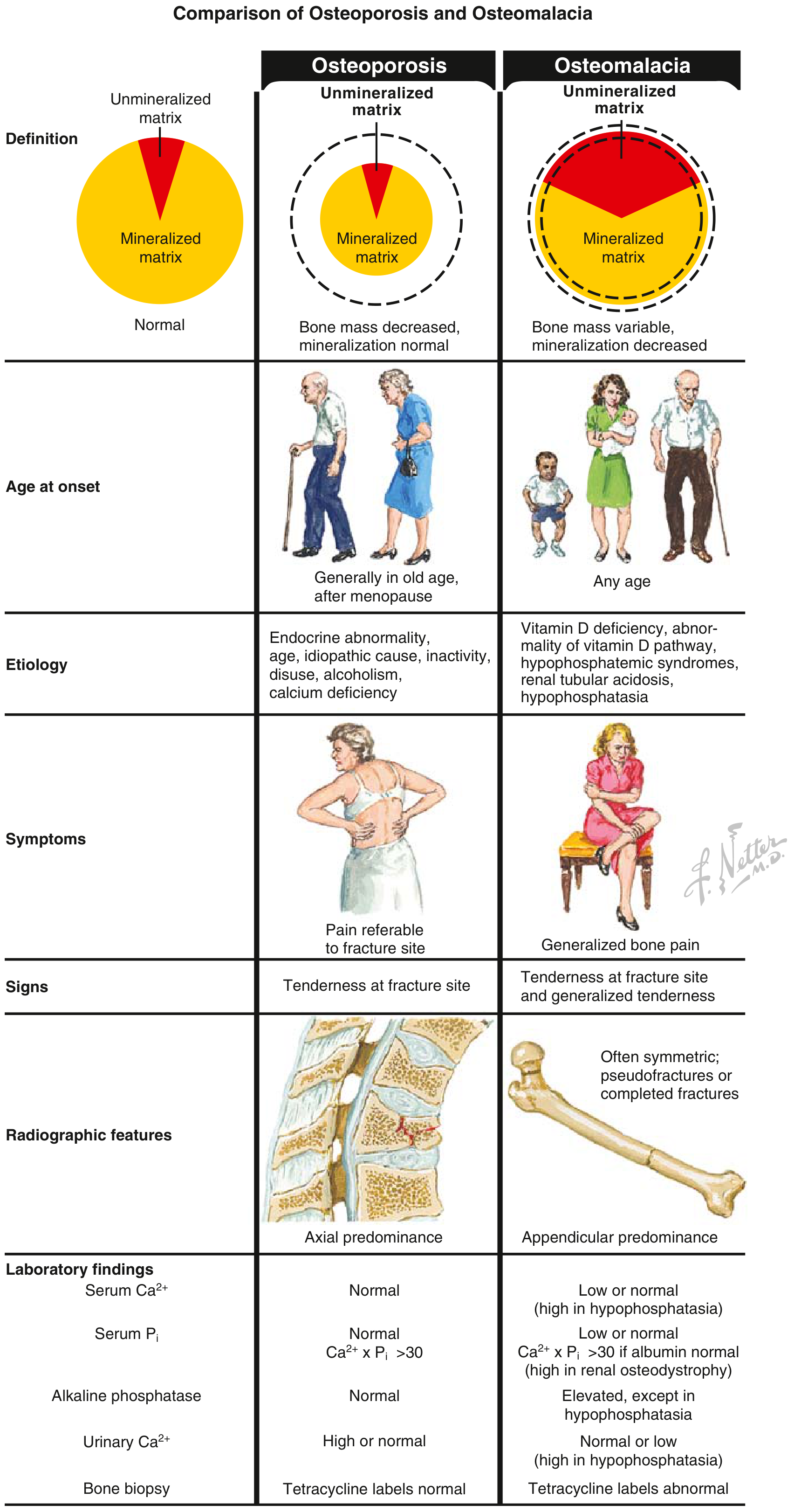
This Netter diagram is an important exam resource. Note that in osteoporosis:
- Signs = tenderness only at the fracture site (not generalized)
- Labs = all normal (Ca²+, PO₄, ALP, PTH)
- Radiographs = axial predominance, fractures
Whereas osteomalacia shows generalized tenderness and abnormal labs (low Ca, low PO₄, elevated ALP).
Summary for Exams
The most asked-about sign in osteoporosis is the Codfish vertebra (biconcave vertebral bodies on lateral spine X-ray). The classic clinical triad is:
- Kyphosis (Dowager's hump)
- Height loss
- Fragility fractures (especially vertebral, hip, and distal radius)
- Miller's Review of Orthopaedics 9th Edition, p. 46-47
- Grainger & Allison's Diagnostic Radiology, p. 1085
- Goldman-Cecil Medicine, Physical Examination of Metabolic Bone Disease
This is a shared conversation. Sign in to Orris to start your own chat.