
Clinical staging for hodgikins
I'll look this up in the medical library for you.
Searching sources...
Reading File
Finding Sources
Finding Sources
Good results. Let me pull the detailed content from the most authoritative sources - Goldman-Cecil and Harrison's.
Reading File
Reading File
Let me get the rest of the Modified Ann Arbor table and the Cotswolds modifications from Goldman-Cecil, plus the Lugano classification from Fishman's.
Reading File
Reading File
I now have comprehensive information. Let me also fetch the anatomical diagram from Goldman-Cecil which is highly relevant.
Loading Image
I have everything needed for a thorough answer. Here it is:
Clinical Staging of Hodgkin Lymphoma
The Ann Arbor System (with Cotswolds Modification)
This is the standard staging system for Hodgkin lymphoma (HL), originally developed in 1971 and later modified at Cotswolds (1989). The Lugano Classification (2014) is its current practical update used in most modern protocols.
The Four Stages
| Stage | Involvement |
|---|---|
| I | Single lymph node region or a single extralymphatic site (IE) |
| II | Two or more lymph node regions on the same side of the diaphragm (II), or limited contiguous extranodal involvement (IIE) |
| III | Lymph node regions on both sides of the diaphragm (III); may include local extralymphatic extension (IIIE), spleen (IIIS), or both (IIISE) |
| IV | Diffuse or disseminated involvement of one or more extralymphatic organs (bone marrow, liver, lung, bone) with or without lymph node involvement |
The spleen and Waldeyer's ring each count as nodal sites in this system. - Goldman-Cecil Medicine
Suffix Designations
| Suffix | Meaning |
|---|---|
| A | No B symptoms (asymptomatic) |
| B | Presence of at least one B symptom: unexplained fever >38°C, recurrent night sweats, unexplained weight loss >10% of body weight within 6 months before staging |
| E | Limited, contiguous extranodal extension that can be included in an irradiation field |
| S | Splenic involvement |
| X (older) / Bulky | Any tumor mass with largest diameter ≥10 cm, OR mediastinal mass >1/3 the transthoracic diameter |
Disease Classification by Stage
- Limited (early) stage: Stages I and II without bulky disease
- In Europe, further subdivided into favorable vs unfavorable limited
- Advanced stage: Stage III, Stage IV, or any stage with bulky disease
Lymph Node Regions Used in Staging
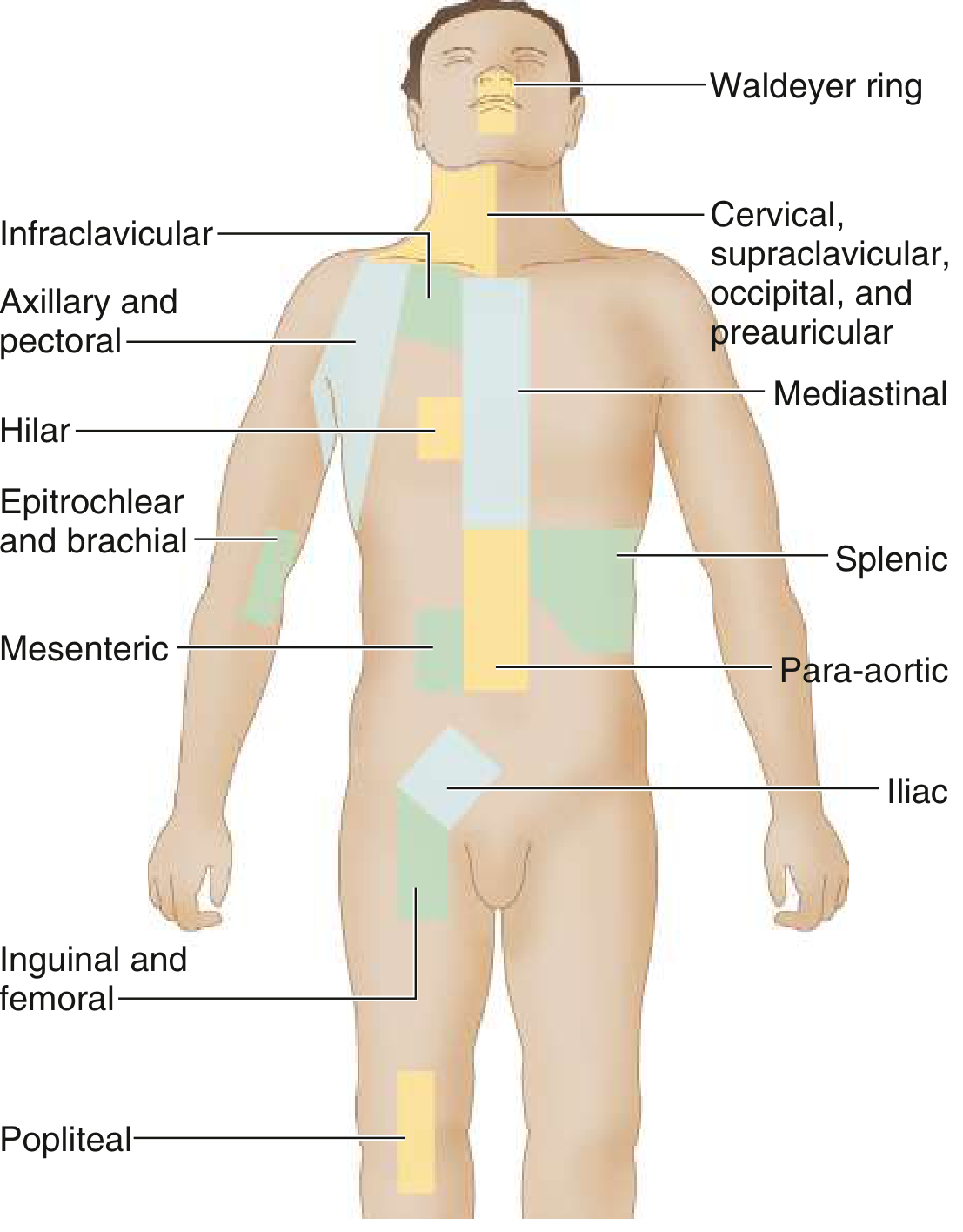
Workup Required for Staging (Table 172-2, Goldman-Cecil)
- Complete history - B symptoms, other constitutional symptoms
- Physical exam - lymphadenopathy, organomegaly
- CBC, ESR, LFTs (bilirubin, ALP, LDH), renal function, serum albumin, protein electrophoresis
- HIV, Hep B and C serology
- Chest X-ray (PA and lateral)
- CT scan - neck, thorax, abdomen, pelvis (contrast-enhanced, ≤1 cm slices)
- FDG-PET/CT - now considered mandatory; more sensitive/specific than CT alone; replaces the need for bone marrow biopsy in staging
Lugano Classification (2014 Update)
The current standard in most guidelines, based on Ann Arbor with Cotswolds modifications:
| Stage | Characteristics |
|---|---|
| I | One lymph node region or group of adjacent nodes; or single extranodal lesion (I |
| II | ≥2 lymph node regions, same side of diaphragm; or limited contiguous extranodal involvement (II |
| III | Lymph node regions or lymphoid structures on both sides of diaphragm |
| IV | Non-contiguous extralymphatic involvement |
| A/B | Absence/presence of B symptoms (for HL only) |
| E | Extranodal involvement |
| Bulky | Mass >10 cm or >1/3 of transthoracic diameter; described by size, no longer labeled "X" |
(Fishman's Pulmonary Diseases, Table 82-7)
Survival by Stage (Approximate)
| Stage | Cure Rate |
|---|---|
| I-II (limited, non-bulky) | >90-95% |
| IIIA | 30-90% |
| IIIB | 60-70% |
| IV | 50-60% |
Key clinical notes:
- Stage IV disease is effectively confined to bone marrow, liver, lung, or bone. HL at any other extranodal site should prompt questioning of the diagnosis or a search for HIV infection.
- B symptoms are a major prognostic marker - stage IIB behaves more like advanced disease than early disease.
- PET/CT is now the gold standard both for initial staging and for interim response assessment during chemotherapy (used to guide consolidation radiotherapy decisions).
Sources: Goldman-Cecil Medicine (Table 172-2, 172-3), Fischer's Mastery of Surgery (Table 188.2), Fishman's Pulmonary Diseases (Table 82-7)
This is a shared conversation. Sign in to Orris to start your own chat.