
Ebola disease
Ebola virus disease
Ebola virus clinical features hemorrhagic fever
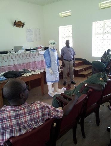
This clinical photograph captures a Personal Protective Equipment (PPE) demonstration during a medical training session focused on Viral Hemorrhagic Fever (VHF) preparedness, specifically for Ebola Virus Disease (EVD). A participant is shown wearing a complete, multi-layered PPE ensemble which includes a white head hood, protective goggles, a light blue long-sleeved isolation gown, a white fluid-resistant apron, white gloves, and high-top white boot covers. The training takes place in a classroom setting with other participants and instructors visible, emphasizing the 'donning and doffing' protocol necessary for infection prevention and control (IPC). The visual illustrates standard barrier nursing precautions and high-level protection required for managing highly infectious pathogens in healthcare or border surveillance contexts. Key educational concepts include contact and droplet precautions, biocontainment procedures, and the role of multi-disciplinary teams in epidemic response.
An infographic-style photograph illustrating the concept of a global health crisis, specifically focusing on Ebola Virus Disease (EVD). The image features a white sheet of paper resting on a dark surface, with the word 'EBOLA' written centrally in bold, crimson-red lettering. The medium used for the text has a fluid, slightly irregular texture resembling blood or liquid pigment, suggesting the visceral nature of the hemorrhagic fever. To the top right of the paper sits a small, transparent glass globe on a gold-toned pedestal, symbolizing the international impact and geographic spread of infectious disease outbreaks. This visual serves as an educational representation of global health security, emerging infectious diseases, and the public health response to viral pathogens with epidemic potential. It is suitable for discussions regarding epidemiology, international disease surveillance, and the sociological impact of high-consequence pathogens.
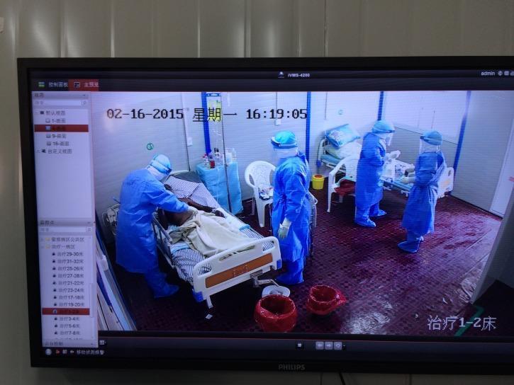
Clinical photograph of a high-containment Ebola Treatment Unit (ETU) ward, captured via a surveillance monitor. The image illustrates advanced infection control measures during the management of a highly infectious viral hemorrhagic fever. Multiple medical personnel are visible wearing full-body Level C/D personal protective equipment (PPE), consisting of fluid-resistant blue coveralls with integrated hoods, full-face shields, and waterproof shoe covers. They are shown providing direct bedside care to a patient on a clinical hospital bed. The environment is partitioned for biocontainment, featuring red-lined biohazard waste receptacles for contaminated materials and intravenous therapy equipment. This visual emphasizes the stringent protocols required for the containment of infectious diseases like Ebola, highlighting the layout of a contaminated zone and the operational requirements of healthcare workers in a specialized treatment center.
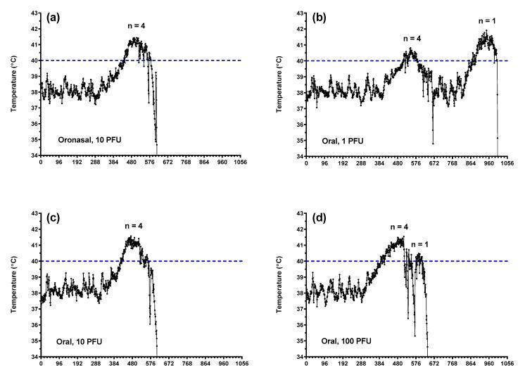
This set of line graphs illustrates core body temperature alterations in a ferret model of Ebola virus (EBOV) disease across different inoculation routes and doses. The graphs (a-d) plot temperature in degrees Celsius (°C) against time (every tick representing 4 hours). Subfigure (a) shows the oronasal route (10 PFU), while (b-d) show the oral route at 1, 10, and 100 PFU doses, respectively. A horizontal dashed line at 40°C indicates the threshold for fever. Common to most groups is an initial baseline fluctuation (37°C–39°C), followed by a progressive febrile response exceeding 41°C. Notably, higher doses (10 and 100 PFU) exhibit more rapid progression to peak fever, while the 1 PFU oral group displays a delayed or biphasic temperature profile. All lethal cases culminate in a sharp terminal decline (hypothermia), with temperatures dropping below 35°C, representing clinical deterioration and the near-terminal phase of the hemorrhagic fever syndrome.
Ebola disease 2024 2025 outbreak treatment guidelines
Ebola Virus Disease (EVD)
Virology & Classification
| Species | Notes |
|---|---|
| Zaire ebolavirus (EBOV) | Highest lethality; responsible for most major outbreaks |
| Sudan ebolavirus (SUDV) | Second most lethal |
| Bundibugyo ebolavirus (BDBV) | Lower CFR (~25-36%) |
| Taï Forest ebolavirus (TAFV) | Single human case reported |
| Reston ebolavirus (RESTV) | Found in Asian pigs/primates; not pathogenic in humans |
| Bombali ebolavirus | Discovered in bats; human pathogenicity unknown |
Epidemiology & History
- First described in 1976 simultaneously in Sudan and Zaire (now DRC), near the Ebola River - hence the name.
- Natural reservoir: Fruit bats (Pteropodidae family) are strongly suspected; the virus has been detected in multiple bat species. The precise spillover mechanism to humans remains under study.
- Index cases in outbreaks are typically linked to handling of dead animals (bats, bushmeat) or exposure to infected body fluids.
Major Outbreaks
| Outbreak | Cases | Deaths | CFR |
|---|---|---|---|
| 1976 DRC (Zaire) | 318 | 280 | 88% |
| 2013-2016 West Africa | 28,652 | 11,325 | ~40-70% |
| 2018-2020 DRC (North Kivu) | ~3,500 | ~2,300 | ~66% |
Transmission
- Direct contact with blood, secretions, organs, or other body fluids of infected humans or animals.
- Fomite transmission: contact with contaminated surfaces/objects.
- NOT airborne under natural conditions (no evidence of droplet or aerosol transmission in community settings).
- Semen: EBOV can persist in semen for months to over a year after recovery - sexual transmission has been documented.
- Burial practices: Unsafe handling of bodies of the deceased is a major transmission route, as viral loads remain high post-mortem.
- Individuals are NOT contagious during the incubation period - only once symptoms begin.
Pathogenesis
- Entry: EBOV GP binds to multiple cell surface receptors (including Niemann-Pick C1/NPC1 within endosomes). Primary targets are monocytes, macrophages, and dendritic cells.
- Immune evasion: VP35 and VP24 proteins inhibit interferon signaling, enabling the virus to replicate unchecked in early infection.
- Cytokine storm: Infected macrophages release massive amounts of pro-inflammatory cytokines (TNF-α, IL-6, IL-8, IL-1β), triggering systemic inflammatory response.
- Vascular damage: Infection of endothelial cells leads to increased permeability, leading to fluid extravasation and shock.
- Coagulopathy: DIC develops due to endothelial injury, tissue factor expression, and hepatic dysfunction - driving the hemorrhagic manifestations.
- Multi-organ failure: Progressive hepatic, renal, and adrenal necrosis culminates in fatal shock.
Clinical Features
Incubation Period
- 2-21 days (most commonly 5-7 days)
Phase 1 - Early/Prodromal (Days 1-5)
- High fever (often >38.5°C)
- Severe headache
- Myalgia and fatigue
- Sore throat
- Nausea, profuse vomiting and diarrhea
- Abdominal pain
Phase 2 - Late/Hemorrhagic (Days 5-10+)
- Hemorrhagic manifestations: petechiae, ecchymoses, mucosal bleeding (gums, nose), gastrointestinal bleeding, conjunctival injection
- Maculopapular rash: erythematous, non-pruritic, eventually desquamates
- Hypovolemia from massive GI fluid losses
- Jaundice (hepatic involvement)
- CNS involvement: confusion, seizures, coma
- Progressive shock and multi-organ failure in fatal cases
Note: Not all patients develop frank hemorrhage. The hallmark is actually the profound GI syndrome.

Laboratory Findings
| Test | Typical Finding |
|---|---|
| CBC | Leukopenia (early), thrombocytopenia |
| Coagulation | Prolonged PT/aPTT, elevated D-dimer (DIC) |
| LFTs | Elevated AST, ALT (transaminitis) |
| Renal | Elevated creatinine (AKI) |
| Electrolytes | Hypokalemia, hypocalcemia, hyponatremia |
| Serum amylase | Elevated (pancreatitis) |
Diagnosis
- RT-PCR (reverse transcriptase polymerase chain reaction) on plasma/blood is the gold-standard confirmatory test. Sensitivity is highest after symptom day 3.
- Rapid antigen point-of-care tests: 15-minute turnaround, good sensitivity/specificity vs. RT-PCR - useful in field settings.
- ELISA for IgM/IgG antibodies: useful for seroprevalence studies and retrospective diagnosis.
- Testing criteria: patient must have both epidemiological exposure history AND compatible symptoms before testing is indicated.
Differential Diagnosis
- Malaria (most important to exclude in endemic areas)
- Typhoid fever
- Meningococcemia
- Marburg virus disease
- Other viral hemorrhagic fevers (Lassa, Crimean-Congo, Rift Valley)
- Leptospirosis
- Septicemia from bacterial causes
Management
1. Infection Control (Paramount)
- Isolate immediately in a single-patient room with negative air pressure if available.
- Full PPE: surgical hood covering head and neck, full-face shield, N95 or PAPR respirator, fluid-resistant gown, double gloves, boot covers. No skin should be exposed.
- Rigorous donning and doffing protocols with trained monitor - the CDC mandates demonstrated competency.
- CDC tiered system: Frontline facilities → Ebola Assessment Hospitals → Ebola Treatment Centers.
2. Supportive Care (Cornerstone of Treatment)
- Aggressive IV fluid and electrolyte resuscitation - combats hypovolemia from GI losses.
- Empirical antimalarial treatment (malaria coinfection common).
- Broad-spectrum antibiotics for secondary bacterial infections.
- Antipyretics (acetaminophen preferred over NSAIDs due to platelet effects).
- Organ support: renal replacement therapy (RRT) for AKI, mechanical ventilation, vasopressors for shock, blood product transfusion (FFP, platelets, PRBCs for DIC/hemorrhage).
3. Specific Antiviral Therapies
| Agent | Type | Status |
|---|---|---|
| Inmazeb (REGN-EB3) | Triple monoclonal antibody (atoltivimab + maftivimab + odesivimab) | FDA approved Oct 2020 for Zaire ebolavirus in adults and children |
| Ebanga (MAb114 / ansuvimab) | Single monoclonal antibody | FDA approved Dec 2020 for Zaire ebolavirus |
| Remdesivir | Nucleotide analogue | Studied; not approved for EVD |
| Convalescent plasma | Polyclonal antibodies | No proven clinical benefit in RCTs |
| ZMapp | Triple monoclonal antibody | Earlier compound; superseded by above |
Prognosis & Mortality
- CFR ranges 25-90% depending on the species, outbreak setting, and available care.
- Zaire EBOV historically: ~60-80% without care; ~37% with optimized supportive + specific therapy (PALM trial).
- Patients treated in high-resource settings (US, Europe) during 2014-2016: mortality ~18.5%, far lower than African outbreak rates.
- Predictors of poor prognosis include: high viral load at presentation, coma, severe hemorrhage, AKI, very young or very old age. (Per the 2026 meta-analysis [PMID: 42163115], high viral load and neurological involvement are the strongest mortality predictors.)
Post-Ebola Syndrome
- Uveitis and other ocular complications (EBOV can persist in the aqueous humor)
- Arthralgia and myalgia
- Fatigue, sleep disturbances
- Neuropsychiatric symptoms (PTSD, depression)
- Reproductive health: A 2025 meta-analysis ([PMID: 39689367]) documented significant impacts on female reproductive health including menstrual irregularities and adverse pregnancy outcomes post-EVD.
- Viral persistence in immune-privileged sites (eye, CNS, testes, placenta) for months to years.
Prevention & Vaccines
Ervebo (rVSV-ZEBOV)
- Live attenuated recombinant vaccine: vesicular stomatitis virus (VSV) vector expressing the Ebola Zaire glycoprotein.
- FDA approved in December 2019 - first approved Ebola vaccine.
- Single intramuscular dose.
- Indications: persons ≥18 years who are outbreak responders, staff at federally designated Ebola treatment centers, or biosafety level-4 facility workers.
- Adverse effects: injection site reactions, headache, fever, arthralgia/arthritis (more common in women and those with prior arthritis history).
- Post-vaccination precautions: avoid blood donation for 6 weeks; use barrier contraception for 2 months; avoid sharing utensils/razors for 2 weeks (live virus shedding possible).
- Deployed with ring vaccination strategy in the 2018-2020 DRC outbreak with considerable success.
Zabdeno + Mvabea (Two-dose regimen)
- Ad26.ZEBOV prime + MVA-BN-Filo boost (8 weeks apart).
- Approved in Europe; used in DRC and neighboring countries.
General Prevention
- Avoid contact with blood/body fluids of infected persons or animals.
- Safe burial practices with full PPE.
- Contact tracing and 21-day surveillance of exposed contacts.
- No travel to active outbreak areas without appropriate preparation.
Public Health Response Framework
- Case surveillance and contact tracing (21-day monitoring period for contacts)
- Laboratory diagnosis network
- Dedicated Ebola Treatment Centers (ETCs)
- Safe and dignified burials
- Ring vaccination of contacts and contacts-of-contacts
- Community engagement and risk communication
- International coordination (WHO, MSF, CDC, etc.)
Key Numbers to Remember
| Parameter | Value |
|---|---|
| Incubation period | 2-21 days (typically 5-7) |
| CFR range | 25-90% |
| CFR with modern care | ~18-37% |
| Contagious period | From symptom onset onwards |
| Semen viral persistence | Up to 12+ months post-recovery |
| Vaccine (Ervebo) dose | Single IM injection |
Recent evidence note: A 2026 systematic review and meta-analysis ([PMID: 42163115], BMC Infectious Diseases) identified high viral load at presentation and neurological features as the strongest independent predictors of mortality in EVD - reinforcing the importance of early diagnosis and rapid initiation of monoclonal antibody therapy.