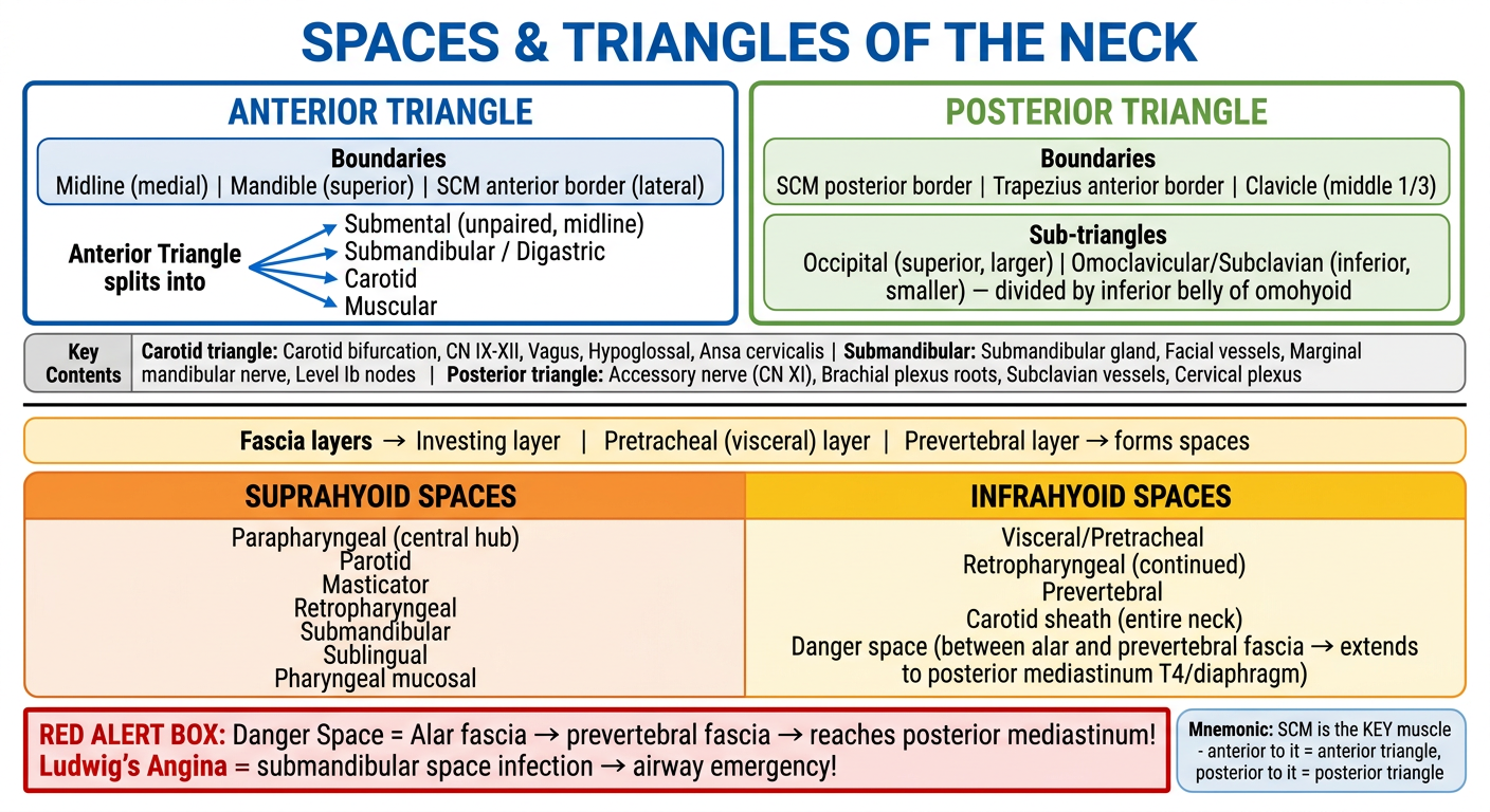
I have all the content I need. Here is the complete, high-yield short note:
🏥 Spaces & Triangles of the Neck
5-Mark Short Note | 2nd Year Resident Edition
🔑 THE ONE RULE TO REMEMBER FIRST
SCM (Sternocleidomastoid) is the master divider.
Anterior to SCM = Anterior Triangle | Posterior to SCM = Posterior Triangle
PART 1: TRIANGLES OF THE NECK
OVERVIEW MINDMAP
                    NECK
                      |
          ____________|____________
         |                         |
  ANTERIOR TRIANGLE          POSTERIOR TRIANGLE
  (medial to SCM)             (lateral to SCM)
         |                         |
  ┌──────┴────────┐          ┌─────┴──────┐
  Boundaries:          Boundaries:
  • Midline (medial)   • SCM (ant. border)
  • Mandible (sup.)    • Trapezius (post.)
  • SCM ant. border    • Clavicle mid-1/3
         |                         |
  Divided by                 Divided by
  Digastric + Omohyoid     Inf. belly omohyoid
         |                         |
  ┌──────┴──────────┐      ┌────────┴────────┐
  4 sub-triangles:         2 sub-triangles:
  1. SUBMENTAL*            1. OCCIPITAL (large, sup)
  2. SUBMANDIBULAR         2. OMOCLAVICULAR
  3. CAROTID                  = Subclavian (small, inf)
  4. MUSCULAR
  (*unpaired, midline)
ANTERIOR TRIANGLE - SUBTRIANGLES AT A GLANCE
| Triangle | Boundaries | Key Contents | Clinical |
|---|
| Submental | Ant. bellies digastric (lat), hyoid (inf), mylohyoid (floor) | Level Ia nodes, small veins | Ludwig's angina |
| Submandibular (= Digastric) | Ant. belly + post. belly digastric, mandible (sup), mylohyoid + hyoglossus (floor) | Submandibular gland, facial A&V, marginal mandibular nerve (CN VII), Level Ib nodes | SMG excision risk: marginal mandibular nerve |
| Carotid | Post. belly digastric (sup), sup. belly omohyoid (inf-med), post. border SCM (lat) | Carotid bifurcation, ECA, ICA, Vagus (X), Hypoglossal (XII), Ansa cervicalis, Superior laryngeal nerve | Carotid endarterectomy, carotid body tumor |
| Muscular | Sup. belly omohyoid (sup-lat), SCM (inf-lat), midline | Strap muscles, thyroid, parathyroid, trachea, lower carotid sheath | Thyroid/parathyroid surgery |
POSTERIOR TRIANGLE - HIGH YIELD CONTENTS
POSTERIOR TRIANGLE CONTENTS (roof = investing fascia, floor = prevertebral muscles)

Superficial → Deep:

Nerve layer:
  • CN XI (Accessory nerve) crosses obliquely ← MOST TESTED
     (exits SCM, crosses triangle, enters trapezius)
  • Cutaneous branches of cervical plexus (C2-C4)
     — Lesser occipital, Great auricular, Transverse cervical, Supraclavicular

Deeper layer:
  • Brachial plexus roots (emerge between scalenus ant. & med.)
  • Subclavian vessels (in omoclavicular sub-triangle)
  • Lymph nodes (Level V)

⚠️ CN XI injury in posterior triangle → trapezius palsy → "winging of scapula" + shoulder drop
PART 2: SPACES OF THE NECK
THE FASCIAL LAYERS FIRST (foundation)
DEEP CERVICAL FASCIA
         |
    ┌────┴──────────────┐
    |                   |
INVESTING LAYER    MIDDLE (VISCERAL)    DEEP (PREVERTEBRAL)
(surrounds SCM,     LAYER               LAYER
trapezius,          (surrounds           (covers prevertebral
masseter,           thyroid, trachea,    muscles, spine)
parotid)            esophagus)
SUPRAHYOID SPACES (above hyoid bone)
                  PARAPHARYNGEAL SPACE (PPS)
                  ↑ THE CENTRAL HUB ↑
                  Communicates with ALL spaces below
                         |
         ┌───────────────┼────────────────┐
         |               |                |
  PHARYNGEAL        MASTICATOR         PAROTID
  MUCOSAL SPACE     SPACE (MS)         SPACE (PS)
  (tonsils,         (masseter,         (parotid gland,
  adenoids)         pterygoids,        facial nerve,
                    mandible,          ECA, retromandibular
                    lower alveolar n.) vein)
         |
  ┌──────┴──────┐
  RETROPHARYNGEAL  SUBMANDIBULAR     SUBLINGUAL
  SPACE (RPS)      SPACE (SMS)       SPACE (SLS)
  (posterior to                       (above mylohyoid)
  pharynx, nodes
  present here!)
Parapharyngeal Space - the "grand central station": displaces with mass lesions - fat displacement pattern on CT/MRI tells you which space the lesion is in.
INFRAHYOID SPACES (below hyoid bone)
| Space | Key Feature |
|---|
| Visceral/Pretracheal | Thyroid, trachea, esophagus; infection tracks to anterior mediastinum |
| Retropharyngeal | Below hyoid = only fat (no nodes); "bow-tie" configuration on CT |
| Carotid sheath | Entire neck, from skull base to aortic arch; CN IX-XII + carotid + jugular |
| Prevertebral | Surrounds vertebral column; infection = cold abscess (TB) |
| Danger Space | Between alar and prevertebral fascia ← THE MOST IMPORTANT |
⚡ THE DANGER SPACE - HIGH YIELD EXAM TRAP
DANGER SPACE

Location: Between ALAR fascia (ant.) and PREVERTEBRAL fascia (post.)
Extent: Skull base → POSTERIOR MEDIASTINUM (down to T4 / even diaphragm)

Why "Danger"?
  Infection here → travels with gravity → descending necrotizing mediastinitis
  → HIGH MORTALITY if missed!

Source of infection: Retropharyngeal abscess ruptures posteriorly into danger space
SPACES SUMMARY FLOWCHART (Spread of Infection)
Dental/Tonsillar Infection
        |
        ↓
Submandibular/Sublingual Space
        |
        ├──→ LUDWIG'S ANGINA (if bilateral submandibular + sublingual + submental)
        |         → Tongue elevation → Airway EMERGENCY
        ↓
Parapharyngeal Space
        |
        ├──→ Retropharyngeal Space
        |         ↓
        |    DANGER SPACE
        |         ↓
        |    POSTERIOR MEDIASTINUM ← Descending Necrotizing Mediastinitis
        |
        └──→ Carotid Space
                  → Jugular vein thrombosis (Lemierre syndrome)
                  → Carotid artery erosion
🔴 HIGH-YIELD EXAM POINTS (Rapid Fire)
| Fact | Answer |
|---|
| Only unpaired anterior sub-triangle | Submental |
| Muscle dividing posterior triangle | Inferior belly of omohyoid |
| Nerve crossing posterior triangle obliquely | CN XI (Accessory) |
| Floor of submental triangle | Mylohyoid |
| Floor of posterior triangle | Prevertebral muscles (splenius capitis, levator scapulae, scalenes) |
| "Central hub" space of neck | Parapharyngeal space |
| Space with no lymph nodes (infrahyoid) | Infrahyoid retropharyngeal space |
| Dangerous potential space | Danger space (alar→prevertebral fascia) |
| Ludwig's angina involves | Submandibular + Sublingual + Submental spaces bilaterally |
| CN XI injury → | Trapezius palsy + shoulder drop + scapular winging |
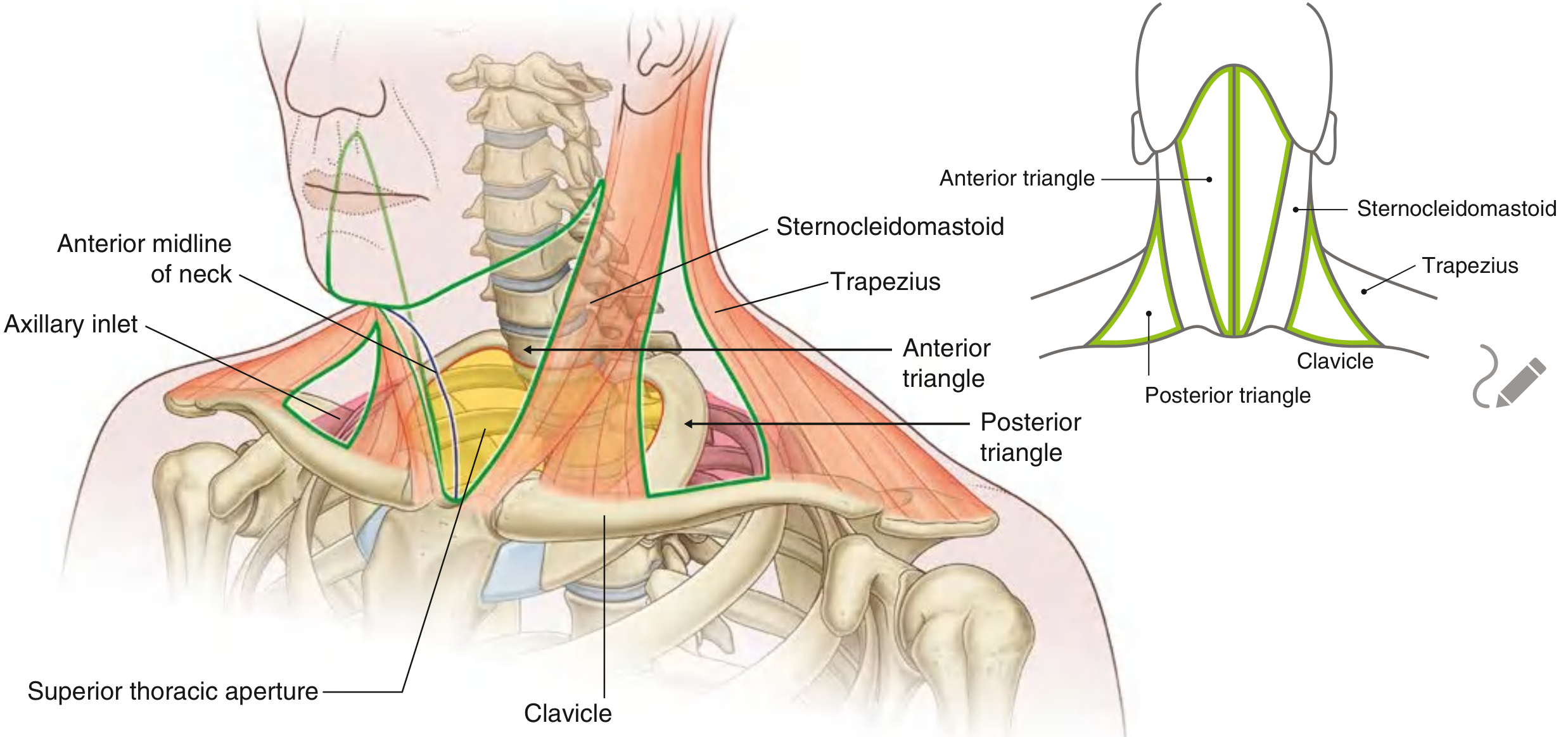
📸 TEXTBOOK ILLUSTRATION (Gray's Anatomy)
Anterior and Posterior Triangles - Gray's Anatomy for Students
🃏 EXAM FLASHCARD (Revise Before Exam)
MNEMONIC BANK
- "SCM is King" - divides neck into anterior (medial) and posterior (lateral)
- "Dead Cows Make Stupid Mistakes" - sub-triangles: Digastric=Submandibular, Carotid, Muscular, Submental, (+Middle = the 4)
- "Danger = D for Deadly Descent" - Danger space descends to posterior mediastinum
- "Ludwig is a Lunatic" - bilateral, involves 3 spaces, lifts the tongue, life-threatening
- "PPS = Grand Central Station" - Parapharyngeal space connects to everything
Sources: Gray's Anatomy for Students; Scott-Brown's Otorhinolaryngology Head & Neck Surgery; Fischer's Mastery of Surgery 8th ed.; Cummings Otolaryngology Head & Neck Surgery