
Explain lipoma complete
lipoma subcutaneous benign fatty tumor gross pathology
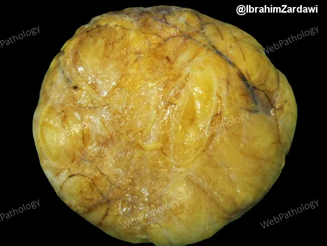
Gross pathology photograph of a superficial subcutaneous lipoma. The specimen is a soft, yellow, well-circumscribed fatty tumor surrounded by a thin fibrous capsule. Size ranges typically from less than 1 centimeter to about 5 centimeters in greatest dimension, with a median near 3 cm; deeper lipomas tend to be larger and more irregular, yet they retain a thin capsule that separates them from adjacent tissues. The surface is smooth to faintly lobulated; the cut surface is homogeneous, yellow-orange with delicate fibrous septa. The lesion is encapsulated and noninfiltrative, composed of mature adipocytes arranged in lobules. The gross appearance is characteristic of benign adipose tumors, but differentiation from liposarcoma relies on histology, especially in large, deep, or atypical lesions. This image, courtesy of Dr. Ibrahim Zardawi, illustrates classic macroscopic features used in medical education to distinguish benign lipomas from other soft tissue neoplasms. Clinically, lipomas present as soft, mobile, painless nodules in adults, with minimal inflammation. In pathology practice, this photograph supports gross specimen handling, capsule presence, adipocyte morphology, and gross differential considerations. Potential clinical applications include education, radiology-pathology correlation, gross pathology benchmarking, and informing surgical pathology reporting. This educational image supports accurate gross diagnosis and consistent documentation standards in practice.
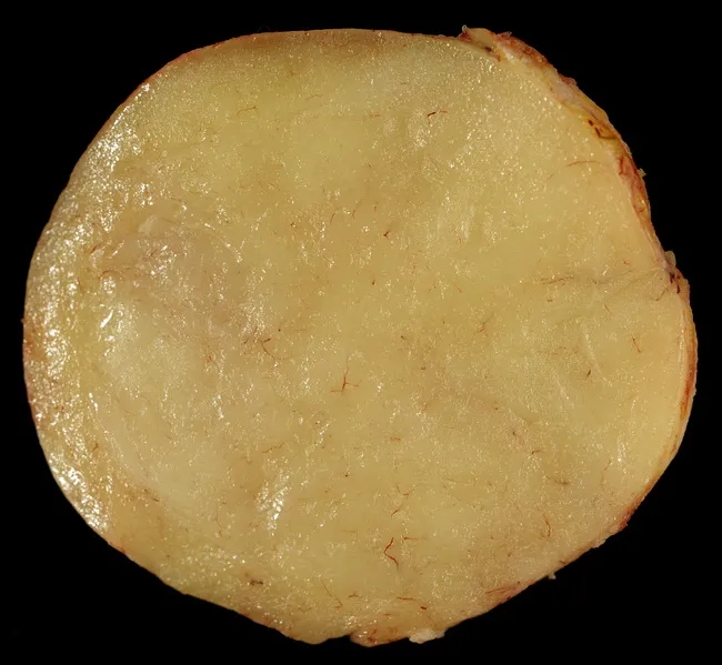
Gross pathology photograph of a soft tissue lipoma, captured on a transverse or cross-sectional cut surface. Anatomy: subcutaneous adipose tissue lesion arising within superficial soft tissue; location may be head, neck, trunk, or extremities, typically in the hypodermis. Visual features: lobulated, homogeneous, greasy appearance with a pale yellow to orange color; surface is often smooth and well circumscribed, reflecting a benign adipocytic tumor with little inflammatory change. Internal architecture may appear uniformly fatty with fine fibrous septa; occasional fat necrosis or hemorrhage can create focal discoloration or nodular heterogeneity, especially in longstanding lesions. Pathology: composed of mature adipocytes arranged in lobules, with minimal cytologic atypia and a fibrous capsule in many cases; variants such as fibrolipoma or angiolipoma may show additional stromal or vascular components altering texture and color. Diagnostic significance: gross features strongly support lipoma, but definitive diagnosis requires histopathology to exclude liposarcoma or other adipocytic neoplasms; correlate with clinical growth pattern and imaging findings. Clinical relevance: benign, slow-growing, often amenable to simple excision; symptoms and depth influence management. Educational value: provides reference for gross morphology of adipose tumors, differential diagnosis of fatty soft tissue masses, and correlation with radiologic fat density on MRI/CT. Educational use in pathology teaching.
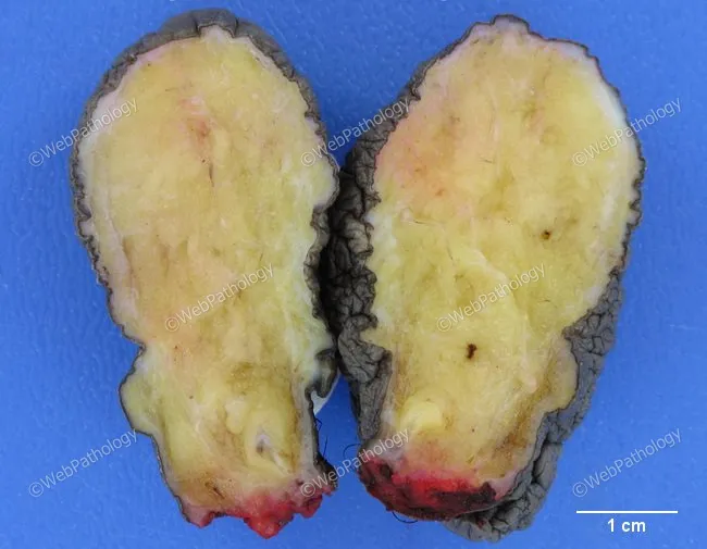
Gross pathology photograph of a fibrolipoma, a lipoma variant characterized by intimate admixture of mature adipose tissue and dense fibrous connective tissue. The image presents a subcutaneous soft tissue lesion in two symmetrical halves with a yellow-taint fatty core and delicate pink-tan fibrous bands extending through cut surface. The outer contour appears encapsulated by a translucent capsule, while the cut surface reveals lobulated adipose tissue partitioned by fibrous septa. The scale bar indicates 1 cm, showing a lesion of a few centimeters in dimension. The specimen displays a well-circumscribed, benign lipomatous tumor with fibrous stroma, consistent with fibrolipoma. Microscopically, the lesion would demonstrate mature adipocytes arranged in lobules separated by thick fibrous septa; absence of lipoblasts, cellular atypia, or necrosis; lack of significant vascular proliferation. This entity represents a benign mesenchymal tumor variant caused by fibrous connective tissue within lipoma; it can present similarly to conventional lipoma but with firmer consistency or increased firmness due to fibrous components. Clinical significance includes differentiation from liposarcoma; surgical excision is curative in typical cases, with rare recurrence. Potential clinical use cases include educational demonstrations of lipoma variants, gross pathology recognition, and differential diagnosis between benign adipocytic tumors and malignant liposarcomas in soft tissue.
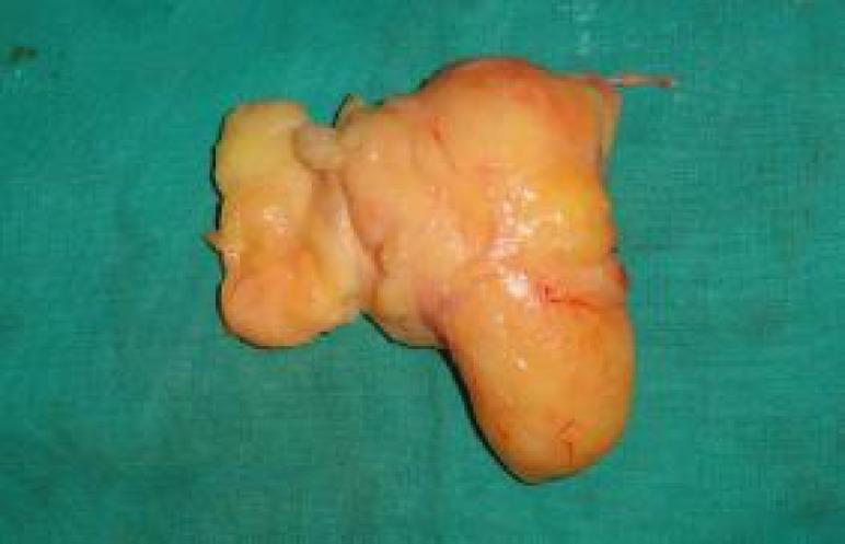
This clinical photograph displays a gross surgical specimen of a resected lipoma placed on a green sterile surgical drape. The specimen exhibits classic features of a benign fatty tumor, characterized by a distinct yellowish-orange color, consistent with mature adipose tissue. The morphology is lobulated and irregular, composed of a larger primary mass and smaller interconnected lobes. The surface texture varies from smooth to mildly irregular, with a thin, glistening translucent capsule visible. Fine red markings across the surface indicate minor superficial vascularity or remnants from the surgical dissection. The image serves as an educational example of soft tissue pathology, specifically illustrating the macroscopic appearance of a large subcutaneous or deep-seated lipoma following surgical excision from a distal forearm.
lipoma soft tissue tumor management
Lipoma - Complete Overview
Definition
Epidemiology
-
Occurs predominantly in adults, typically in the 5th to 7th decades
-
No sex predilection (male = female)
-
Rare in children; multiple lesions are uncommon
-
May be sporadic or part of a syndrome (see below)
-
Grainger & Allison's Diagnostic Radiology; Fitzpatrick's Dermatology
Anatomy and Location
| Type | Location | Features |
|---|---|---|
| Superficial (subcutaneous) | Trunk, neck, proximal extremities | Small (<5 cm), mobile, well-defined |
| Intramuscular / intermuscular | Deep soft tissues | Larger, poorly circumscribed, infiltrative |
| Parosteal | Adjacent to bone surface | May stimulate periosteal reaction |
| Lipoma arborescens | Synovial membrane | Villous lipomatous proliferation |
| Dermal lipoma | Within dermis | Unencapsulated, ill-defined |
Clinical Features
- Painless, slowly growing mass
- Soft and mobile on palpation
- Compressible with light pressure
- Usually <5 cm (superficial); deeper lesions may be considerably larger
- Occasional pain when nerve compression occurs (carpal tunnel, tarsal tunnel, cubital tunnel)
- Appearance is classically described as a "slipping sign" - the lesion can be gently squeezed to make it bulge forward through a small incision - Pye's Surgical Handicraft
Gross Pathology
Histology
-
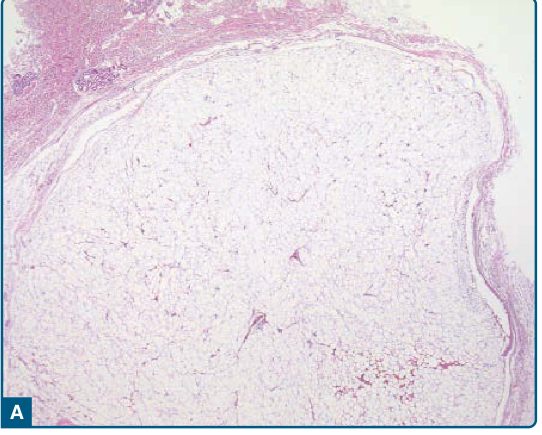
Well-circumscribed, encapsulated, lobulated lesion
-
Composed of mature adipocytes with thin, hypocellular fibrous septa containing thin-walled capillaries
-
Only slight variation in size and shape of adipocytes
-
No nuclear atypia, no lipoblasts
-
Immunohistochemistry: Tumor cells are negative for p16, MDM2, and CDK4 (positivity for these markers suggests atypical lipomatous tumor/well-differentiated liposarcoma)
-
Fitzpatrick's Dermatology, p. 2202-2203
Etiology and Pathogenesis
- Aberrations at 12q13-15 - involving the HMGA2 gene (at 12q14.3), which plays an important role in lipoma pathogenesis. Recombination of HMGA2 with several partner genes has been reported.
- Rearrangements at 6p21-23
- Deletions at 13q
- Fitzpatrick's Dermatology, p. 2202
Variants
| Variant | Defining Feature | Clinical Note |
|---|---|---|
| Angiolipoma | Increased blood vessels with fibrin thrombi | Often painful, frequently multiple |
| Fibrolipoma | Abundant fibrous tissue admixed with fat | Firmer consistency than typical lipoma |
| Chondrolipoma | Cartilaginous metaplasia within fat | Uncommon |
| Osteolipoma | Osseous metaplasia (post-trauma/ischemia) | Contains calcifications on imaging |
| Myxolipoma | Extensive myxoid change | Heterogeneous on imaging |
| Intramuscular lipoma | Infiltrative, within skeletal muscle | Higher recurrence rate after excision |
| Adenolipoma | Fat + sweat ducts/glands | Dermal location |
| Lipoma arborescens | Villous synovial proliferation | Intra-articular |
Lipomatosis Syndromes
| Syndrome | Key Features |
|---|---|
| Gardner syndrome | FAP, desmoid fibromatosis, osteomas, cysts |
| Bannayan-Riley-Ruvalcaba syndrome | Macrocephaly, lipomas, lymphangiomas, hamartomatous intestinal polyposis, lentigines |
| Cowden syndrome | Hamartomas, trichilemmomas, oral papillomas, palmoplantar keratoses |
| Madelung disease (symmetric lipomatosis) | Middle-aged Mediterranean men; neck, shoulder, proximal upper limbs; peripheral neuropathy |
| Diffuse lipomatosis | Children; diffuse subcutaneous and intramuscular overgrowth |
| Pelvic lipomatosis | More frequent in Black males |
| CLOVE syndrome | Congenital lipomatous overgrowth, vascular malformations, epidermal nevi |
| Steroid lipomatosis | Exogenous steroids or Cushing syndrome |
| HIV-associated lipodystrophy | Protease inhibitor therapy; cervical/breast/visceral fat deposition |
- Fitzpatrick's Dermatology, Table 122-1
Investigations and Imaging
Radiograph (X-ray)
- Large lipomas visible as areas of relative lucency compared to soft tissue
- Small lesions are not visible
- Foci of mineralisation (calcification, ossification) visible in ~11%
Ultrasound
- Elliptical mass, often well-defined, with long axis and septa parallel to the skin surface
- Variable reflectivity (often relatively hyperechoic)
- Compressible with probe pressure
- May appear encapsulated or blend with surrounding tissue without a visible capsule
CT
- Mass of density similar to subcutaneous fat (Hounsfield units: approximately -65 to -120 HU)
- Non-encapsulated lipomas may be occult
- Deeper lesions more likely to show heterogeneity
MRI (gold standard for deep lesions)
- Isointense to subcutaneous fat on both T1 and T2
- Low signal on STIR, suppressed on fat-saturation sequences
- Fine septa (<2 mm) are often present - no gadolinium enhancement in benign lesions
-
Incomplete fat signal suppression
-
Thick, nodular septa (especially if they enhance)
-
Focal non-lipomatous areas
-
Grainger & Allison's Diagnostic Radiology, pp. 1063-1064
Differential Diagnosis
| Condition | Differentiating Features |
|---|---|
| Atypical lipomatous tumor (WDL) | Larger, deeper; MDM2/CDK4 positive on IHC; thick nodular septa on MRI |
| Liposarcoma (myxoid/pleomorphic) | Non-fatty elements, aggressive growth, atypia on histology |
| Sebaceous cyst (epidermal cyst) | Attached to skin, has punctum, contains cheesy material |
| Lipodermoid | Subconjunctival, head/neck in children |
| Angiolipoma | Painful, multiple, vascular component with fibrin thrombi |
| Ganglion cyst | Fluctuant, related to joint/tendon, contains synovial fluid |
Treatment
Surgical Excision (Mainstay)
- Most lipomas are cured by simple excision - Robbins & Kumar Basic Pathology
- Indications: symptomatic lesions, cosmetic concern, rapid growth, uncertain diagnosis, large or deep lesions
- Technique (Pye's Surgical Handicraft):
- Solitary round lipoma: squeeze the tissues to make it bulge, incise down to the lesion, then enucleate
- Large/multilocular type: establish plane of cleavage, remove with blunt and sharp dissection
- Meticulous hemostasis required; close dead space or insert a small suction drain if dissection is extensive
- Axillary and supraclavicular lipomas may be more extensive than they appear superficially - may require local infiltration or general anesthesia
Intramuscular Lipomas
- Higher recurrence rate due to infiltrative nature - curative excision is more difficult
- Fitzpatrick's Dermatology
Liposuction
- Used for cosmetically problematic lipomas, especially larger or multiple lesions - less frequently performed for definitive excision
Observation
- Asymptomatic, small, clinically obvious lipomas may be safely observed
Prognosis
- Conventional lipoma: excellent prognosis, cured by excision, very low recurrence
- Intramuscular lipoma: higher recurrence rate
- No malignant transformation in true lipoma (contrary to popular belief - lipomas do NOT transform into liposarcomas)
- Liposarcomas arise de novo, not from pre-existing lipomas
Key Distinguishing Summary
| Feature | Lipoma | Liposarcoma |
|---|---|---|
| Behavior | Benign | Malignant |
| Age | Any adult | 5th-6th decade |
| Atypia | None | Present (varies by subtype) |
| MDM2/CDK4 IHC | Negative | Positive (well-differentiated) |
| MRI septa | Thin (<2 mm), no enhancement | Thick, nodular, may enhance |
| Treatment | Simple excision, curative | Wide excision ± radiotherapy |
What is the difference between lump and a swelling
clinical difference between lump and swelling surgery examination
Lump vs. Swelling - The Clinical Distinction
Core Definitions
| Term | Definition |
|---|---|
| Lump | A vague mass of body tissue - a discrete, palpable, localized solid mass |
| Swelling | A vague term denoting any enlargement or protuberance in the body due to any cause |
| Tumour / Neoplasm | A growth of new cells that proliferate independent of the body's needs |
"A 'Lump' is a vague mass of body tissue. A 'Swelling' is a vague term which denotes any enlargement or protuberance in the body due to any cause."
- S Das, Manual on Clinical Surgery
Key Differences
| Feature | Lump | Swelling |
|---|---|---|
| Nature | Discrete, localized, solid or semi-solid mass | Any enlargement, diffuse or localized, from any cause |
| Scope | More specific - refers to a palpable, defined mass | Broader term - includes fluid, inflammation, edema, gas, neoplasm |
| Cause | Usually neoplastic or structural (cyst, lipoma, lymph node) | Congenital, traumatic, inflammatory, neoplastic, or miscellaneous |
| Clinical usage | Used when the mass is well-circumscribed and palpable | Used for any visible/palpable enlargement of a body part |
| Examples | Lipoma, cyst, lymph node, abscess | Edema, goiter, joint effusion, abscess, hematoma |
Practical Clinical Points
-
A lump implies a discrete, palpable mass - you can define its edges, size, and shape. It suggests a structural lesion (neoplastic, cystic, lymphoid).
-
A swelling is broader - it can be diffuse (like pitting edema of a limb) or localized (like a joint effusion). It does not imply a discrete mass.
Clinical aid from S Das:
- Short duration + pain → likely inflammatory (acute)
- Long duration + no pain → likely neoplastic (benign)
- Long duration + slight pain → possibly chronic inflammatory
- Short duration, rapid growth → consider malignancy
Classification of Swellings
| Category | Examples |
|---|---|
| Congenital | Cystic hygroma, meningocele, branchial cyst |
| Traumatic | Hematoma, seroma |
| Inflammatory | Abscess, lymphadenitis, cellulitis |
| Neoplastic | Lipoma, fibroma, carcinoma, sarcoma |
| Miscellaneous | Goiter, hernia, aneurysm, ganglion |
Summary
- "Lump" = a patient's or clinician's description of something you can feel as a discrete mass
- "Swelling" = a broader, more general term for any visible or palpable enlargement, regardless of whether it has a discrete edge
Examination of a lump and a swelling
Examination of a Lump and a Swelling
Structure of Examination
- General Survey
- Local Examination (Inspection → Palpation → Special Tests → Auscultation)
- General Examination (systemic, lymph nodes, metastases)
Phase 1 - General Survey
- Cachexia / malnutrition - suggests underlying malignancy
- Attitude and posture - a swelling pressing on a nerve (e.g. osteosarcoma) may cause paresis of the distal limb; a displaced fracture causes abnormal limb attitude
- Raised temperature and pulse rate - always associated with an inflammatory swelling
- Build, nutrition, mental state, facies - all give diagnostic clues
Phase 2 - Local Examination
A. INSPECTION
| Feature | What to Look For | Significance |
|---|---|---|
| Site | Exact anatomical location | Guides differential diagnosis |
| Size | Estimate dimensions visually | Large vs. small |
| Shape | Hemispherical, elongated, irregular | Cysts are usually hemispherical |
| Surface | Smooth vs. irregular/nodular | Benign = smooth; malignant = irregular |
| Colour of overlying skin | Red/inflamed, normal, pigmented | Red = inflammatory; normal = neoplastic |
| Skin changes | Stretched, shiny, dilated veins, punctum, ulceration, peau d'orange | Ulcer over a swelling suggests malignancy; dilated veins = venous obstruction |
| Movements | With respiration, on swallowing, on tongue protrusion | Thyroid moves on swallowing; thyroglossal cyst moves on tongue protrusion |
| Pulsatility | Visible pulsation | Aneurysm or vascular swelling |
A good clinician always spends time in observation before palpation.
B. PALPATION
1. Temperature
- Feel with the back of the fingers (more sensitive to heat)
- Increased temperature → inflammatory swelling (abscess, acute lymphadenitis)
- Normal temperature → neoplastic or chronic swelling
2. Tenderness
- Press gently and watch the patient's face for a wince
- Tender → inflammatory, or rapid stretching of a capsule
- Non-tender → neoplastic (benign or malignant)
- Exception: Osteosarcoma - mild pain is usually the first symptom and precedes the swelling
3. Site and Size
- Confirm with palpation; measure dimensions (length × breadth in cm)
4. Shape
- Palpate the overall form - hemispherical, ovoid, irregular
5. Surface
- Smooth surface → benign (lipoma, cyst, fibroma)
- Irregular or nodular → malignant tumor, multinodular goiter
6. Edge / Margin
- Feel around the periphery with the finger-tips
- Well-defined margins → neoplastic or chronic inflammatory
- Ill-defined / indistinct margins → acute inflammatory swelling
- Benign tumors = smooth margins; Malignant = irregular margins
Slip Sign: The key to differentiating a solid lump (e.g. lipoma) from a cyst:
- Solid lump (lipoma): the margin slips away from the palpating finger but does not yield to it
- Cyst: the margin yields to the palpating finger and does not slip away
7. Consistency
| Consistency | Example |
|---|---|
| Very soft | Lipoma |
| Cystic (fluctuant) | Cysts, chronic abscess, hydrocele |
| Firm | Fibroma, lymph node |
| Hard but yielding | Chondroma |
| Bony hard | Osteoma |
| Stony hard | Carcinoma |
| Crepitus on palpation | Gas gangrene, surgical emphysema |
In case of gaseous swellings (e.g. gas gangrene, surgical emphysema), a crepitus may be felt on palpation.
8. Fluctuation Test
- Place two "watching fingers" (W) on opposite poles of the swelling
- Press with a "displacing finger" (D) at the centre
- Positive: fluid wave is transmitted to the watching fingers in both planes
- Must be done in two planes at right angles to each other
- A fleshy muscle (e.g. quadriceps) may show false fluctuation along one plane only - not diagnostic
- Very soft solid swellings (lipoma) may give a false positive
- Paget's Test (for small swellings): press the centre - a fluid-filled swelling is softer at the centre and firmer at the periphery; a solid swelling is firmer at the centre
- For small swellings where two fingers cannot fit: use two watching fingers on the sides and one displacing finger in the middle
9. Pulsatility
- Place two fingers on the swelling and feel for movement
- Expansile pulsation (fingers pushed apart in all directions) → aneurysm (true vascular lesion)
- Transmitted pulsation (swelling moves as a whole without expansion) → swelling overlying a vessel (e.g. psoas abscess overlying aorta)
10. Compressibility and Reducibility
| Feature | Definition | Example |
|---|---|---|
| Compressible | Swelling decreases in size on compression and does not return when pressure is removed | Haemangioma, lymphangioma |
| Reducible | Swelling disappears on pressure and returns when pressure is released or patient stands/coughs | Hernia |
11. Transillumination Test
- Darken the room
- Press a torch firmly against one side of the swelling
- Positive (glowing red light transmitted through swelling) → contains clear fluid (e.g. hydrocele, cystic hygroma, ganglion)
- Negative → solid tumor, blood-filled cyst (haematocele), abscess
"Brilliantly transilluminant" → cystic hygroma (lymphangioma). Hydrocele also transilluminates. A haematocele does not.
12. Mobility and Fixation
- Move the skin over the swelling - if it does not move, the swelling is fixed to skin
- Pinch up the skin over the swelling - if it cannot be pinched, it is adherent
- If a lipoma is pushed sideways, the skin will pucker slightly due to fibrous strands from the capsule to the overlying skin
- With muscle relaxed - move the swelling sideways
- Then ask the patient to contract the muscle against resistance (makes it taut)
- If the swelling cannot be moved when muscle is taut → fixed to underlying muscle
- If swelling moves with the muscle → it arises from the muscle
- If swelling is unaffected by muscle contraction → it is superficial to the muscle
- Feel for the underlying bony contour - swelling arising from bone will be fixed to it
13. Transverse and Longitudinal Mobility
- Move the swelling side to side and up and down
- A nerve tumor (neurofibroma) is mobile transversely but not longitudinally (along the nerve trunk)
14. Relation to Skin (Fixation Test)
- Move the skin over the swelling and also try to pinch the skin
- Normal skin over a swelling → subcutaneous/deep origin
- Stretched, adherent, or dimpled skin → involvement by tumor or inflammation
C. PERCUSSION
- A dull note → solid or fluid-filled swelling
- Resonant note → gas-containing swelling (e.g. hernia containing bowel, hydatid cyst occasionally)
- Over the skull, percussion of a dermoid cyst may reveal bony erosion
D. AUSCULTATION
- Bruit → arteriovenous fistula, vascular tumor (thyroid goiter, renal cell carcinoma)
- Bowel sounds → hernia containing bowel
- Machinery murmur → AV fistula
E. SPECIAL TESTS
Fluctuation (detailed above)
Transillumination (detailed above)
Paget's Test
Brilliance Test
F. EXAMINATION FOR PRESSURE EFFECTS
- Arterial pulse distal to the swelling - weak/absent pulse if main artery is compressed
- Neurological exam - wasting, paresis, paralysis, sensory loss distal to swelling if nerve is compressed
- Bony erosion - feel the bone beneath the swelling (dermoid on skull, aneurysm eroding vertebrae)
- Joint involvement - check range of movement of adjacent joint
Phase 3 - General Examination
| Concern | What to Examine |
|---|---|
| Malignancy | Lungs (consolidation, effusion), liver (metastases), abdomen (peritoneal secondaries), spine/pelvis/skull (bony metastases) |
| Lymph nodes | All regional node groups - enlarged nodes indicate infection or malignancy |
| Syphilitic swelling | Look for other syphilitic stigmas systemically |
| Hernia | Examine all hernial orifices |
Special Investigations
| Test | Indication |
|---|---|
| Blood count (TC, DC, Hb, ESR) | Inflammatory swellings → leucocytosis; malignancy → anaemia, raised ESR |
| X-ray | Bony involvement, calcification within swelling, chest for metastasis |
| Ultrasound | Solid vs. cystic, vascular flow (Doppler) |
| CT / MRI | Deep or complex swellings, staging |
| FNAC / Biopsy | Tissue diagnosis - mandatory when malignancy is suspected |
| Aspiration | Fluid from cysts - for cytology and culture |
Quick Reference: What Each Sign Tells You
| Sign | Positive Meaning |
|---|---|
| Fluctuation | Fluid-containing swelling |
| Transillumination | Clear fluid (cyst, hydrocele, cystic hygroma) |
| Expansile pulsation | Aneurysm |
| Transmitted pulsation | Swelling overlying a vessel |
| Compressible (no return) | Haemangioma / lymphangioma |
| Reducible (returns) | Hernia |
| Slip sign | Solid tumor (e.g. lipoma) vs. cyst |
| Moves on swallowing | Thyroid or related structure |
| Moves on tongue protrusion | Thyroglossal cyst |
| Transverse mobility only | Nerve sheath tumor (neurofibroma) |
| Crepitus | Gas-containing swelling (surgical emphysema) |
| Bruit | Vascular tumor or AV fistula |
| Bowel sounds | Hernia with bowel content |
Pallor and icterus grades
pallor grading clinical examination 1+ 2+ 3+ 4+ anaemia conjunctiva tongue
https://ihatepsm.com/blog/pallor-and-its-grading
icterus grading clinical examination grade 1 2 3 4 scleral jaundice bilirubin levels
https://www.ncbi.nlm.nih.gov/books/NBK544252
scleral icterus jaundice yellow eyes clinical sign
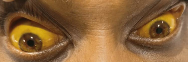
This clinical photograph displays a close-up view of a patient's eyes and upper face, illustrating severe scleral icterus. Both eyes exhibit deep, uniform yellow pigmentation of the sclera and bulbar conjunctiva, a hallmark sign of hyperbilirubinemia. The surrounding facial skin, particularly on the forehead and periorbital regions, also shows a subtle yellowish hue consistent with jaundice, though less pronounced than the ocular findings. The image serves as a clear educational example of the physical manifestations of liver dysfunction, biliary obstruction, or systemic toxicity—in this context, following rodenticide (zinc phosphide) ingestion. The visibility of the distinct yellowing against the patient's natural skin tone provides a significant diagnostic reference for internal medicine and gastroenterology students and practitioners.

Clinical photograph comparison of a patient's eyes demonstrating the resolution of jaundice. Panel (a) shows bilateral scleral icterus, characterized by a distinct yellowing of the sclera and bulbar conjunctiva, marked with blue arrows. This appearance is secondary to hyperbilirubinemia often associated with hepatobiliary disease. Panel (b) shows the same patient after two weeks of treatment, demonstrating a marked reduction in the yellow hue. The sclera has returned to a near-normal white color, indicating a decrease in systemic bilirubin levels and successful therapeutic intervention. This side-by-side comparison serves as an educational tool for identifying physical signs of liver dysfunction and monitoring clinical progression during treatment.
pallor conjunctiva anaemia lower eyelid pale
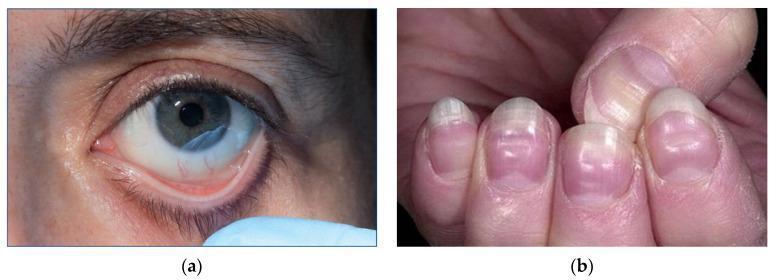
This composite of clinical photographs illustrates physical manifestations of severe anemia. Image (a) shows a close-up of a patient's eye with the lower eyelid retracted to reveal marked conjunctival pallor. The typically pink or reddish palpebral conjunctiva appears significantly pale and porcelain-white, a sign classically associated with a hemoglobin concentration below 90 g/L. Image (b) presents the fingernails of a patient demonstrating pronounced nail bed pallor. The distal nail beds lack the normal healthy pink hue. Additionally, horizontal white bands, known as Muehrcke's lines, are visible across the nail plates; these non-palpable transverse lines are associated with systemic conditions such as hypoalbuminemia and severe anemia. Together, these images demonstrate key external clinical signs used in the bedside assessment of hematological disorders and circulatory efficiency. The content is suitable for medical education regarding the physical examination findings of iron deficiency and other severe anemias.
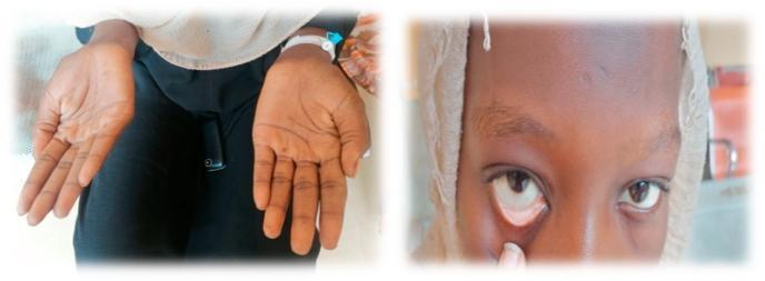
This composite clinical photograph illustrates common physical examination findings associated with severe anemia. The left panel shows the bilateral palms of a patient, demonstrating marked palmar pallor. The skin on the palms and creases appears significantly lighter and lacks the typical pinkish hue of healthy vascularized tissue, suggesting reduced hemoglobin concentration. The right panel is a close-up of the patient's face with the lower eyelid retracted to reveal the palpebral conjunctiva. Instead of the normal vibrant red or pink color, the conjunctiva exhibits conjunctival pallor, appearing whitish or very pale pink. These visual signs are critical diagnostic markers in hematology and general medicine for identifying systemic conditions such as iron-deficiency anemia or other forms of chronic blood loss. The image serves as a teaching tool for medical students and clinicians to recognize mucosal and cutaneous indicators of decreased red blood cell mass or oxygen-carrying capacity.
Pallor and Icterus - Definition, Examination, and Grading
PALLOR
Definition
Causes
- Anaemia (most common cause of pallor)
- Massive haemorrhage
- Shock
- Intense emotion / vasovagal syncope
- Hypothyroidism, renal failure, malignancy
Sites to Examine for Pallor
| Site | How to Examine |
|---|---|
| Lower palpebral conjunctiva | Pull down the lower eyelid - the inner rim should be bright pink/red. If pale = anaemia |
| Mucous membranes (lips, buccal mucosa) | Look at the inner lip and cheeks |
| Tongue (dorsum) | Ask patient to protrude - tongue should be pink; pallor indicates anaemia |
| Nail beds | Press on the nail tip and release - capillary refill time + colour |
| Palmar creases | Flatten the palm fully - if creases are pale = significant anaemia |
The conjunctiva is the most sensitive and commonly used site. The tongue has been shown to have the highest sensitivity for severe anaemia (Hb <7 g/dL). - Kalantri et al., PMC2797134
Grading of Pallor
| Grade | Description | Approximate Hb (g/dL) |
|---|---|---|
| 0 (Absent) | Normal pink conjunctiva; no pallor at any site | >10 (normal) |
| 1+ (Mild) | Slight paleness of conjunctiva; palmar creases still pink | ~9-10 |
| 2+ (Moderate) | Definite pallor of conjunctiva AND mucous membranes; palmar creases pale | ~7-9 |
| 3+ (Severe) | Marked pallor at all sites - conjunctiva, tongue, palms, nail beds; patient looks visibly pale | ~5-7 |
| 4+ (Very severe / Extreme) | Ghost-white pallor at all sites; associated features of severe anaemia (tachycardia, dyspnoea, oedema) | <5 |
Key Hb thresholds:
- Absence of conjunctival pallor at Hb <7 g/dL virtually rules out Hb <7 g/dL (high negative predictive value)
- Severe pallor of the tongue is most specific for Hb <7 g/dL
Visual reference - Conjunctival pallor and palmar pallor in severe anaemia:
Lemon-Yellow Pallor
Seen in: Pernicious anaemia (Vitamin B12 deficiency) - ineffective erythropoiesis causes both pallor (low Hb) and mild jaundice (haemolysis of defective RBCs).
ICTERUS (JAUNDICE)
Definition
"The icteric tint varies from the faint yellow of viral hepatitis to the dark olive greenish yellow of obstructive jaundice." - S Das, Manual on Clinical Surgery
Sites to Examine for Icterus
| Site | Significance |
|---|---|
| Sclera (eyeball) | First site to become yellow; sclerae have high elastin content with high affinity for bilirubin. Ask patient to look downward while you hold the eyelid open. |
| Under-surface of tongue | Second most sensitive site |
| Lobule of the ear | Useful in dark-skinned patients |
| Tip of the nose | |
| Nail bed | |
| Skin (face, trunk) | Appears later, at higher bilirubin levels |
Scleral icterus: detectable at bilirubin >2.5-3 mg/dL Skin yellowing: detectable at bilirubin >4-5 mg/dL
Grading of Icterus
| Grade | Bilirubin Level (approx.) | Clinical Appearance |
|---|---|---|
| 1+ (Mild) | 2-4 mg/dL | Icterus detectable only in the sclera (scleral icterus). Skin not yet yellow. |
| 2+ (Moderate) | 4-8 mg/dL | Yellow discolouration of sclera + skin of the face and trunk. Urine begins to darken. |
| 3+ (Marked) | 8-15 mg/dL | Deep yellow skin + sclera. Dark urine, pale stools. Pruritus may appear (bile acids). |
| 4+ (Severe / Deep) | >15 mg/dL | Greenish-yellow or olive-green skin and sclera due to biliverdin accumulation. Associated with longstanding obstructive jaundice. Marked pruritus (scratch marks). |
Deep, long-standing jaundice → skin and sclerae turn green (biliverdin) - particularly seen in obstructive jaundice (cholestasis).
- S Das, Manual on Clinical Surgery; StatPearls - Hyperbilirubinemia
Visual reference - Scleral icterus (deep jaundice):
Comparing before and after treatment:
Associated Features in Icterus
| Feature | Cause |
|---|---|
| Dark (tea-colored) urine | Conjugated bilirubin (water-soluble) excreted in urine |
| Pale (clay-colored) stools | Absent bile in intestine (obstructive jaundice) |
| Pruritus + scratch marks | Bile acid retention in skin (obstructive jaundice) |
| Greenish tinge | Biliverdin accumulation in longstanding obstruction |
| Bradycardia (Bradycardia of jaundice) | Bile salts slow the heart |
Quick Comparison Summary
| Feature | Pallor | Icterus |
|---|---|---|
| Colour | Pale / white | Yellow / green-yellow |
| Best site to detect | Lower palpebral conjunctiva | Sclera |
| Caused by | Low Hb / vasoconstriction | Excess bilirubin |
| Lab threshold | Hb <10 g/dL (mild) | Bilirubin >3 mg/dL |
| Grading | 0 to 4+ (absent to very severe) | 1+ to 4+ (scleral → green skin) |
| Combined appearance | Lemon-yellow = pallor + mild icterus | Pernicious anaemia, haemolytic anaemia |
Lump nide examination and lump nodes
lymph node regions groups examination neck axilla groin diagram
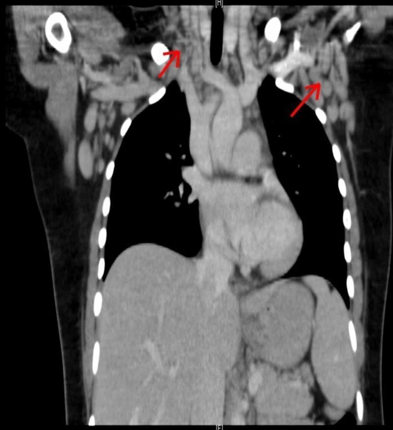
This diagnostic image is a coronal Computed Tomography (CT) scan of the human chest and lower neck, demonstrating extensive bilateral lymphadenopathy. The scan reveals multiple pathologically enlarged lymph nodes in several anatomical regions, including the cervical (neck), supraclavicular, axillary (axilla), and mediastinal compartments. The lymph nodes are characterized by their rounded or ovoid morphology and significant size increase relative to adjacent vascular and soft tissue structures. Anatomical landmarks clearly visible include the air-filled lungs (low-attenuation/dark), the central heart and mediastinal great vessels, and the thoracic skeleton including the clavicles and ribs. Red arrows are used to annotate specific clusters of enlarged nodes in the supraclavicular and axillary regions. This clinical imaging illustrates systemic lymph node involvement, which is essential for diagnosing conditions such as lymphoma, sarcoidosis, or systemic inflammatory syndromes like Hemophagocytic Lymphohistiocytosis (HLH). The imaging modality and presentation are typical for evaluating thoracic and systemic lymphadenopathy in an internal medicine or oncology context.
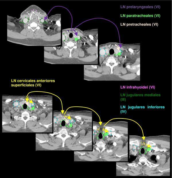
This diagnostic image consists of a series of seven axial computerized tomography (CT) slices of the human neck, illustrating the anatomical delineation of cervical lymph node regions for clinical indexing and radiotherapy planning. The slices are arranged chronologically to show descending anatomical levels. Color-coded contouring identifies specific lymph node (LN) groups: LN cervicales anteriores superficiales (yellow) and LN cervicales anteriores profundi, which are subdivided into LN infrahyoidei (pink), LN prelaryngeales (violet), LN pretracheales (light pink), and LN paratracheales (light green). Additionally, LN jugulares mediales (dark green) and LN jugulares inferiores (cyan) are marked. Key anatomical landmarks visible across the axial plane include the trachea (central air-filled void), the thyroid gland, the carotid arteries and jugular veins (lateral to the trachea), and the cervical vertebral bodies (posterior). The image highlights Level VI (anterior compartment) nodes in relation to Level III and IV (jugular) chains, providing a spatial guide for localizing lymphadenopathy or defining clinical target volumes in head and neck oncology.
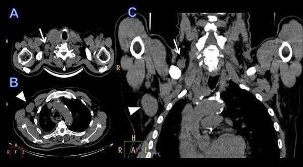
This diagnostic imaging composite displays contrast-enhanced Computed Tomography (CT) scans of the neck, chest, and shoulder regions in axial (a, b) and coronal (c) planes. The images demonstrate significant pathological findings characteristic of metastatic disease, specifically involving the lymphatic system. In the axial view at the level of the lower neck (a), a white arrow identifies an enlarged, heterogeneously dense lymph node in the right supraclavicular fossa. Further inferiorly in the axial chest view (b), a white arrowhead indicates a prominent, rounded, and enlarged lymph node in the right axilla. The coronal reconstruction (c) provides a comprehensive view of both regions, highlighting the spatial relationship and abnormal morphology of the supraclavicular (arrow) and axillary (arrowhead) lymphadenopathy. These findings are clinically consistent with regional lymph node metastasis, as seen in cases of advanced primary thyroid carcinoma. The images serve as an educational example of oncological staging and the identification of distant nodal involvement in head and neck malignancies.
Lymph Node Examination - Complete Guide
Why Examine Lymph Nodes?
"It cannot be impressed too strongly that no examination of a swelling is complete without the examination of the draining lymph nodes."
- S Das, Manual on Clinical Surgery, 13th Edition
- They drain the region where a lump is located
- Enlargement indicates infection, inflammation, or spread of malignancy
- Their characteristics help determine the nature of the primary lesion
- Generalized lymphadenopathy suggests a systemic disease
Step 1 - Know the Drainage Anatomy
| Region of Lump | Draining Lymph Node Group |
|---|---|
| Scalp, face, ear | Pre-auricular, post-auricular, occipital, submandibular, submental |
| Lip, tongue, floor of mouth | Submental, submandibular, deep cervical |
| Thyroid, larynx | Deep cervical, pretracheal |
| Upper limb, breast (lateral) | Axillary |
| Breast (medial) | Internal mammary |
| Lower limb, genitalia, perineum | Inguinal |
| Testes | Para-aortic (NOT inguinal) |
| Stomach, GI tract | Para-aortic, mesenteric |
| Lung, retroperitoneum | Supraclavicular, scalene |
| GI primary tumors | Virchow's node (left supraclavicular) |
Step 2 - Examination of Lymph Nodes (Systematic Approach)
A. INSPECTION
- Look for visible enlargement over the node-bearing regions
- Note any skin changes over the node: redness (acute lymphadenitis), sinus/discharging tract (TB, actinomycosis), puckering
B. PALPATION - Parameters to Assess
1. Site
- Identify the exact anatomical group involved
- Note whether it is a single group (regional) or multiple groups (generalized)
2. Size
- Measure in two dimensions (e.g., 2 × 2 cm)
- Nodes <1 cm² (1 cm × 1 cm) are almost always due to benign/reactive causes
- Nodes >2 cm diameter in young patients - biopsy warranted to exclude malignancy/granulomatous disease
- Nodes >2.25 cm² (1.5 cm × 1.5 cm) - best threshold for distinguishing malignant from benign on clinical grounds - Harrison's Principles of Internal Medicine
3. Number
- Single enlarged node → likely regional cause (infection, malignancy in drainage zone)
- Multiple groups → systemic disease (lymphoma, TB, EBV, HIV, sarcoidosis, leukaemia)
4. Shape
- Oval / bean-shaped → normal architecture retained (reactive)
- Spherical / globular → suggests pathological infiltration
5. Surface
- Smooth → reactive or lymphoma
- Irregular, nodular → metastatic carcinoma
6. Consistency / Texture
| Consistency | Clinical Diagnosis |
|---|---|
| Soft | Acute non-specific lymphadenitis |
| Firm / Rubbery | Lymphoma |
| Hard (stony) | Metastatic carcinoma |
| Fluctuant | Abscess (pyogenic or TB cold abscess) |
| Rubbery-firm, discrete | Lymphoma |
| Matted (fused together) | Tuberculosis; occasionally lymphoma |
"Nodes involved by lymphoma tend to be large, discrete, symmetric, rubbery, firm, mobile, and nontender. Nodes containing metastatic cancer are often hard, nontender, and nonmovable because of fixation to surrounding tissues."
- Harrison's Principles of Internal Medicine, 22nd Ed.
7. Tenderness
- Tender → inflammatory cause (acute infection - bacteria/virus); capsule stretched by rapid enlargement
- Non-tender → neoplastic (benign or malignant)
- Exception: Acute leukaemia may cause painful rapidly enlarging nodes despite malignancy
8. Mobility / Fixation
| Mobility | Significance |
|---|---|
| Mobile, discrete | Reactive or lymphoma |
| Matted (fixed to each other, move together) | Tuberculosis (periadenitis), occasionally lymphoma |
| Fixed to skin | Inflammation (periadenitis), malignant infiltration |
| Fixed to deep structures | Malignant invasion of surrounding tissues |
| Fixed + hard + nontender | Classic triad of metastatic carcinoma |
9. Relationship to Overlying Skin
- Normal skin overlying → subcutaneous lymphoma, metastasis
- Red, warm, tethered skin → acute lymphadenitis, TB with collar-stud abscess
- Collar-stud abscess (TB): fluctuant swelling connected through deep fascia - "collar-stud" shape
10. Discharging Sinus
- TB lymphadenitis → chronic discharging sinus with thin watery discharge (pus)
- Actinomycosis → multiple discharging sinuses with "sulfur granules"
Step 3 - Regional Groups and Their Clinical Significance
Cervical Lymph Nodes
| Group | Location | Significant Causes |
|---|---|---|
| Submental | Below chin | Infections of lower lip, floor of mouth |
| Submandibular | Below jaw | Oral, dental, tongue infections; floor of mouth cancer |
| Pre-auricular | In front of ear | Conjunctival infections, cat-scratch disease |
| Post-auricular | Behind ear | Scalp infections, rubella |
| Occipital | Back of skull | Scalp infections, rubella, toxoplasmosis |
| Anterior cervical | Along anterior triangle | Tonsillar, pharyngeal infections |
| Posterior cervical | Along posterior triangle | TB, lymphoma, EBV |
| Supraclavicular | Above clavicle | Always abnormal - malignancy until proven otherwise |
Virchow's node: Left supraclavicular node enlarged by metastatic carcinoma from GI tract (stomach, colon, pancreas). Also called Troisier's sign when positive.
Supraclavicular and scalene nodes: Drain the lung and retroperitoneum - reflect lymphoma, metastatic cancer, TB, sarcoidosis. - Harrison's Principles
Axillary Lymph Nodes
| Group | Drains |
|---|---|
| Anterior (pectoral) | Breast (lateral), chest wall |
| Posterior (subscapular) | Back, shoulder |
| Lateral | Upper limb |
| Central | Receives from all others |
| Apical | Main outflow → subclavian trunk |
Inguinal Lymph Nodes
| Group | Drains |
|---|---|
| Horizontal (superficial) | Perineum, genitalia, lower abdominal wall, anus |
| Vertical (along long saphenous vein) | Lower limb (medial) |
Important: Testicular tumors drain to para-aortic nodes (NOT inguinal), because the testes descend from the abdomen.
Step 4 - Distinguishing Features of Common Causes
| Feature | Acute Lymphadenitis | TB Lymphadenitis | Lymphoma | Metastatic Carcinoma |
|---|---|---|---|---|
| Tenderness | Yes (very tender) | Mild | No | No |
| Consistency | Soft | Firm → fluctuant (cold abscess) | Rubbery | Stony hard |
| Matting | No | Yes (classic) | Sometimes | Sometimes |
| Fixation | Fixed to skin (periadenitis) | May form sinus | Mobile | Fixed to deep |
| Number | Few (regional) | Multiple groups | Multiple groups | Regional (primary site) |
| Skin | Red, warm | Sinus, collar-stud | Normal | May be fixed |
| Systemic signs | Fever, leucocytosis | Night sweats, weight loss, fever | B-symptoms | Primary tumor signs |
Step 5 - Generalized Lymphadenopathy
| Category | Examples |
|---|---|
| Infections | EBV (infectious mononucleosis), CMV, HIV, TB, brucellosis, toxoplasmosis |
| Neoplastic | Lymphoma (Hodgkin's and Non-Hodgkin's), ALL, CLL |
| Autoimmune | SLE, rheumatoid arthritis |
| Granulomatous | Sarcoidosis |
| Miscellaneous | Drug reactions (phenytoin, allopurinol), hyperthyroidism |
Coexistence of splenomegaly + lymphadenopathy strongly suggests: infectious mononucleosis, lymphoma, leukaemia, SLE, sarcoidosis, or toxoplasmosis. - Harrison's Principles
Step 6 - Investigations
| Investigation | Purpose |
|---|---|
| CBC with differential | Leukaemia, EBV (atypical lymphocytes), infection |
| ESR, CRP | Inflammation vs. malignancy |
| Monospot / Paul-Bunnell | EBV mononucleosis |
| Chest X-ray | TB (hilar adenopathy), lymphoma (mediastinal nodes) |
| Ultrasound | Size, shape, cortex thickness, L/S ratio (L/S <2.0 = malignant, sensitivity 95%) |
| CT / MRI | Deep nodes, staging, 65-90% accuracy for metastatic cervical nodes |
| FNAC | First-line tissue diagnosis |
| Excision biopsy | Definitive diagnosis when FNAC inconclusive |
Quick Summary - "SSSSTE MFFD" Mnemonic for Lymph Node Exam
| Letter | Parameter |
|---|---|
| S | Site |
| S | Size |
| S | Shape |
| S | Surface |
| T | Tenderness |
| E | Edge / margins |
| M | Mobility |
| F | Fixation (to skin / deep) |
| F | Fluctuation (abscess?) |
| D | Discharging sinus |
All lymph nodes location and groups
lymph node groups body map superficial cervical axillary inguinal anatomy diagram
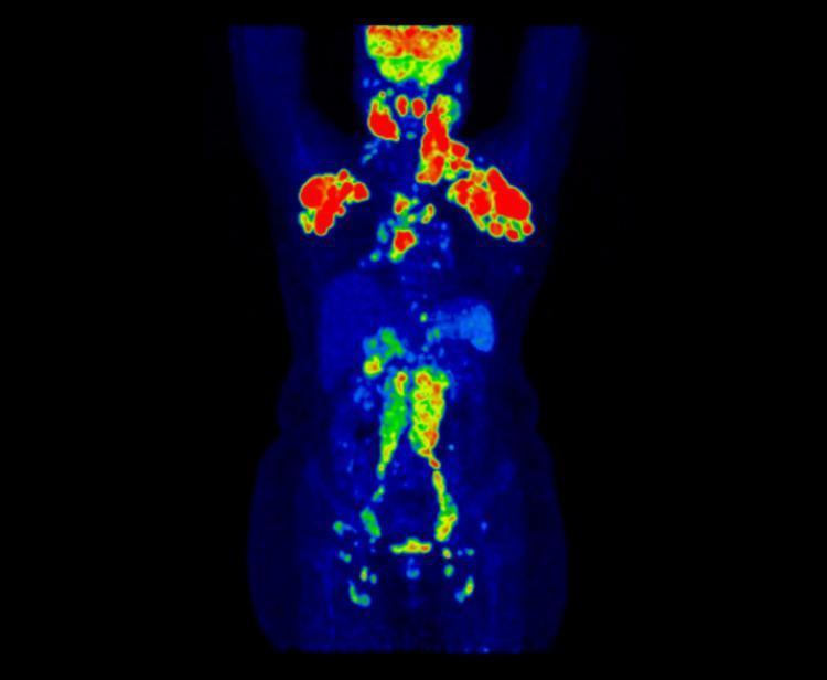
This diagnostic image is a whole-body Maximum Intensity Projection (MIP) from an 18F-FDG PET-CT scan, demonstrating widespread lymphadenopathy. The coronal projection utilizes a false-color heat map where red and yellow represent high metabolic activity (SUVmax) against a blue background of low tracer uptake. There is significant, bilateral hypermetabolic activity in the cervical and supraclavicular regions, axillary lymph nodes, and mediastinum. Furthermore, the scan reveals increased tracer uptake in the retroperitoneal, pelvic, and bilateral inguinal lymph node chains, spanning both sides of the diaphragm. The intensity of FDG avidity is most pronounced in the upper body (cervical and axillary regions) and decreases in the lower abdominal and pelvic regions. This presentation is characteristic of systemic inflammatory or neoplastic processes involving the lymphatic system, such as lymphoma or inflammatory myofibroblastic tumors, requiring histological correlation.
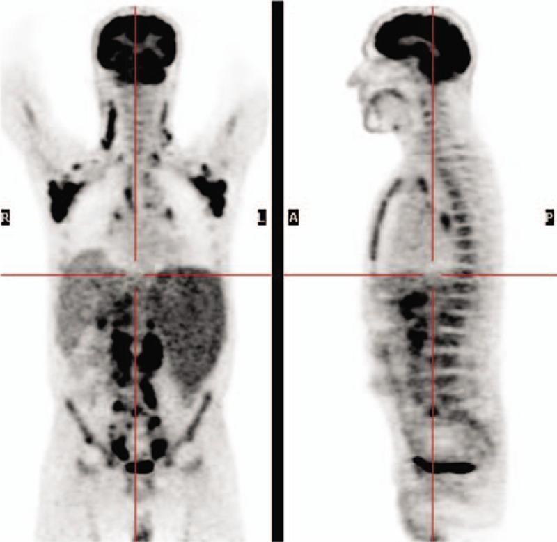
This diagnostic image consists of 18F-FDG PET/CT scans showing anterior (Maximum Intensity Projection - MIP) and sagittal views of a whole-body study at the time of diagnosis. The scans demonstrate widespread pathological fluorodeoxyglucose (FDG) accumulation in multiple anatomical regions. Distinct hypermetabolic foci are visible in several superficial and deep lymph node groups, including the cervical (neck), bilateral axillary, abdominal, and pelvic/inguinal regions (SUV max range 2–7.7). Physiological FDG uptake is noted in the brain, heart, and urinary bladder. The distribution pattern is consistent with Stage IV follicular non-Hodgkin lymphoma (fNHL), illustrating extensive lymphatic involvement both above and below the diaphragm. This imaging modality is used in oncology for baseline staging, evaluating disease burden, and identifying optimal sites for biopsy.
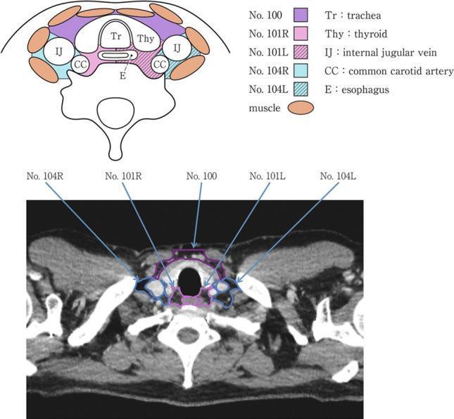
This educational graphic consists of a diagnostic CT image and a corresponding anatomical diagram illustrating cervical lymph node stations in the axial plane at the level of the thyroid gland. The anatomical diagram (top) uses color-coded regions and labels to identify the trachea (Tr), thyroid (Thy), internal jugular veins (IJ), common carotid arteries (CC), and esophagus (E). The accompanying legend classifies specific nodal stations: No. 100 (superficial cervical), No. 101R/L (upper esophageal/paratracheal), and No. 104R/L (supraclavicular). The axial CT scan (bottom) displays these anatomical structures in situ with corresponding color-coded overlays and pointers that map the nodal station classification onto the clinical image. The trachea is centrally located, anterior to the collapsed esophagus and vertebral body. The thyroid lobes flank the trachea, while the carotid sheaths containing the common carotid arteries and internal jugular veins are positioned laterally. This resource is designed for surgical oncology and radiology education, specifically for staging esophageal cancer and identifying cervical lymph node zones.
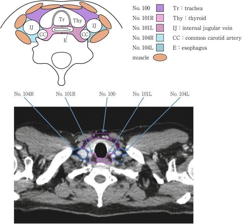
This educational graphic combines an anatomical diagram and a corresponding axial CT scan to illustrate the transverse anatomy of the neck, specifically focused on cervical lymph node stations and surrounding visceral structures. The diagram (top) and contrast-enhanced CT image (bottom) demonstrate the central, air-filled trachea (Tr, labeled No. 100) and the posterior, slightly left-deviated esophagus (E, labeled No. 104L). The thyroid gland (Thy, labeled No. 101R/L) is shown in a characteristic butterfly configuration, wrapping laterally and anteriorly around the trachea. Lateral to the thyroid lobes are the major neurovascular bundles, containing the internal jugular veins (IJ) and the common carotid arteries (CC, labeled Nos. 104R/L). Superficial muscle groups are indicated in orange. The resource is designed for medical students and radiology residents to master anatomical landmarks in the cervical region, facilitating the accurate classification of lymph node levels and the assessment of visceral pathology such as thyroid nodules or esophageal displacement.
All Lymph Node Locations and Groups
Overview Diagram
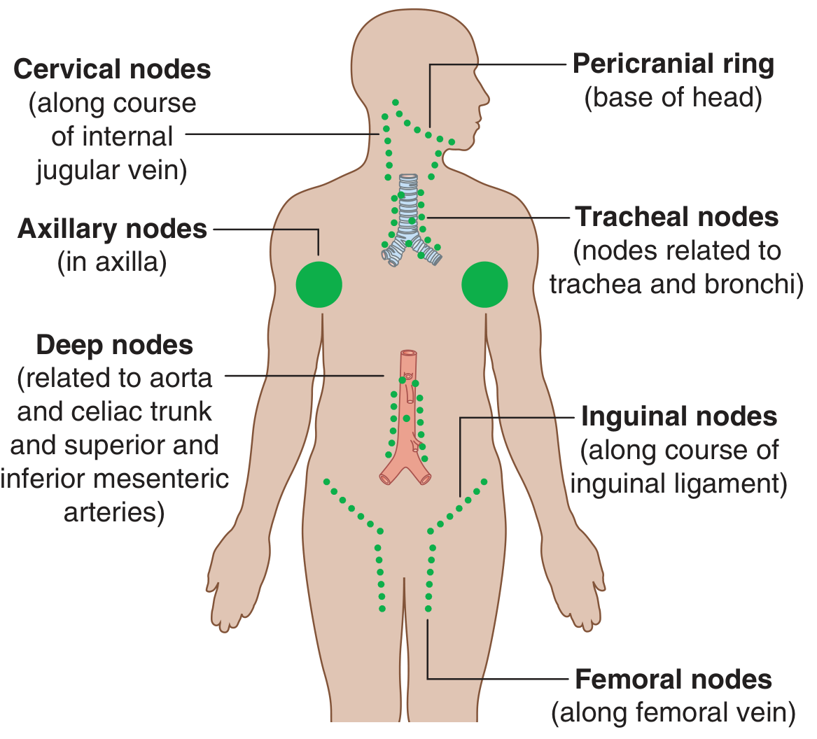
Lymph nodes are abundant and palpable in the axilla, groin/femoral region, and neck. Deep nodes (thoracic, abdominal) are not palpable and are detected on imaging. - Gray's Anatomy for Students
Lymphatic Drainage Rule
- Right side of head/neck, right upper limb, right thorax → Right lymphatic duct → Right subclavian vein junction
- Everything else (rest of body) → Thoracic duct → Left subclavian vein junction
1. HEAD AND NECK
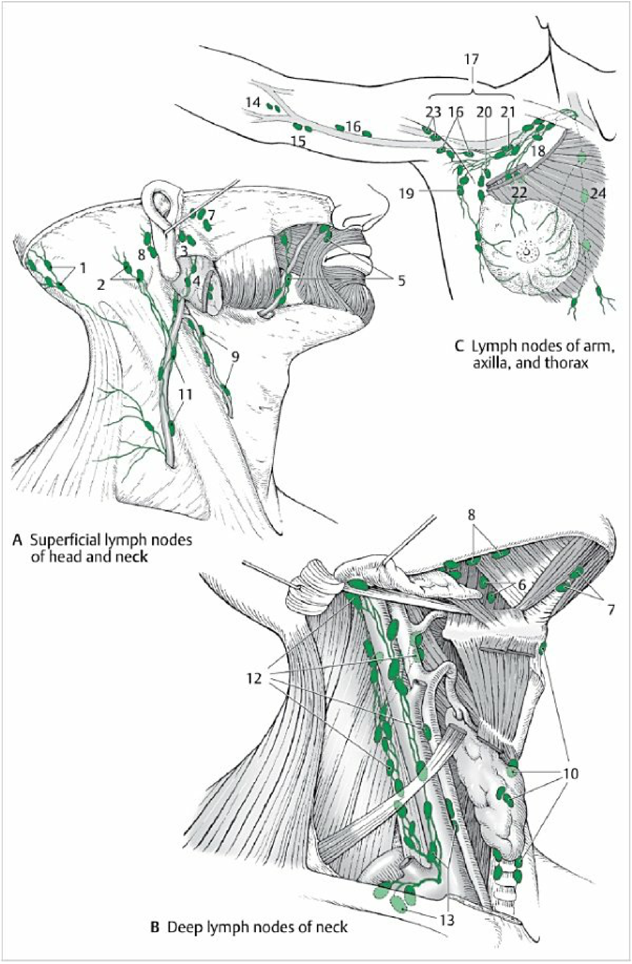
A. Pericranial Ring (Superficial nodes at base of skull)
| Node Group | Location | Drains |
|---|---|---|
| Occipital | Border of trapezius at occiput | Occiput, posterior scalp |
| Mastoid (Post-auricular) | Over mastoid process, behind ear | Posterior scalp, back of ear, external auditory canal |
| Superficial parotid (Pre-auricular) | On parotid fascia, in front of ear | Parotid gland, eyelids, external auditory canal, external nose |
| Deep parotid | Deep to parotid fascia | Deeper parotid, infratemporal fossa |
B. Face and Oral Cavity Nodes
| Node Group | Location | Drains |
|---|---|---|
| Facial nodes | Along facial artery (inconstant) | Eyelids, nose, palate, pharynx |
| Lingual nodes | Near tongue | Tongue |
| Submental nodes | Below chin (between two digastric bellies) | Tip of tongue, floor of mouth, lower lip, chin |
| Submandibular nodes | Between mandible and submandibular gland | Medial angle of eye, cheek, nose, lips, gingiva, parts of tongue; also receive from submental nodes |
C. Cervical Nodes
Anterior Cervical Group
| Node Group | Location | Drains |
|---|---|---|
| Superficial anterior cervical | Along anterior jugular vein | Superficial anterior neck |
| Deep anterior cervical | Along trachea/larynx (prelaryngeal, pretracheal, paratracheal, infrahyoid) | Larynx, trachea, thyroid, esophagus |
Lateral Cervical Group
| Node Group | Location | Drains |
|---|---|---|
| Superficial lateral cervical | Along external jugular vein | Auricle, inferior parotid |
| Superior deep cervical | Along internal jugular vein (upper part), under SCM | Second station for all head nodes; includes jugulodigastric node (tonsillar node - largest, drains tonsil and tongue) |
| Inferior deep cervical | Along internal jugular vein (lower part) | Second station for neck nodes; last filter for head nodes; includes jugulo-omohyoid node (drains tongue) |
| Supraclavicular | Above clavicle (lowest of deep cervical chain) | Lung, retroperitoneum, mediastinum; always abnormal |
Virchow's node = enlarged left supraclavicular node = metastasis from GI primary (Troisier's sign)
Cervical Levels Classification (Used in ENT/Oncology)
| Level | Location | Key Contents |
|---|---|---|
| I | Submental (Ia) + Submandibular (Ib) | Oral cavity |
| II | Upper jugular (jugulodigastric) | Oropharynx, larynx |
| III | Middle jugular | Hypopharynx, larynx |
| IV | Lower jugular | Hypopharynx, cervical esophagus |
| V | Posterior triangle | Nasopharynx, scalp |
| VI | Anterior compartment (pretracheal, paratracheal) | Thyroid, larynx, esophagus |
2. UPPER LIMB AND AXILLA
| Node Group | Location | Drains |
|---|---|---|
| Superficial cubital nodes | At elbow (superficial) | Forearm, hand |
| Deep cubital nodes | At elbow (deep) | Deep forearm structures |
| Supratrochlear nodes | Medial to brachial vein, above elbow | Medial forearm, ring/little fingers |
| Brachial nodes | Along brachial vessels | Arm |
| Deltopectoral nodes | In deltopectoral groove | Upper arm, shoulder |
Axillary Nodes (Most Important Clinically)
| Group | Location | Drains |
|---|---|---|
| Anterior (Pectoral) | Inferior border of pectoralis minor, medial axillary wall | Breast (lateral and central), anterior chest wall |
| Posterior (Subscapular) | Posterior axillary wall (subscapularis) | Back, posterior neck, shoulder |
| Lateral (Brachial) | Along axillary/brachial artery | Upper limb (all except superficial medial) |
| Central | Central axillary fat | Receives from all 3 above groups |
| Apical | Above pectoralis minor, apex of axilla | Receives from all other axillary groups; drains into subclavian trunk |
Parasternal (Internal mammary) nodes: Along internal thoracic vessels on inner chest wall - drain medial breast, intercostal spaces, liver surface.
3. THORAX
Parietal Groups (Drain the chest wall)
| Node Group | Location | Drains |
|---|---|---|
| Intercostal nodes | Posterior intercostal spaces | Pleura, intercostal muscles |
| Parasternal nodes | Along internal thoracic vessels | Breast (medial), intercostal spaces, diaphragm, liver |
| Superior diaphragmatic nodes | At diaphragmatic openings | Diaphragm, liver |
| Prevertebral nodes | Between esophagus and vertebral column | Surrounding structures |
Mediastinal Groups (Visceral)
| Node Group | Location | Drains |
|---|---|---|
| Anterior mediastinal nodes | Anterior to aortic arch | Thymus, pericardium |
| Prepericardial nodes | Between sternum and pericardium | Heart, pericardium |
| Lateral pericardial nodes | Between mediastinal pleura and pericardium | Heart area |
| Posterior mediastinal nodes (including tracheobronchial, paratracheal) | Posterior mediastinum along trachea/bronchi | Lungs, bronchi, trachea, esophagus, pericardium, diaphragm, liver |
Mediastinal/hilar adenopathy → TB, lymphoma, sarcoidosis, lung cancer, histoplasmosis
4. ABDOMEN
Parietal Groups (Drain abdominal wall and retroperitoneum)
| Node Group | Location | Drains |
|---|---|---|
| Left lumbar nodes (para-aortic) | Along abdominal aorta | Adrenal glands, kidneys, ureters, testes/ovaries, uterine fundus |
| Right lumbar nodes (para-caval) | Along inferior vena cava | Same as left side |
| Intermediate lumbar nodes | Between aorta and IVC | Same organs |
| Inferior diaphragmatic nodes | Inferior diaphragm surface | Diaphragm |
| Inferior epigastric nodes | Along inferior epigastric artery | Abdominal wall |
Visceral Groups (Drain GI organs)
| Node Group | Location | Drains |
|---|---|---|
| Celiac nodes | Around celiac trunk | Common collecting station for upper GI organs |
| Gastric nodes (right + left) | Along lesser curvature | Stomach |
| Gastro-omental nodes | Along greater curvature | Stomach, greater omentum |
| Pyloric nodes | Behind pylorus | Pyloric region |
| Pancreatic nodes | Along superior/inferior pancreas borders | Pancreas |
| Splenic nodes | At splenic hilum | Spleen |
| Pancreaticoduodenal nodes | Between pancreas and duodenum | Pancreas, duodenum |
| Hepatic nodes | Near porta hepatis | Liver, gallbladder, bile ducts |
| Mesenteric nodes (100-150) | Along root of mesentery | Small intestine (drain via celiac nodes) |
| Ileocolic nodes | Along ileocolic artery | Terminal ileum, cecum |
| Appendicular nodes | Along appendicular artery | Appendix |
| Mesocolic nodes | Along mesocolon | Large intestine |
| Inferior mesenteric nodes | Along inferior mesenteric artery | Descending colon, sigmoid colon, rectum |
Key point: Testicular tumor → para-aortic (lumbar) nodes, NOT inguinal (because testes descend from retroperitoneum)
5. PELVIS
Parietal Groups
| Node Group | Location | Drains |
|---|---|---|
| Common iliac nodes | Along common iliac vessels | Most pelvic viscera, inner abdominal wall, gluteal muscles (second filter) → drain to lumbar trunk |
| External iliac nodes | Surrounding external iliac vessels | Lower limb (via inguinal nodes), bladder (parts), vagina |
| Internal iliac nodes | Along internal iliac vessels | Pelvic viscera, perineum, pelvic walls |
Visceral Groups
| Node Group | Location | Drains |
|---|---|---|
| Paravesical nodes | Around urinary bladder | Bladder, prostate |
| Parauterine nodes | Adjacent to uterus | Cervix of uterus |
| Paravaginal nodes | Adjacent to vagina | Vagina |
| Pararectal nodes | Adjacent to rectum | Rectum |
6. LOWER LIMB
| Node Group | Location | Drains |
|---|---|---|
| Superficial inguinal nodes | Subcutaneous fat of inguinal region, along inguinal ligament | Superficial vessels of leg, anus, perineum, external genitalia, lower abdominal wall → drain to external iliac nodes |
| Deep inguinal nodes | Deep to fascia lata, medial to femoral vein | Deep vessels of leg; uppermost = Rosenmüller's node (in femoral canal) |
| Superficial popliteal nodes | At proximal end of small saphenous vein, popliteal fossa | Heel, lateral foot |
| Deep popliteal nodes | Along popliteal artery | Deep structures of leg, knee joint |
| Anterior/posterior tibial nodes | Along tibial vessels (inconstant) | Foot, leg |
Complete Summary Table
| Region | Superficial/Palpable Nodes | Deep/Impalpable Nodes |
|---|---|---|
| Head | Pericranial ring: occipital, mastoid, pre/post-auricular | Deep parotid, facial |
| Oral cavity | Submental, submandibular | Lingual |
| Neck | Superficial anterior/lateral cervical | Deep cervical chain (levels I-VI), supraclavicular |
| Upper limb | Supratrochlear, cubital | Brachial |
| Chest wall/breast | Axillary (anterior, posterior, lateral, central, apical) | Parasternal (internal mammary) |
| Thorax (deep) | - | Mediastinal, paratracheal, tracheobronchial, hilar |
| Abdomen (deep) | - | Para-aortic (lumbar), celiac, mesenteric, hepatic |
| Pelvis (deep) | - | Common iliac, external iliac, internal iliac |
| Groin | Superficial inguinal | Deep inguinal (Rosenmüller's), popliteal |
Clinical Significance by Site
| Enlarged Node | Think First Of |
|---|---|
| Submental | Lower lip, floor of mouth cancer, dental infection |
| Submandibular | Oral/tongue/facial infection, oral cancer |
| Jugulodigastric | Tonsillitis, oropharyngeal cancer |
| Posterior triangle | Nasopharyngeal carcinoma, lymphoma, TB |
| Left supraclavicular (Virchow's) | GI malignancy (stomach, colon, pancreas) |
| Right supraclavicular | Lung/esophageal cancer |
| Axillary | Breast cancer, upper limb infection, melanoma |
| Para-aortic (retroperitoneal) | Testicular cancer, lymphoma, renal cancer |
| Inguinal | STIs, lower limb infection, anal/vulvar/penile cancer, melanoma |
Where to palpate in neck for posterior traingle lymph nodes
posterior triangle neck lymph node palpation examination technique SCM trapezius
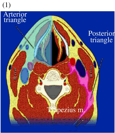
**Imaging Modality:** Anatomical cross-sectional diagram (schematic). **Anatomical Region:** Axial view of the neck at the level of the cervical vertebrae. **Key Landmarks:** - **Musculature:** Sternocleidomastoid (SCM) muscle, trapezius muscle, and prevertebral muscles are clearly delineated. - **Skeletal Structures:** Cervical vertebra with visible spinal canal. - **Vascular/Soft Tissue:** Internal jugular vein and carotid artery are represented. **Characteristic Visual Features:** The diagram uses color-coded overlays to differentiate the primary fascial spaces and anatomical compartments of the neck. - **Anterior Triangle:** Highlighted on the left side of the diagram, bounded by the midline, the anterior border of the SCM, and the mandible (superiorly). - **Posterior Triangle:** Highlighted on the right side of the diagram, bounded by the posterior border of the SCM, the anterior border of the trapezius, and the clavicle (inferiorly). **Clinical/Diagnostic Relevance:** This schematic is used for clinical indexing of neck anatomy, providing a topographical reference for identifying cervical lymph node levels and localizing pathology such as masses, abscesses, or lymphadenopathy within specific anatomical boundaries.
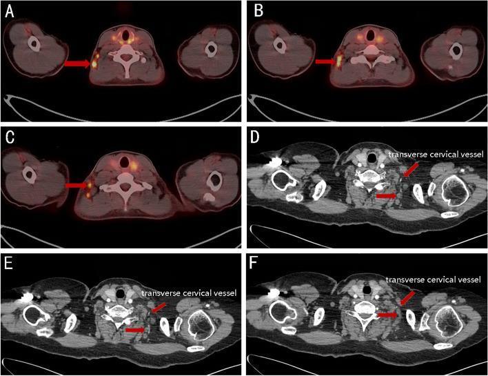
This composite of diagnostic images demonstrates the anatomical localization and metabolic activity of metastatic lymph nodes in the neck. Panels A-C show fused PET/CT axial slices where red arrows highlight hypermetabolic (FDG-avid) nodes in the posterior triangle (Level V, lateral supraclavicular group). These nodes are positioned laterally to the cervical vertebrae and deep to the trapezius muscle. Panels D-F are axial CT scans at slightly lower levels, using red arrows and text labels to identify the transverse cervical vessels. These vascular structures serve as critical surgical and radiological landmarks in the lateral neck, situated between the scalene muscle bundle medially and the omohyoid muscle laterally. The comparison illustrates the distribution of supraclavicular lymph node metastasis, often occurring in patients with extensive axillary nodal involvement from breast cancer. The images emphasize the relationship between metastatic deposits and the regional neurovascular bundle within the supraclavicular fossa, aiding in clinical target volume (CTV) delineation for radiotherapy.
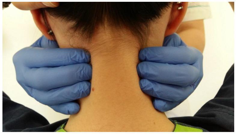
A procedural clinical photograph demonstrating the palpation of the posterior neck muscles, specifically the upper trapezius, splenius cervicis, and levator scapulae. The image is taken from a posterior view of the patient's head and neck. An examiner, wearing blue nitrile gloves, is shown with hands positioned symmetrically on both sides of the patient’s cervical region. The examiner’s fingers are gripping the muscular bulk lateral to the cervical spine, while the thumbs are likely positioned anteriorly to provide stabilization. Key anatomical landmarks visible include the occipital base, the auricles (ears), and the posterior cervical midline. This clinical maneuver is used to assess for muscle tension, trigger points, tenderness, or structural abnormalities often associated with temporomandibular disorders (TMD) or cervicalgia. The procedure illustrates a standardized physical examination technique for evaluating referred pain or compensatory muscle strain in the masticatory and cervical systems.
Palpation of Posterior Triangle Lymph Nodes
Anatomy of the Posterior Triangle
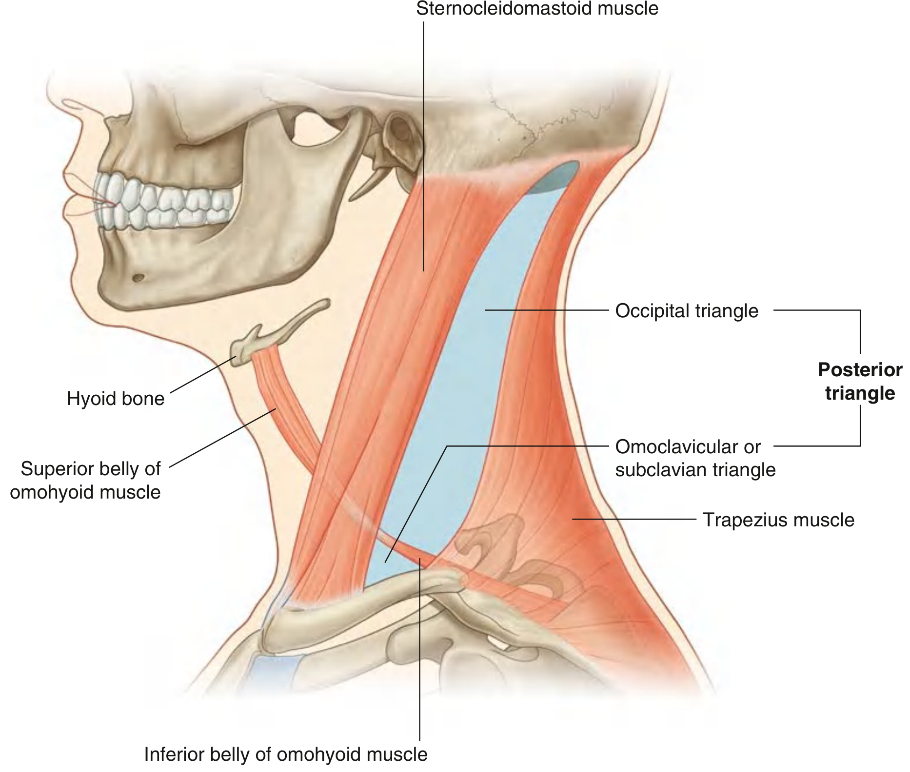
Boundaries of the Posterior Triangle
| Border | Structure |
|---|---|
| Anterior | Posterior border of Sternocleidomastoid (SCM) |
| Posterior | Anterior border of Trapezius |
| Base (inferior) | Middle one-third of the Clavicle |
| Apex | Occipital bone just posterior to the mastoid process (where SCM and trapezius attachments converge) |
- Occipital triangle (larger, upper part)
- Omoclavicular / Subclavian triangle (smaller, lower part - near clavicle)
Where Exactly to Palpate
Step-by-Step Palpation Technique
Zone 1 - Along the Posterior Border of SCM (Most Common Site)
- Place your fingers along the entire posterior edge of the SCM from mastoid tip superiorly down to the clavicle inferiorly
- The majority of posterior triangle nodes lie just behind this border
- Run fingertips anteroposteriorly into the triangle from this edge
- These correspond to Level IIb (upper, behind SCM) and Level Va/Vb nodes
Zone 2 - Occipital Triangle (Upper Posterior Triangle)
- Palpate below and behind the mastoid process
- Move fingers in the space between the posterior SCM edge and the anterior trapezius edge, in the upper neck
- Slide fingers along the floor of the triangle (over splenius capitis and levator scapulae muscles)
- Nodes here (Level Va) drain: nasopharynx, scalp, posterior neck
Zone 3 - Along the Accessory Chain (Spinal Accessory Nerve)
- The spinal accessory nerve (CN XI) runs diagonally through the posterior triangle from the upper SCM to the trapezius
- Lymph nodes cluster along this nerve course - follow an oblique line from the junction of the upper-third and lower-third of the SCM posteriorly down toward the trapezius
- This is where Level Vb (lower posterior triangle) nodes lie
Zone 4 - Supraclavicular Fossa / Base of Triangle (Level Vb + Vc)
- Palpate just above the clavicle, in the hollow between SCM and trapezius
- Hook fingers down into the supraclavicular fossa
- This is the omoclavicular triangle - where Virchow's-equivalent supraclavicular nodes sit
- Always abnormal - suggests lung, breast, GI, or lymphoma
Visual Summary - Where to Place Your Fingers
Mastoid process
↓
┌─────────────────────────────┐
│ APEX of posterior triangle │
│ │
│ ← SCM │ Zone 1 (nodes) │ ← Zone 2 (occipital triangle)
│ posterior │
│ border Zone 3 (accessory │ Trapezius
│ chain nodes) │ anterior
│ │ border →
│ ← Zone 4 (supraclavicular │
│ fossa, base of triangle) │
└─────────────────────────────┘
Middle 1/3 of Clavicle
Palpation Order for All Cervical Nodes (S Das Sequence)
- Supraclavicular group (base of posterior triangle)
- Posterior triangle (Level V nodes - along posterior SCM border, accessory chain)
- Jugulo-omohyoid (anterior triangle, Level III-IV)
- Jugulodigastric (Level II, behind angle of jaw)
- Submandibular (under jaw)
- Submental (below chin)
- Pre-auricular (in front of ear)
- Occipital (back of skull)
"A system should be maintained to palpate all the groups of lymph nodes in the neck. It may be started from below with the supraclavicular group, then moving upwards palpating the lymph nodes in the posterior triangle..."
- S Das, Manual on Clinical Surgery
What Posterior Triangle (Level V) Nodes Drain
| Source | Node Level |
|---|---|
| Nasopharynx, oropharynx | Level Va (upper posterior triangle) |
| Thyroid, cervical esophagus | Level Vb (lower posterior triangle) |
| Scalp, posterior neck | Level Va |
| Skin of posterior neck/ear | Level Va/Vb |
Clinical Significance of Posterior Triangle Enlargement
| Cause | Features |
|---|---|
| Nasopharyngeal carcinoma | Hard, fixed, non-tender; often first sign |
| Lymphoma (NHL/HL) | Rubbery, mobile, discrete |
| Tuberculosis | Matted, may be fluctuant/discharging |
| EBV / Infectious mononucleosis | Soft, tender, bilateral |
| Scalp/posterior neck infection | Tender, soft |
Posterior triangle enlargement in a middle-aged or older patient = nasopharyngeal carcinoma until proven otherwise