
Cerebral venous thrombosis (CVT) IN SIMPLE EASY TO REPORODUCE IN DNB MEDICINE FINAL EXAM 10MARK Questions
"cerebral venous thrombosis" AND treatment
cerebral venous thrombosis DNB medicine exam short note 10 marks
cerebral venous sinus thrombosis MRI venography empty delta sign
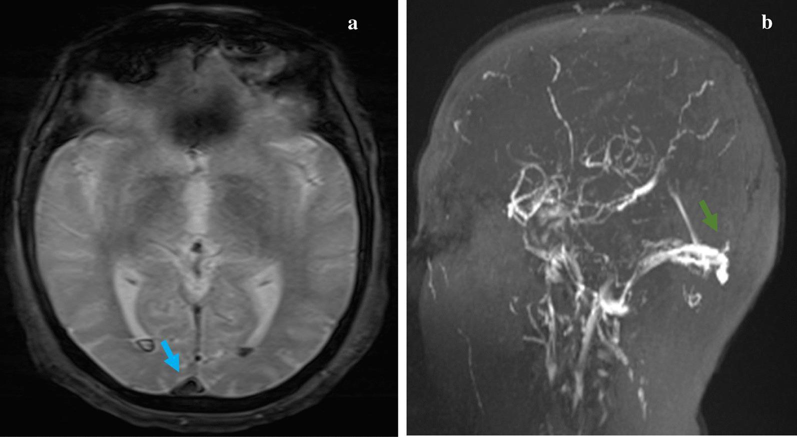
Diagnostic neuroimaging panel demonstrating Superior Sagittal Sinus Thrombosis (SSST) and associated complications. Image (a) is an axial T2*-weighted gradient echo (T2*GE) MRI sequence at the level of the lateral ventricles. It shows a distinct signal void and hypointense thrombus within the posterior superior sagittal sinus (blue arrow), creating a classic 'empty delta' sign. Additionally, hypointense blooming artifacts are visible within the occipital horns of both lateral ventricles, consistent with dependent intraventricular hemorrhage or subarachnoid blood. Image (b) is a sagittal magnetic resonance venography (MRV) reconstruction. It demonstrates a significant filling defect in the posterior segment of the superior sagittal sinus (green arrow), where the normal bright flow signal is absent or interrupted, confirming the venous sinus thrombosis. These findings are critical for diagnosing cerebral venous thrombosis, a condition that can present with intracranial hypertension or hemorrhage.
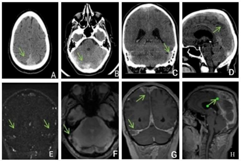
This composite diagnostic image illustrates Cerebral Venous Sinus Thrombosis (CVST) through multiple neuroimaging modalities. Top row (A-D) shows unenhanced CT scans in axial (A, B), coronal (C), and sagittal (D) planes. These images demonstrate pathognomonic hyperdensity (dense sinus sign) within the dural venous sinuses, including the superior sagittal, straight, transverse, and sigmoid sinuses, indicating acute clot formation. Bottom row (E-H) presents MRI sequences: (E) Coronal Time-of-Flight (TOF) MR venography reveals a loss of flow signal in the affected sinuses; (F) Axial T2*-weighted gradient-echo (hemo) sequence shows hypointense 'blooming' artifacts indicative of thrombus or hemorrhage; (G, H) Post-contrast T1-weighted coronal and sagittal images display the 'empty delta sign' or filling defects where contrast fails to fill the thrombosed venous lumina. The image provides a comprehensive educational overview of how CVST manifests across different radiological views and sequences, highlighting the importance of multi-modal assessment for diagnosing extensive intracranial venous thrombosis.
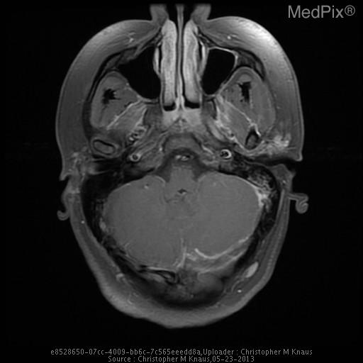
**Imaging Modality:** Contrast-enhanced Magnetic Resonance Imaging (MRI), specifically a post-contrast T1-weighted axial section or MR venography (MRV) source image.
Cerebral Venous Thrombosis (CVT) — 10 Mark Answer
1. Definition
2. Etiology / Risk Factors (mnemonic: think Virchow's triad - Hypercoagulability, Stasis, Vessel wall injury)
- Pregnancy and puerperium (peripartum state)
- Oral contraceptive pills, hormone replacement therapy
- Inherited thrombophilia: Factor V Leiden, prothrombin gene mutation, protein C/S and antithrombin deficiency
- Antiphospholipid antibody syndrome
- Malignancy (especially adenocarcinomas), paroxysmal nocturnal hemoglobinuria
- Polycythemia, thrombocytosis, nephrotic syndrome
- Behçet disease
- Infection: otitis media, mastoiditis, sinusitis, meningitis (classic infective route to lateral/cavernous sinus)
- Head injury, neurosurgical procedures, lumbar puncture
- Direct tumor invasion of sinus
3. Pathophysiology
- Venous sinus occlusion → impaired CSF absorption via arachnoid villi → raised intracranial pressure → headache, papilledema, VI nerve palsy
- Cortical/deep vein occlusion → local venous hypertension → vasogenic and cytotoxic edema → venous infarction, often hemorrhagic, that does not respect arterial territories
4. Clinical Features
- Headache - commonest symptom (>90%), often diffuse, progressive, worse lying down
- Raised ICP: papilledema, vomiting, blurred vision, VI nerve palsy
- Seizures (focal or generalized) - much more common than in arterial stroke
- Focal neurological deficits: hemiparesis, aphasia (may fluctuate, or be bilateral in sagittal sinus thrombosis - bilateral leg weakness)
- Altered sensorium - coma if deep venous system involved (poor prognosis)
- Site-specific syndromes:
- Superior sagittal sinus - bilateral leg weakness/sensory loss, seizures
- Cavernous sinus - painful ophthalmoplegia, proptosis, chemosis (usually septic, from facial/sinus infection)
- Lateral/sigmoid sinus - mastoid pain, VI/VII/VIII palsies
- Cortical vein - focal seizures, hemorrhagic infarct
- Deep venous system (straight sinus, internal cerebral veins) - bilateral thalamic involvement, coma
5. Investigations
- CT head (plain): may show "cord sign" (hyperdense thrombosed cortical vein) or "dense triangle sign"; may be normal in 30%
- CT venography / MR venography - investigation of choice: filling defect, "empty delta sign" on contrast CT/MRI (triangular filling defect in superior sagittal sinus)
- MRI brain with venogram: T2*/SWI shows blooming due to thrombus; venous infarcts often hemorrhagic and don't match arterial territory
- Digital subtraction angiography (DSA) - gold standard, rarely needed now
- D-dimer - sensitive but not fully reliable to exclude CVT (can be normal, especially in isolated headache presentations)
- Thrombophilia workup: protein C/S, antithrombin, factor V Leiden, antiphospholipid antibodies, homocysteine (send after acute phase/anticoagulation, not during)
- Baseline: CBC, coagulation profile, LP (if no mass effect, to check opening pressure and exclude infection)
6. Treatment
- Anticoagulation - mainstay, started even in presence of hemorrhagic venous infarct
- Low molecular weight heparin (body-weight adjusted, subcutaneous) preferred over unfractionated heparin (associated with lower mortality in trials) OR dose-adjusted IV UFH if renal impairment/rapid reversal needed
- Followed by oral anticoagulation (warfarin, INR target 2-3) for at least 3-6 months; longer/indefinite if strong persistent prothrombotic state
- DOACs (e.g., dabigatran) show comparable efficacy/safety to warfarin in recent meta-analyses (PMID: 38772568) - increasingly used
- Treat raised ICP: head elevation, mannitol/hyperventilation (temporizing); acetazolamide; therapeutic LP if impending visual loss and no mass effect; decompressive hemicraniectomy in malignant edema with herniation
- Seizure control: antiepileptics for those with seizures (prophylaxis debated)
- Endovascular thrombectomy/thrombolysis - reserved for patients deteriorating despite adequate anticoagulation ("medically refractory" CVT); systematic reviews show no added benefit over medical therapy alone as first-line (PMID: 38329489, 36471504)
- Treat underlying cause: antibiotics if septic (mastoiditis/sinusitis-related), stop offending drug/OCP, manage malignancy
- Decompressive surgery for large hemorrhagic infarcts with herniation risk