
show me thewhere we can find medial palpebral ligament
Reading File
Now let me search for the medial palpebral ligament in the medical library and web simultaneously.
Finding Sources
Searching the Web
medial palpebral ligament anatomy location attachments
Good results. Now let me read the textbook section and fetch the most relevant anatomy images in parallel.
Reading File
Loading Image
Loading Image
Now let me fetch the textbook diagrams showing the medial palpebral ligament directly.
Loading Image
Loading Image
Excellent - I have everything needed. Here is a complete answer with the textbook diagrams.
Medial Palpebral Ligament - Location & Anatomy
The medial palpebral ligament (also called the medial canthal tendon) is found at the medial angle (inner corner) of the eye, at the medial canthus. It is a fibrous band that anchors the eyelids to the medial orbital wall.
Location
It lies at the medial end of the tarsal plates, connecting them to the bones of the medial orbit. Specifically:
- Laterally - it arises from the medial ends of both the superior tarsus (upper eyelid) and inferior tarsus (lower eyelid)
- Medially - its anterior (superficial) part attaches to the anterior lacrimal crest of the frontal process of the maxilla; its posterior (deep) part attaches to the posterior lacrimal crest
The ligament sits anterior to the lacrimal sac, which lies between the anterior and posterior lacrimal crests, just deep to the ligament.
Diagram 1 - Orbicularis Oculi & Medial Palpebral Ligament (Fig. 8.81, Gray's Anatomy for Students)
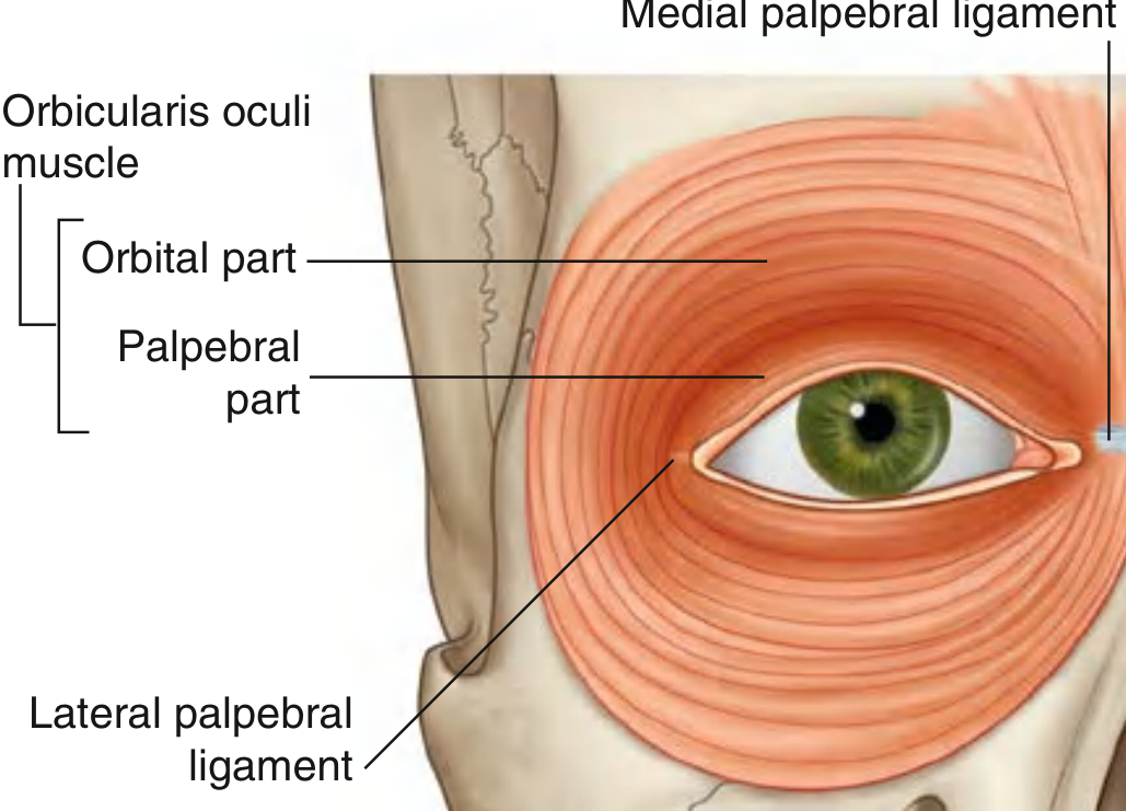
The medial palpebral ligament is labeled at the medial corner of the eye (upper right of the diagram). The palpebral part of orbicularis oculi is anchored to it medially.
Diagram 2 - Tarsal Plates & Palpebral Ligaments (Fig. 8.83, Gray's Anatomy for Students)
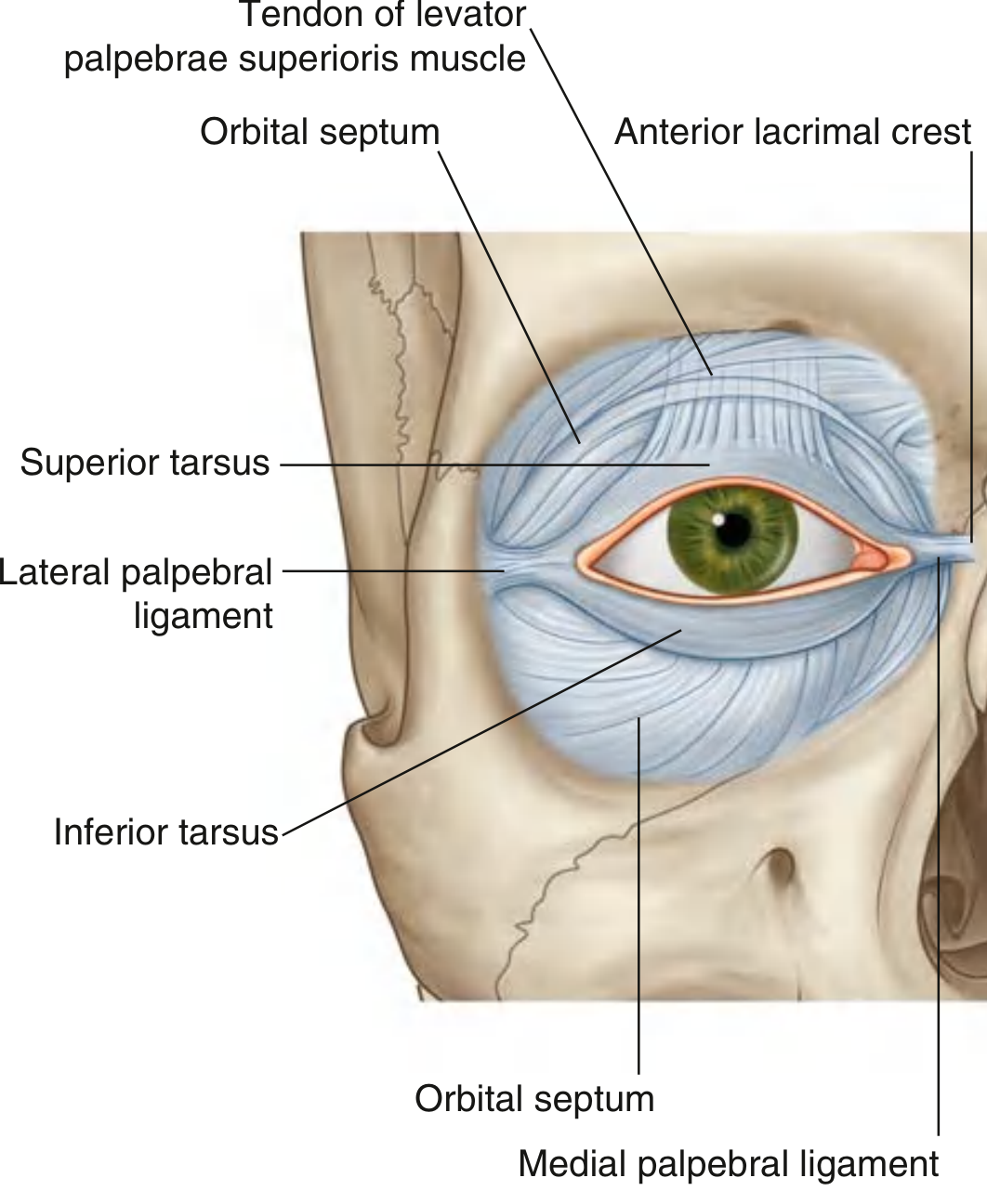
This shows the medial palpebral ligament at the lower right, connecting the superior and inferior tarsi to the anterior lacrimal crest medially.
Diagram 3 - Overall orbital anatomy with medial palpebral ligament in context
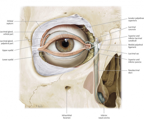
Key Relations
| Structure | Relationship to medial palpebral ligament |
|---|---|
| Lacrimal sac | Lies posterior to the ligament |
| Anterior lacrimal crest (maxilla) | Bone attachment (main insertion) |
| Posterior lacrimal crest | Attachment of the deep head |
| Orbicularis oculi - palpebral part | Anchored medially to it |
| Lacrimal canaliculi | Pass posterior to the ligament to reach the lacrimal sac |
Clinical Note
The medial palpebral ligament is an important landmark in telecanthus (increased intercanthal distance) and medial canthal tendon laxity, which can cause epiphora (tearing) or ectropion. It is also at risk in naso-orbital-ethmoid fractures.
- Gray's Anatomy for Students, Figs. 8.81, 8.83 (Tarsus and levator palpebrae superioris section)
This is a shared conversation. Sign in to Orris to start your own chat.