
lupus perinio and different high yield images for sacrcoidosis
lupus pernio sarcoidosis skin facial plaques
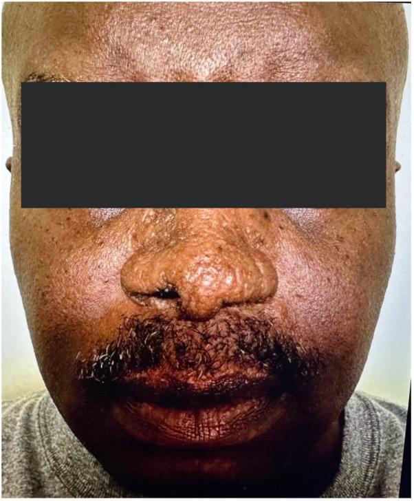
This clinical photograph shows the frontal view of a patient with cutaneous sarcoidosis, specifically lupus pernio, following systemic treatment. The image focuses on the mid-face region, including the nose, cheeks, and upper lip. While there is evidence of post-inflammatory texture changes and some subtle residual thickening, there is a marked absence of the highly characteristic indurated, violaceous, or reddish-brown shiny nodules and plaques typically seen in active lupus pernio. The nasal bridge and alae appear relatively smooth and the previous bulbous distortion has largely resolved. The skin tone on the cheeks and perioral area is more uniform, though some mild follicular prominence and chronic scarring may persist. This image serves as an educational example of the therapeutic response of cutaneous sarcoidosis to Janus kinase (JAK) inhibitor therapy, illustrating the significant reduction in granulomatous infiltration and the restoration of normal facial contours.
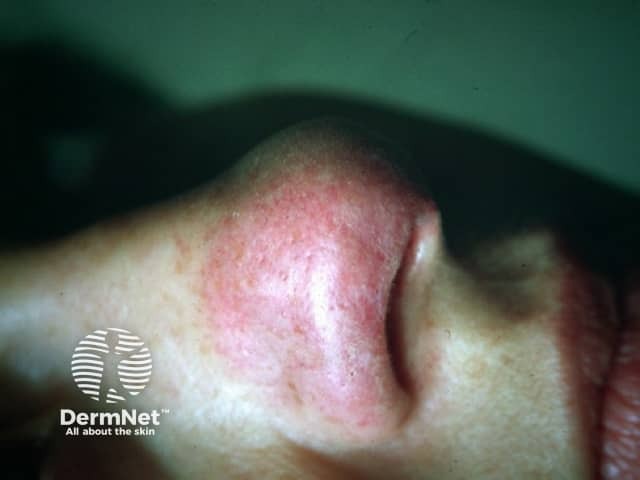
This high-resolution clinical photography shows cutaneous lupus pernio, the characteristic violaceous, indurated plaques of sarcoidosis involving the nasal dorsum and surrounding integument. Acquisition modality: bedside clinical photography, full-face anterior view, with close-up focus on the nose (nasal pyramid, alar rims) to document contour irregularities and color. The lesion appears as erythematous-violaceous plaques with firm, raised induration, subtle scaling, and blending erythema into adjacent normal skin. Dermal granulomatous inflammation in lupus pernio typically yields palpable plaques and nodules; in this image, the lesion is non-ulcerated, symmetric along the nasal region, with mild edema and vascular prominence. In the clinical context, lupus pernio strongly suggests sarcoidosis and often indicates chronic systemic involvement, particularly of the lungs and upper airways; dermatologic findings may precede or accompany pulmonary disease. Differential diagnoses include granulomatous rosacea, tuberculoid granulomatous lesions, and other granulomatous dermatoses; biopsy would show noncaseating granulomas with tight epithelioid granulomas and multinucleated giant cells. This image supports diagnostic correlation, educational case illustration, and differential diagnosis discussion for dermatology, rheumatology, pulmonology, and pathology. It has clinical relevance for prognosis and monitoring response to therapy, including corticosteroids or steroid-sparing agents, and for documenting treatment outcomes. Correlation with biopsy and systemic evaluation remains essential for definitive diagnosis.
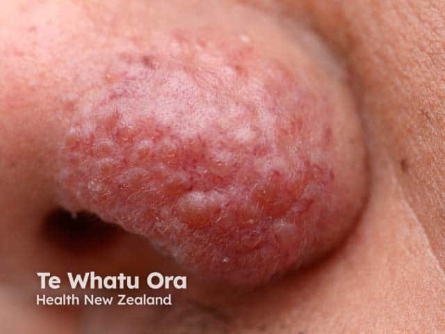
Imaging modality: Clinical dermatologic photography of a localized cutaneous plaque on the left nasal ala. The lesion is an indurated, erythematous-to-violaceous plaque with a rough surface and subtle nodularity, coalescing into a conspicuous patch along the ala nasi. The appearance is classic for lupus pernio-like cutaneous sarcoidosis, though biopsy is often required for definitive confirmation. The image emphasizes surface texture with increased dermal density and faintly infiltrated margins against surrounding normal facial skin. Involvement is confined to the external nose region; no adjacent mucosal ulceration or necrosis is evident in this view. The lesion color ranges from reddish to purplish pink, reflecting granulomatous inflammation beneath the epidermis. Clinically, such plaques may precede systemic sarcoidosis or accompany pulmonary, ocular, or multiorgan involvement; thus, this photo is valuable for differential diagnosis and educational case discussion. When compared with other granulomatous dermatoses, this presentation helps distinguish sarcoid skin lesions from infectious etiologies or rosacea variants. Potential clinical applications include guiding biopsy planning, monitoring therapeutic response to corticosteroids or steroid-sparing agents, and prompting systemic evaluation for sarcoidosis. This image serves as a teaching example for dermatologists, radiologists, and medical trainees studying cutaneous markers of systemic granulomatous disease. Clinical relevance includes monitoring progression and response.
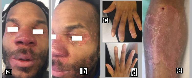
This composite clinical photograph displays the multi-systemic cutaneous and musculoskeletal manifestations of chronic sarcoidosis. Figures (a) and (b) show the patient's face with characteristic lupus pernio, presenting as indurated, violaceous (purplish) plaques and nodules affecting the nose, cheeks, and periorbital regions. Figure (e) illustrates a large, well-demarcated, erythematous to purplish indurated plaque on the medial aspect of the right leg. Figures (c) and (d) demonstrate clinical dactylitis ('sausage digits') of the hands, characterized by diffuse swelling of the fingers involving the proximal interphalangeal (PIP) and distal interphalangeal (DIP) joints. The image serves as an educational resource for identifying the classic visual signs of sarcoidosis, specifically the association between lupus pernio skin lesions and sarcoid dactylitis or inflammatory arthritis. It is intended for medical students and clinicians specializing in dermatology and rheumatology.
sarcoidosis chest X-ray bilateral hilar lymphadenopathy
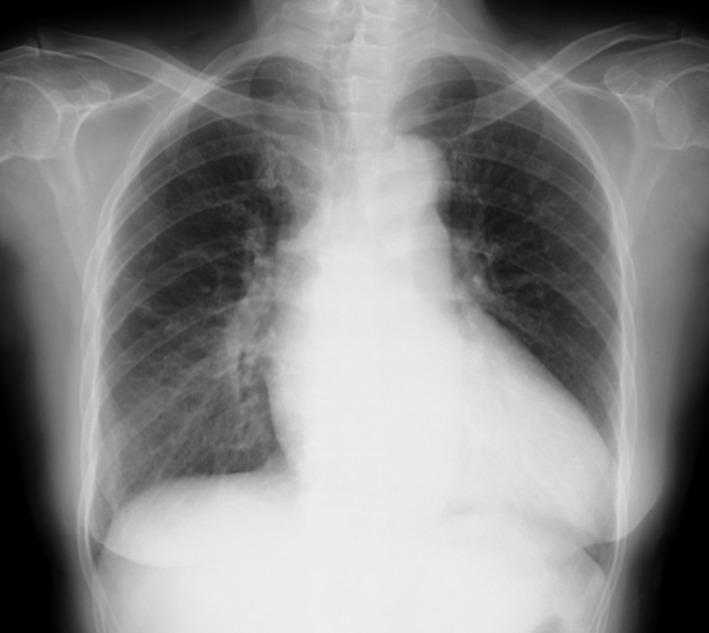
A posterior-anterior (PA) chest X-ray demonstrating a combination of thoracic findings. The image reveals prominent bilateral hilar lymphadenopathy (BHL), characterized by well-defined, bulky opacities in both hilar regions, consistent with Stage II sarcoidosis. Additionally, there is evidence of pulmonary edema, visualized as increased perihilar haziness and interstitial markings radiating from the central lung zones, which obscures the sharp margins of the pulmonary vasculature. The cardiac silhouette appears slightly enlarged, and there is a mild reduction in the clarity of the costophrenic angles. The musculoskeletal structures and overlying soft tissues are unremarkable. This radiographic presentation is significant for assessing systemic conditions such as cardiac sarcoidosis, where lymphadenopathy and acute congestive heart failure findings may coexist.
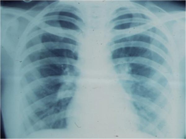
This diagnostic image is an anteroposterior chest X-ray demonstrating a classic presentation of bilateral hilar lymphadenopathy. The primary finding is the symmetrical enlargement of the hilar regions, appearing as lobulated, increased-density masses where the major bronchi and pulmonary vessels enter the lung parenchyma. This 'potato-node' appearance is a hallmark of pulmonary sarcoidosis. The lung fields are generally clear without obvious parenchymal infiltrates or consolidation, though the hilar margins are ill-defined due to the lymphatic enlargement. The mediastinal borders are slightly widened, but the cardiac silhouette and diaphragmatic contours remain distinguishable. The ribcage and clavicles are visualized and provide anatomical orientation. Clinically, this imaging is characteristic of Stage I sarcoidosis, often used to evaluate systemic granulomatous disease in both radiology and internal medicine education.
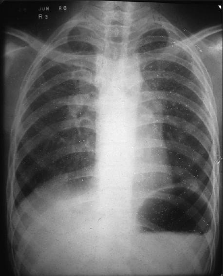
This diagnostic image is a posteroanterior (PA) chest X-ray illustrating classic features of stage II sarcoidosis. The primary finding is significant bilateral hilar lymphadenopathy, characterized by symmetrical enlargement and lobulated contours of both lung roots. Additionally, there is prominent soft tissue density in the left paratracheal region, indicating paratracheal lymphadenopathy. The lung parenchyma exhibits a diffuse reticulonodular pattern, with granular infiltrates most concentrated in the upper and middle lung zones bilaterally. These small, ill-defined opacities represent granulomatous inflammation within the interstitial spaces. The combination of symmetric hilar nodes and upper-lobe predominant parenchymal changes is highly characteristic of sarcoidosis. The image serves as a clinical reference for pulmonary manifestations of systemic granulomatous disease and the radiological assessment of mediastinal lymphadenopathy.
sarcoidosis non-caseating granuloma histology pathology
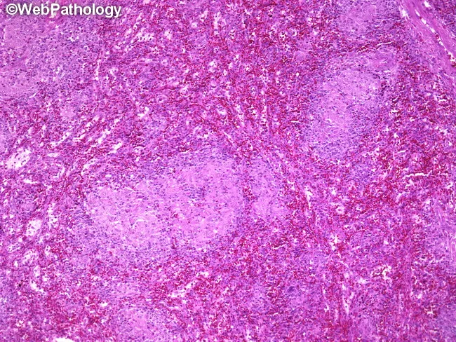
This is a photomicrograph of a splenic parenchyma section from a splenectomy specimen, prepared for light microscopy and stained with hematoxylin and eosin. The specimen shows multiple subcentimeter nodules within the splenic parenchyma, with well-formed, non-caseating granulomas composed of tightly arranged epithelioid macrophages and multinucleated giant cells surrounded by a lymphocytic rim. The granulomas lack central necrosis, which, along with histiocytic microarchitecture, is highly suggestive of granulomatous inflammation due to Toxoplasma gondii infection. Serologic tests confirmed toxoplasmosis in this patient, correlating with disseminated disease or focal splenic involvement associated with lymphadenopathy and splenomegaly. In the spleen, toxoplasmosis can produce discrete nodules or focal granulomatous lesions that may mimic lymphoma or other granulomatous diseases on imaging or gross examination. Key differential diagnoses include sarcoidosis, fungal infections such as histoplasmosis, mycobacterial infections, and other parasitic infestations; clinical correlation with serology and immune status is essential. This image is relevant for teaching granulomatous splenitis due to toxoplasmosis, assessing granuloma morphology, distinguishing non-caseating from caseating granulomas, and correlating histology with systemic infection. Appropriate clinical scenarios include immunocompromised patients with fever, cytopenias, or organomegaly and routine pathology review in splenectomy specimens. This image exemplifies infectious granulomatous disease companion to serology and morphology in clinical practice.
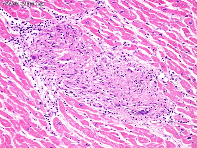
Histology, light microscopy of cardiac tissue, reveals classic non-caseating granulomatous inflammation characteristic of cardiac sarcoidosis within the full thickness of the myocardium. The predominant architectural feature is well-demarcated granulomas composed of epithelioid histiocytes with multinucleated giant cells, embedded in a mononuclear lymphocytic infiltrate. Rare eosinophils may be present, but necrosis is minimal or absent. Granulomas are distributed along lymphatic channels and are frequently associated with the cardiac conduction system tracts, explaining potential electrical disturbances. The surrounding myocytes show preserved morphology with only focal interstitial fibrosis in many areas, reflecting chronicity and prior inflammatory injury. This histologic pattern differentiates sarcoid granulomas from other granulomatous myocarditis such as tuberculosis or hypersensitivity reactions, where caseation or different inflammatory profiles may occur. The tissue shows extensive lymphatic involvement and perivascular localization, contributing to impaired conduction and remodeling. The specimen, stained with Hematoxylin and Eosin, highlights the pink cytoplasm of myocardium and the dark nuclei of inflammatory cells, facilitating recognition of granulomatous foci, giant cells, and fibrotic change. Clinically, these findings support a diagnosis of cardiac sarcoidosis and carry implications for prognosis, arrhythmia risk, and indications for immunosuppressive therapy and device management. This description enhances searchability for cardiac pathology, granulomatous myocarditis, and sarcoid research applications.
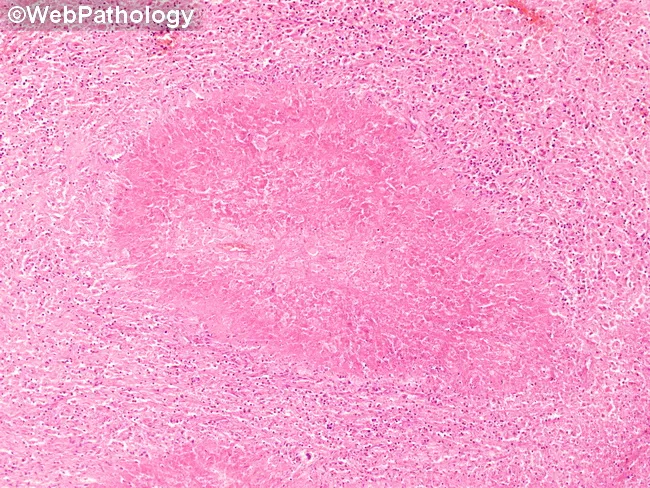
This is a low-magnification brightfield histology image of a tissue section stained with Hematoxylin and Eosin (H&E). The specimen shows granulomatous inflammation with aggregates of epithelioid macrophages forming a rounded granuloma, often with multinucleated giant cells, surrounded by a lymphocytic cuff. The central focus is well circumscribed within native parenchyma, suggesting a chronic, organized immune response to a persistent antigen. Necrosis is not clearly evident at this magnification, though subtle caseation cannot be excluded. Morphology is compatible with tuberculoid or non-caseating granulomas, and infectious versus noninfectious etiologies must be distinguished with ancillary studies. This pattern can occur in lymph nodes or solid organs and prompts a differential that includes tuberculous lymphadenitis, sarcoidosis, fungal granulomatous infections (Histoplasma, Coccidioides), and foreign-body reaction. Clinically, recognition of granulomas guides testing: acid-fast bacilli staining (Ziehl-Neelsen), fungal stains (GMS/PAS), cultures, PCR panels, and radiologic correlation. The image is educational for medical trainees, illustrating granuloma architecture, macrophage differentiation, and the spectrum of chronic inflammatory responses. Educational context.
sarcoidosis eye anterior uveitis ocular manifestation
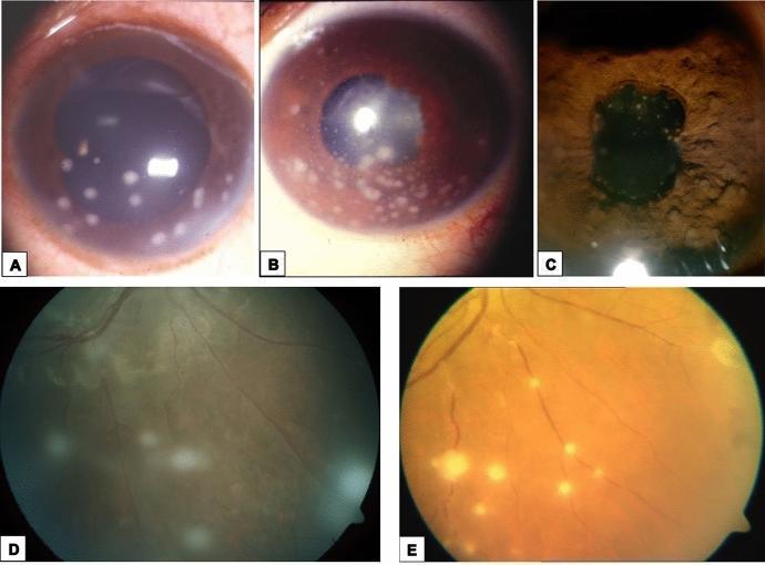
This composite of clinical ophthalmic photographs illustrates the ocular manifestations of systemic sarcoidosis across different anatomical segments. Panels (a), (b), and (c) are slit-lamp photographs of the anterior segment. Panels (a) and (b) demonstrate granulomatous anterior uveitis, characterized by mutton-fat keratic precipitates (large, greasy-appearing inflammatory deposits on the corneal endothelium). Panel (c) shows the iris stroma featuring multiple Busacca nodules, which are inflammatory granulomas located away from the pupillary margin. Panels (d) and (e) are fundus photographs demonstrating posterior segment involvement. Panel (d) illustrates intermediate uveitis with 'snowball' opacities—distinct, rounded inflammatory aggregates suspended in the vitreous humor. Panel (e) depicts sarcoid retinal vasculitis, specifically segmental periphlebitis with thick, yellowish-white perivenous exudates resembling 'candle wax drippings.' This collection serves as an educational reference for identifying characteristic granulomatous inflammatory signs in the eye, which are critical for the diagnosis and classification of ocular sarcoidosis.
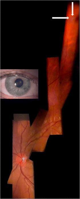
This diagnostic visual is a composite digital fundus mosaic of a right eye, created by stitching six slit-lamp video frames together. The image demonstrates clinical features of ocular sarcoidosis, specifically highlighting peripheral pre-retinal 'snowballs.' The main visualization shows the optic disc and retinal vasculature extending into the periphery. In the far superior-peripheral retina, white arrows indicate small, discrete, whitish-bright lesions—vitreous opacities known as snowballs—which are characteristic of intermediate uveitis. These lesions exhibit a raised or opaque quality, evidenced by adjacent shadows cast onto the underlying fundus. The composite illustrates the topographical relationship between the central optic nerve and far-peripheral inflammatory findings. An inset photograph of the anterior segment shows a narrow, undialated pupil, demonstrating that the fundus examination and videography were performed through a small aperture using a slit lamp and a +90D lens at 12x magnification. This imaging technique provides an alternative for capturing peripheral retinal pathology when full mydriasis is absent or clinically challenging.
sarcoidosis erythema nodosum lower legs tender nodules
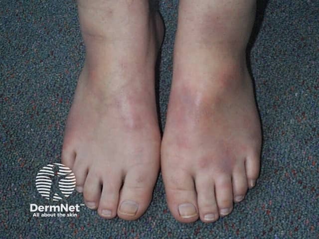
Clinical photography of erythema nodosum on the bilateral lower legs and ankles. The image demonstrates multiple tender, erythematous nodules with raised, firm, subcutaneous plaques located along the pretibial region and extending to the ankles. Color and texture provide contrast against the pale skin of the dorsal feet, highlighting the typical nodular morphology, subtle edema, and differential shading from acute inflammation. The lesions are non-ulcerated, with no purpura, necrosis, or vesiculation evident. This dermatologic presentation aligns with septal panniculitis confined to the subcutaneous adipose tissue, commonly associated with infections (streptococcal pharyngitis), medications, pregnancy, inflammatory diseases (sarcoidosis, inflammatory bowel disease), or hypersensitivity reactions. The image is intended for educational reference, illustrating the characteristic distribution pattern—bilateral, symmetric pretibial nodules on the lower extremities—and the brisk clinical correlation with systemic symptoms such as fever or arthralgia that may accompany erythema nodosum. Radiologic or histopathologic correlation would be pursued if underlying etiologies are suspected. In practice, this finding prompts clinicians to evaluate recent infections, medication exposure, or systemic disorders, and to initiate targeted investigations or empiric therapy while observing for resolution over weeks. Overall, the representation provides a concise visual guide for recognition, differential diagnosis, and management planning in dermatology and internal medicine. Reference resources.
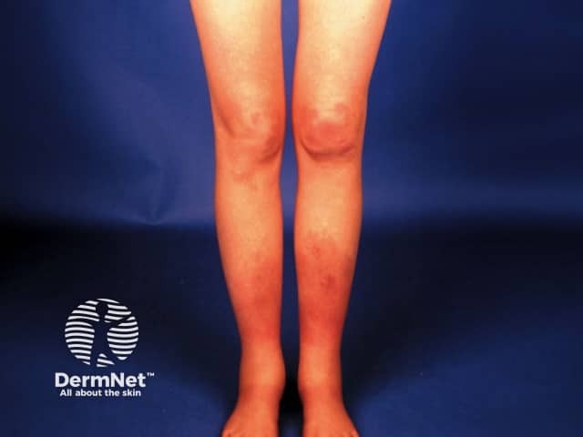
Clinical photograph of the anterior lower legs (bilateral shins) in frontal view, acquired as a color digital image for dermatologic assessment. The image depicts multiple erythematous, raised nodules concentrated over the pretibial regions, symmetrically distributed on the shins with mild surrounding edema and no ulceration. The nodules are tender to palpation in typical erythema nodosum patterns and lie within the subcutaneous tissue just below the dermis. This presentation is characteristic of erythema nodosum, aseptal panniculitis without vasculitis, which may accompany infectious (streptococcal pharyngitis), medication-related, or systemic conditions such as sarcoidosis and inflammatory bowel disease. The clinical diagnosis is supported by the symmetry and localization to the anterior lower legs, a common site. In differential diagnosis, erythema induratum (nodular vasculitis), lipodermatosclerosis from venous insufficiency, cellulitis, and other panniculitides should be considered. The image serves educational purposes and aids teledermatology assessment, patient triage, and documentation of cutaneous nodules. If biopsy were performed, histology would typically show septal panniculitis without vasculitis; occasional Miescher radial granulomas may be observed in EN, correlating with chronicity. Management emphasizes treating underlying cause and symptomatic relief with NSAIDs and leg elevation. Documentation of lesion evolution over time and response to therapy can guide assessment of etiologic triggers and inform prognosis.
sarcoidosis asteroid body Schaumann body histology giant cell
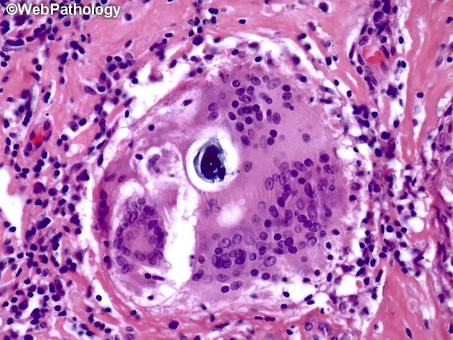
Histopathology image showing a granulomatous lesion composed of epithelioid histiocytes with a surrounding rim of lymphocytes and occasional Langhans-type multinucleated giant cells. Within a giant cell, lamellar inclusions known as Schaumann bodies are evident as concentric rings of protein and mineral material; calcium oxalate crystals may also be present. Asteroid bodies may be seen as stellate inclusions, though they are not required for diagnosis. The granuloma is noncaseating, lacking central necrosis, which favors sarcoidosis over infectious granulomatous processes such as tuberculosis. The tissue architecture shows well-formed granulomas with distinct borders, often adjacent to blood vessels in this section. The image is stained with Hematoxylin and Eosin (H&E), highlighting eosinophilic granulomatous material and basophilic nuclei within giant cells. Clinically, the presence of Schaumann bodies and Langhans giant cells supports a granulomatous inflammatory syndrome but is not pathognomonic for sarcoidosis. Differential considerations include berylliosis, hypersensitivity pneumonitis, fungal or mycobacterial infections, and foreign body reactions. Correlation with chest imaging, serum ACE levels, and clinical features is essential for diagnosis. This slide serves an educational role in recognizing granulomatous histology, giant cell inclusions, and lamellar calcific bodies, informing diagnostic workflows and teaching pathology trainees about sarcoidosis-related granulomatous disease.
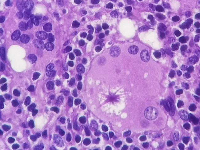
Histology of a silicone lymph node biopsy examined by light microscopy using Hematoxylin and Eosin staining. The specimen shows a granulomatous lymphadenopathy with a prominent giant cell reaction. Within the cytoplasm of epithelioid and multinucleated giant cells sits a large, eosinophilic asteroid body characterized by a centralized core and radiating, star‑like filamentous arms. The asteroid inclusions stain intensely with hematoxylin, and their radiating processes extend toward surrounding macrophages. Surrounding tissue displays silicone-induced foreign body granulomas with vacuolated spaces and surrounding chronic inflammatory cells. The asteroid body itself is a distinctive cytoplasmic inclusion thought to contain complex lipoproteins and mineral components such as calcium, phosphorus, silicon, and aluminum. This morphological finding is most classically described in silicone lymphadenopathy but can also be observed in sarcoidosis and other granulomatous diseases. Clinically, asteroid bodies support a silicone exposure–related process when correlated with implant history or imaging, but their presence is not pathognomonic. Differential diagnosis includes sarcoidosis, foreign body granulomatosis, mycobacterial, and fungal granulomatous infections. The observation aids diagnostic stratification, guides further testing for systemic granulomatous disease, and informs patient counseling regarding implant-associated lymphadenopathy and granulomatous inflammation. This description emphasizes asteroid body recognition as a valuable histopathologic clue in implant-era lymphadenopathy for clinical correlation.
sarcoidosis CT scan pulmonary lymphadenopathy nodules