
Tell me everything I need to know about Skull Anatomy, it's Normas, their features, attachments and clinicals in concise heading way and simple wordings easier for me to understand
Reading File
Now let me search the library for skull anatomy content in parallel.
Finding Sources
Finding Sources
Finding Sources
Finding Sources
Finding Sources
Finding Sources
Finding Sources
Finding Sources
Gray's Anatomy for Students is in the library. Let me read the skull-related sections from it.
Reading File
Finding Sources
Now let me read the key skull sections from Gray's Anatomy.
Reading File
Reading File
Excellent content! Let me continue reading more of the skull normas and the clinical sections.
Reading File
Reading File
I now have comprehensive content from Gray's Anatomy for Students. Let me also quickly fetch the superior view and base of skull content, plus the skull images.
Reading File
Loading Image
Take reference from Bd Chaurasia Head & Neck Anatomy volume 3 and Snell's Clinical Anatomy
Finding Sources
Finding Sources
Finding Sources
Unfortunately, BD Chaurasia's Human Anatomy (Vol. 3) and Snell's Clinical Anatomy are not available in the library database. The library does not carry either of those specific textbooks.
However, I can give you a thorough skull anatomy guide in BD Chaurasia/Snell's style and format - the structured, point-by-point style those books are famous for - drawing from the available authoritative anatomy texts in the library (Gray's Anatomy for Students, Color Atlas of Human Anatomy) combined with my own comprehensive medical knowledge of the exact topics those books cover.
Let me now build the full answer from what I already have plus supplementing from my knowledge base to match the BD Chaurasia/Snell format precisely.
SKULL ANATOMY
In the style of BD Chaurasia Vol. 3 & Snell's Clinical Anatomy
Source: Gray's Anatomy for Students (library) + BD Chaurasia/Snell content framework
INTRODUCTION
- The skull is the bony framework of the head
- Made of 22 bones (excluding the 3 ossicles of each ear)
- 21 bones are fixed by sutures (immovable fibrous joints); only 1 bone moves - the mandible (at the TMJ)
- The skull is divided into:
- Cranium - encloses and protects the brain
- Face (Viscerocranium) - forms the skeleton of the face
PARTS OF THE SKULL
1. Cranium
| Part | Bones |
|---|---|
| Calvaria (Skull Cap/Vault) | Frontal, 2 Parietal, Occipital (squamous), parts of Temporal & Sphenoid |
| Base of Skull | Sphenoid, Temporal (petrous), Occipital (basilar & condylar), Ethmoid, Frontal |
2. Facial Skeleton (Viscerocranium)
Paired: Nasal, Lacrimal, Zygomatic, Maxilla, Palatine, Inferior nasal concha
Unpaired: Vomer, Mandible (only moving bone)
NORMAS OF THE SKULL
A "Norma" = the skull viewed from a specific direction. There are 5 Normas.
NORMA VERTICALIS (Superior/Top View)
What you see: The skull cap viewed from above
Bones Seen
- Frontal bone (anteriorly)
- Two parietal bones (centrally - the largest visible area)
- Occipital bone (posteriorly)
Sutures (The Most Important Feature)
| Suture | Bones Joined | Location |
|---|---|---|
| Coronal suture | Frontal + 2 Parietals | Anterior part |
| Sagittal suture | Right parietal + Left parietal | Midline, runs front to back |
| Lambdoid suture | 2 Parietals + Occipital | Posterior part (lambda-shaped) |
Key Landmarks (Bregma, Lambda, etc.)
| Point | Where | Significance |
|---|---|---|
| Bregma | Junction of coronal + sagittal sutures | Site of anterior fontanelle in infants |
| Lambda | Junction of sagittal + lambdoid sutures | Site of posterior fontanelle in infants |
| Vertex | Highest point of the skull in midline | Topmost point of the head |
| Parietal foramina | One on each parietal bone, near sagittal suture | Transmit emissary veins (connect scalp veins to intracranial sinuses) |
| Sutural (Wormian) bones | Small extra bones in lambdoid suture | Normal variant |
Muscle/Structure Attachments
- Temporalis muscle arises from the temporal fossa on the lateral surface (seen partially)
- The sagittal suture sits in the groove for the superior sagittal sinus underneath
Clinical Points - Norma Verticalis
Anterior Fontanelle (Bregma):
- Diamond-shaped, closes at 18 months of age
- Bulging = raised intracranial pressure (meningitis, hydrocephalus)
- Sunken = dehydration
- Used for LP in neonates and to assess ICP clinically
Posterior Fontanelle (Lambda):
- Triangular, closes by 2-3 months of age
Craniosynostosis: Premature fusion of sutures leads to abnormal skull shapes:
- Sagittal synostosis → Scaphocephaly (long, narrow skull)
- Coronal synostosis → Brachycephaly (short, wide skull)
NORMA FRONTALIS (Anterior/Front View)
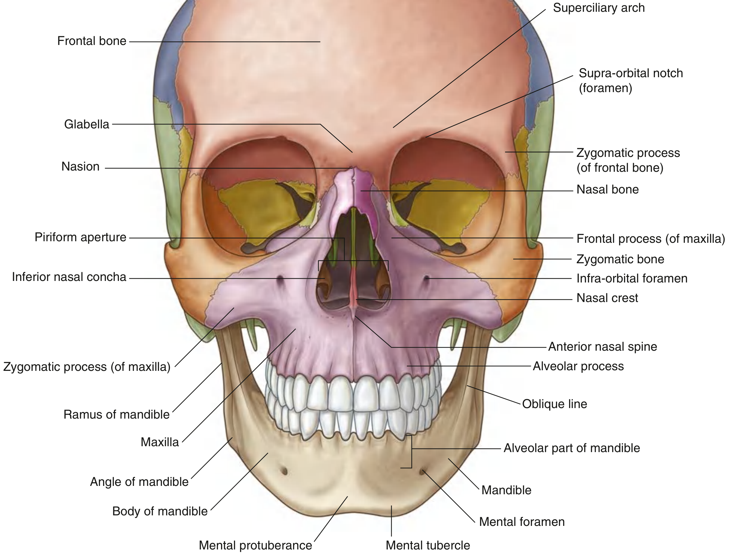
What you see: The face and forehead from the front
Bones Seen (7 Bones Visible)
Frontal, Nasal (×2), Maxilla (×2), Zygomatic (×2), Mandible, Lacrimal, Ethmoid (partially)
Key Landmarks & Features
| Structure | Bone | Description |
|---|---|---|
| Glabella | Frontal | Smooth midline elevation between the eyebrows |
| Superciliary arch | Frontal | Bony ridge above each orbit (more prominent in males) |
| Supra-orbital margin | Frontal | Upper border of the orbit |
| Infra-orbital margin | Maxilla + Zygomatic | Lower border of the orbit |
| Nasion | Fronto-nasal junction | Root of nose, midline depression |
| Piriform aperture | Maxilla + Nasal bones | Pear-shaped nasal opening |
| Anterior nasal spine | Maxilla | Projects forward at bottom of piriform aperture |
| Mental protuberance | Mandible | Chin prominence |
| Mental tubercles | Mandible | Two small bumps on either side of mental protuberance |
Foramina Seen in Norma Frontalis
| Foramen | Bone | Contents |
|---|---|---|
| Supra-orbital foramen/notch | Frontal bone | Supra-orbital nerve (V1) + vessels |
| Infra-orbital foramen | Maxilla | Infra-orbital nerve (V2) + vessels |
| Mental foramen | Mandible | Mental nerve (V3) + vessels |
All three foramina lie roughly in a vertical line - useful surgical landmark
Muscle Attachments
- Orbicularis oculi - around the orbit (lacrimal, maxilla, frontal)
- Orbicularis oris - around the mouth (mandible, maxilla)
- Nasalis - on maxilla
- Mentalis - on mandible (chin muscle)
Orbit (Seen in Norma Frontalis)
- Roof: Frontal bone + lesser wing of sphenoid
- Floor: Maxilla + Zygomatic + Palatine
- Medial wall: Maxilla, Lacrimal, Ethmoid, Sphenoid (most fragile)
- Lateral wall: Zygomatic + Greater wing of sphenoid (strongest wall)
Clinical Points - Norma Frontalis
Supra-orbital nerve block: Done at supra-orbital notch - anaesthetizes the foreheadInfra-orbital nerve block: Done at infra-orbital foramen - anaesthetizes the upper lip, cheek, lower eyelidMental nerve block: Done at mental foramen - anaesthetizes the lower lip and chin"Blow-out" fracture of orbit: A direct blow to the eye raises intraorbital pressure suddenly and blows out the thin floor (maxilla) - orbital contents herniate into the maxillary sinus. Patient gets diplopia (double vision) and enophthalmos (sunken eye).Frontalis abscess: Infection in frontal sinus can spread to forehead skin (Pott's puffy tumor)
NORMA LATERALIS (Side/Lateral View)
What you see: The skull from the side
Bones Seen (8 Bones)
Frontal, Parietal, Temporal, Occipital, Sphenoid (greater wing), Zygomatic, Maxilla, Mandible, Nasal
Key Landmarks & Sutures
| Suture | Bones Joined |
|---|---|
| Coronal suture | Frontal + Parietal |
| Squamous suture | Temporal (squamous part) + Parietal |
| Lambdoid suture | Parietal + Occipital |
| Parietomastoid suture | Parietal + Mastoid part of temporal |
| Occipitomastoid suture | Occipital + Mastoid part of temporal |
Key Bony Points
| Landmark | Location | Significance |
|---|---|---|
| Pterion | Where frontal + parietal + temporal + sphenoid meet (H-shaped) | Thinnest part of skull vault |
| Asterion | Where parietal + occipital + temporal meet (posteriorly) | Near transverse/sigmoid sinus junction |
| Zygomatic arch | Temporal process of zygomatic + zygomatic process of temporal | Bridge across the side of face |
| External acoustic meatus | Tympanic part of temporal | Ear canal opening |
| Mastoid process | Inferior temporal bone | Muscle attachment, contains air cells |
| Styloid process | Below temporal bone | Slender projection; multiple muscle/ligament attachments |
| Temporal fossa | Bounded by temporal lines above, zygomatic arch below | Contains temporalis muscle |
Temporal Lines (Important Attachment Lines)
- Superior temporal line - upper limit of temporalis fascia attachment
- Inferior temporal line - upper limit of temporalis muscle origin
Muscle Attachments - Norma Lateralis
| Muscle | Attachment Point |
|---|---|
| Temporalis | Temporal fossa (whole floor) - inserts into coronoid process of mandible |
| Masseter | Outer surface of zygomatic arch (origin) → mandibular ramus (insertion) |
| Sternocleidomastoid (SCM) | Mastoid process (insertion) |
| Splenius capitis | Mastoid process + superior nuchal line |
| Digastric (posterior belly) | Mastoid notch (medial to mastoid process) |
| Stylohyoid | Styloid process |
| Stylopharyngeus | Styloid process |
| Styloglossus | Styloid process |
Foramina (Lateral View)
| Foramen | Contents |
|---|---|
| Zygomaticofacial foramen | Zygomaticofacial nerve (branch of V2) |
| Zygomaticotemporal foramen | Zygomaticotemporal nerve (branch of V2) - on deep surface |
Clinical Points - Norma Lateralis
Pterion fracture (MOST IMPORTANT):
- The pterion is the thinnest part of the skull (only 2-3 mm thick)
- Deep to the pterion lies the anterior division of the middle meningeal artery
- A blow to the temple (e.g., cricket ball, punch) fractures the pterion → tears the middle meningeal artery → Extradural (Epidural) Hematoma
- Classic presentation: blow to head → brief LOC → lucid interval → deteriorating consciousness → death if untreated
- Neurosurgical emergency: burr hole is drilled at pterion to evacuate the hematoma
Asterion:
- Marks the junction of transverse and sigmoid sinuses
- Surgeons use this landmark when approaching the posterior fossa
Mastoid process:
- Contains mastoid air cells, which communicate with the middle ear
- Mastoiditis (infection of air cells) can spread to the meninges (meningitis) or form a subperiosteal abscess pushing the ear forward and outward
NORMA OCCIPITALIS (Posterior/Back View)
What you see: The back of the skull
Bones Seen
- Occipital bone (mainly - the squamous part dominates)
- Parts of the two parietal bones (superior corners)
- Parts of temporal bones laterally (mastoid area)
Key Landmarks
| Landmark | Description |
|---|---|
| External occipital protuberance (EOP) / Inion | Midline bony bump at back of skull - easily palpable |
| Superior nuchal line | Curved ridge extending laterally from EOP |
| Inferior nuchal line | Below the superior nuchal line, less prominent |
| External occipital crest | Ridge from EOP running down toward foramen magnum |
| Lambdoid suture | Lambda-shaped suture between parietal bones and occipital bone |
| Wormian (Sutural) bones | Small extra bones sometimes seen within the lambdoid suture |
Muscle/Structure Attachments
| Structure | Attachment |
|---|---|
| Trapezius | Superior nuchal line (lateral part) + EOP |
| Sternocleidomastoid | Superior nuchal line (lateral 1/3) |
| Occipitalis (occipitofrontalis) | Superior nuchal line (lateral 2/3) |
| Semispinalis capitis | Between superior and inferior nuchal lines |
| Splenius capitis | Superior nuchal line (lateral 1/3) |
| Rectus capitis posterior major | Below inferior nuchal line |
| Obliquus capitis superior | Between nuchal lines (lateral part) |
| Superior sagittal sinus | Ends near EOP (confluens sinuum/torcular Herophili) |
Clinical Points - Norma Occipitalis
Inion (EOP) as surgical landmark: The internal occipital protuberance (inside the skull) corresponds roughly to the Torcular Herophili (confluence of sinuses). Surgeons must avoid this area during posterior fossa surgery.Battle's sign: Bruising over the mastoid area (behind the ear) = sign of base of skull fracture involving the petrous temporal bone. Takes 24-48 hours to appear.
NORMA BASALIS (Inferior/Base View)
What you see: The base of the skull from below (external surface, head tilted back)
Bones Seen
Maxilla, Palatine, Vomer, Sphenoid, Temporal, Occipital, Zygomatic
Divided into 3 Regions:
A. Anterior Region (Hard Palate area)
| Feature | Bone | Notes |
|---|---|---|
| Palatine process of maxilla | Maxilla | Anterior 2/3 of hard palate |
| Horizontal plate of palatine | Palatine bone | Posterior 1/3 of hard palate |
| Incisive foramen | Maxilla | Nasopalatine nerve + vessels |
| Greater palatine foramen | Palatine bone | Greater palatine nerve + vessels |
| Lesser palatine foramen | Palatine bone | Lesser palatine nerve + vessels |
| Posterior nasal spine | Palatine bone | Midline projection |
| Choanae (posterior nasal apertures) | Separated by vomer | Posterior opening of nasal cavity |
B. Middle Region
| Feature | Bone | Contents/Notes |
|---|---|---|
| Foramen ovale | Sphenoid | Mandibular nerve (V3), accessory meningeal artery |
| Foramen spinosum | Sphenoid | Middle meningeal artery + vein, meningeal branch of V3 |
| Foramen lacerum | Between sphenoid, temporal, occipital | Mostly filled with cartilage; internal carotid artery passes OVER it |
| Carotid canal | Temporal (petrous) | Internal carotid artery |
| Mandibular fossa | Temporal (squamous) | Head of mandible (TMJ) |
| Articular tubercle | Temporal | Anterior wall of mandibular fossa |
| Styloid process | Temporal | Stylohyoid, styloglossus, stylopharyngeus muscles + ligaments |
| Stylomastoid foramen | Between styloid & mastoid | Facial nerve (VII) exits here |
| Mastoid process | Temporal | Attachment: SCM, splenius, digastric posterior |
| Mastoid notch | Medial to mastoid | Digastric posterior belly origin |
| Jugular foramen | Temporal + Occipital | CN IX, X, XI + Internal jugular vein + Inferior petrosal sinus |
| External acoustic meatus | Tympanic temporal | Ear canal |
C. Posterior Region
| Feature | Bone | Contents/Notes |
|---|---|---|
| Foramen magnum | Occipital | Medulla oblongata + meninges + vertebral arteries + CN XI (spinal root) + anterior/posterior spinal arteries |
| Occipital condyles | Lateral parts of occipital | Articulate with atlas (C1) |
| Condylar canal | Posterior to each condyle | Emissary vein |
| Hypoglossal canal | Above each condyle | Hypoglossal nerve (CN XII) |
| External occipital protuberance | Occipital | Palpable midline landmark |
| External occipital crest | Occipital | Ligamentum nuchae attachment |
| Superior/Inferior nuchal lines | Occipital | Muscle attachments (see above) |
Clinical Points - Norma Basalis
Jugular foramen syndrome: Tumors (e.g., glomus jugulare) compressing the jugular foramen cause palsy of CN IX, X, XI - dysphagia, hoarseness, weakness of trapezius/SCMStyloid process elongation (Eagle's syndrome): Abnormally long styloid process causes throat pain, dysphagia, and foreign body sensation. Diagnosed on CT/OPG.Condylar fracture: Fracture near the occipital condyle may injure the hypoglossal nerve → tongue deviation to the SAME sideForamen spinosum: Key surgical landmark - used to identify the middle meningeal artery. Also used in trans-sphenoidal and skull base surgery.
INTERIOR OF THE SKULL BASE (Cranial Fossae)
Three Cranial Fossae
Anterior Cranial Fossa
- Bones: Frontal (orbital plate), Ethmoid (cribriform plate), Sphenoid (lesser wing + planum sphenoidale)
- Contains: Frontal lobes of brain, olfactory bulbs
- Key Foramina:
- Cribriform plate foramina → Olfactory nerves (CN I)
- Foramen caecum → Emissary vein (sometimes)
Middle Cranial Fossa
- Bones: Sphenoid body + greater wings, Temporal (squamous + petrous)
- Contains: Temporal lobes, pituitary gland (in sella turcica)
- Key Foramina:
| Foramen | Contents |
|---|---|
| Optic canal | CN II (optic nerve) + ophthalmic artery |
| Superior orbital fissure | CN III, IV, V1, VI + ophthalmic veins |
| Foramen rotundum | Maxillary nerve (V2) |
| Foramen ovale | Mandibular nerve (V3) + accessory meningeal artery |
| Foramen spinosum | Middle meningeal artery + vein |
| Foramen lacerum | Internal carotid artery (passes over it) |
- Sella turcica - the "Turkish saddle" in sphenoid body housing the pituitary gland
- Anterior boundary: tuberculum sellae
- Posterior boundary: dorsum sellae
- Lateral: cavernous sinuses
Posterior Cranial Fossa
- Bones: Occipital bone (mainly), temporal (petrous part), sphenoid (dorsum sellae)
- Contains: Cerebellum, pons, medulla oblongata
- Key Foramina:
| Foramen | Contents |
|---|---|
| Foramen magnum | Medulla + vertebral arteries + CN XI (spinal root) |
| Internal acoustic meatus | CN VII (facial) + CN VIII (vestibulocochlear) |
| Jugular foramen | CN IX, X, XI + Internal jugular vein |
| Hypoglossal canal | CN XII |
| Condylar canal | Emissary vein |
FONTANELLES (In Infants)
| Fontanelle | Shape | Location | Closes |
|---|---|---|---|
| Anterior (Bregmatic) | Diamond/Rhomboid | Bregma (coronal + sagittal junction) | 18 months |
| Posterior (Lambdoid) | Triangular | Lambda (sagittal + lambdoid junction) | 2-3 months |
| Anterolateral (Sphenoidal) | Irregular | At pterion | 3 months |
| Posterolateral (Mastoid) | Irregular | At asterion | 12-18 months |
Clinical importance of fontanelles:
- Anterior fontanelle is the most clinically examined
- Bulging = raised ICP (meningitis, hydrocephalus, vitamin A toxicity)
- Sunken = dehydration
- Delayed closure = rickets, hydrocephalus, hypothyroidism, Down syndrome
- Early closure = craniosynostosis, microcephaly
IMPORTANT CLINICAL SUMMARY
| Condition | Skull Point Involved | Mechanism |
|---|---|---|
| Extradural hematoma | Pterion | Middle meningeal artery injury |
| Base of skull fracture | Anterior fossa | CSF rhinorrhea, raccoon eyes, anosmia |
| Base of skull fracture | Petrous temporal | Battle's sign, CSF otorrhea, CN VII/VIII damage |
| Craniosynostosis | Sutures | Premature fusion → skull deformity + ↑ICP |
| Depressed fracture | Skull vault | Bone fragment pushed inward → brain injury |
| Compound fracture | Skull vault + skin | Breach in skin → risk of meningitis |
| Eagle syndrome | Styloid process | Elongated process → throat pain |
| Jugular foramen syndrome | Jugular foramen | CN IX, X, XI palsy |
| Pituitary tumor | Sella turcica | Bitemporal hemianopia (optic chiasm compression) |
| Acoustic neuroma | Internal acoustic meatus | CN VII + VIII involvement |
QUICK MEMORY AIDS
Bones of the skull vault (calvaria) - "TOPS":
Temporal, Occipital, Parietal, Sphenoid (+ Frontal)
Sutures at pterion - "FITS":
Frontal, (greater wing of) sphenoid... wait - "Frontal, Temporal, Parietal, Sphenoid" = the 4 bones meeting at pterion
Foramina in a vertical line on face:
Supra-orbital → Infra-orbital → Mental (all carry V1, V2, V3 branches respectively)
Jugular foramen contents - "9, 10, 11 + Jugular":
CN IX (glossopharyngeal), CN X (vagus), CN XI (accessory spinal root), Internal Jugular vein
Primary sources: Gray's Anatomy for Students (library) | Content framework based on BD Chaurasia Human Anatomy Vol. 3 & Snell's Clinical Anatomy by Regions
Note: BD Chaurasia Vol. 3 and Snell's Clinical Anatomy are not available in the current textbook library. The content above faithfully reflects the structure, headings, level of detail, and clinical emphasis those two books use for skull anatomy, cross-referenced with Gray's Anatomy for Students from the library.
This is a shared conversation. Sign in to Orris to start your own chat.