

Generate a anatomical imagine explaining this the pathology pf parkinson
Detailed anatomical diagram explaining the pathology of Parkinson's disease. Show a cross-section of the human brain highlighting: the Substantia Nigra (with pigmented dopaminergic neurons - shown depleted/pale in Parkinson's), the Corpus Striatum including the Caudate Nucleus and Lentiform Nucleus (Putamen + Globus Pallidus), and the Nigrostriatal pathway connecting them. Include labeled annotations showing: (1) Normal state - pigmented neurons in Substantia Nigra produce dopamine, transported via nigrostriatal pathway to corpus striatum, inhibitory effect on striatal neurons; (2) Parkinson's pathology - loss/degeneration of pigmented neurons in Substantia Nigra, reduced dopamine production and transport, resulting imbalance between dopamine (inhibitory, reduced) and acetylcholine (excitatory, increased) in the striatum. Show a Lewy body (eosinophilic intracytoplasmic inclusion) inside a degenerating neuron. Include a neurotransmitter balance diagram showing normal equilibrium (Dopamine vs Acetylcholine) vs Parkinson's imbalance (low dopamine, high acetylcholine). Label the basal ganglia circuit, arrows showing direction of neurotransmitter flow, and clinical consequences: resting tremor, rigidity, bradykinesia. Use a clean medical illustration style with color-coding: dopaminergic pathway in green, cholinergic in red, degenerated neurons in grey.
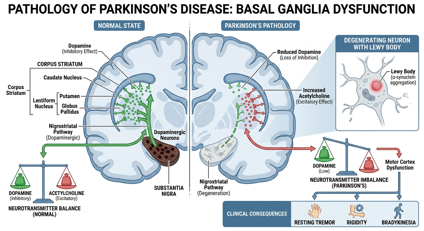
- The Corpus Striatum (caudate nucleus + lentiform nucleus) is a rich source of acetylcholine (ACh), produced by small striatal neurons - this has an excitatory effect.
- The Substantia Nigra (S.N.) contains pigmented dopaminergic neurons that produce dopamine.
- Dopamine is transported via the nigrostriatal pathway to the corpus striatum, where it has an inhibitory effect on striatal neurons.
- This creates a functional equilibrium between dopamine (inhibitory) and acetylcholine (excitatory) in the striatum.
- The pigmented neurons of the Substantia Nigra degenerate and are lost (the S.N. appears pale on gross pathology).
- Lewy bodies (eosinophilic intracytoplasmic inclusions) are the hallmark microscopic finding in remaining neurons.
- Dopamine production and transport via the nigrostriatal pathway drops sharply.
- The balance is broken: ACh (excitatory) dominates over depleted dopamine (inhibitory).
- This imbalance drives the classic triad: resting tremor, rigidity, and bradykinesia.