
Paget disease of bones
Reading File
Finding Sources
Finding Sources
Searching PubMed
"Paget Disease of Bone"[MeSH Terms]
Searching the Web
Paget disease of bone diagnosis management guidelines alkaline phosphatase bisphosphonate
Reading File
Reading File
Searching Images
Paget disease of bone radiograph characteristic bone changes osteolytic sclerotic enlargement
Reading Web Page
https://www.nhs.uk/conditions/pagets-disease-bone/diagnosis
Paget disease of bone
Paget disease of bone, also called osteitis deformans, is a chronic focal disorder of bone remodeling. Osteoclast-mediated bone resorption becomes excessive, followed by disorganized compensatory bone formation. The resulting bone is enlarged but structurally weak, prone to deformity and fracture.
It is distinct from Paget disease of the breast or extramammary Paget disease.
Common sites
- Pelvis
- Femur and tibia
- Lumbar spine
- Skull
It occurs in adults, becomes more frequent with age, and may involve one bone (monostotic) or several bones (polyostotic). Many people have no symptoms. Rheumatology, 2-Volume Set, pp. 498-520.
Cause and pathology
The exact cause is uncertain. There is a familial tendency in some patients, including variants in SQSTM1. Bone turnover has three broad phases:
- Osteolytic phase: marked osteoclast activity and bone resorption
- Mixed phase: resorption plus excessive new bone formation
- Sclerotic phase: dense, enlarged, disorganized bone
Histology shows irregular cement or reversal lines, giving a classic mosaic pattern of lamellar bone.
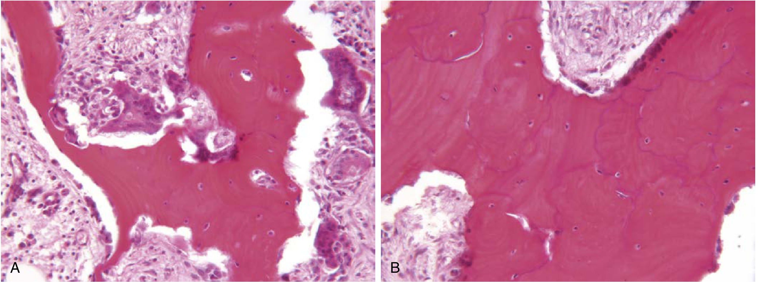
Figure source: Henry's Clinical Diagnosis and Management by Laboratory Methods, p. 260.
Clinical features
- Often asymptomatic and found incidentally from raised alkaline phosphatase or imaging
- Deep aching bone pain, often worse at rest
- Bone enlargement or deformity, such as bowed tibia/femur or increased skull size
- Local warmth over affected bone due to hypervascularity
- Skull involvement: headache, hearing loss, rarely cranial nerve compression
- Spine involvement: spinal stenosis, radiculopathy, or cord compression
- Secondary osteoarthritis near affected joints
Complications
- Pathological fractures
- Bone deformity
- Secondary osteoarthritis
- Hearing loss and other neurological compression syndromes
- Hypercalcemia during prolonged immobilization
- High-output cardiac failure in very extensive active disease, rarely
- Osteosarcoma or other sarcoma, rare but should be considered with new severe pain, swelling, or a destructive lesion. Rheumatology, 2-Volume Set, pp. 498-520.
Investigations
| Test | Typical finding / role |
|---|---|
| Serum alkaline phosphatase (ALP) | Usually raised and used to monitor activity, though it may be normal in limited disease |
| Calcium and phosphate | Usually normal |
| Bone-specific ALP or other turnover markers | Useful when total ALP is hard to interpret, for example with concurrent liver disease |
| X-ray | Bone enlargement, cortical thickening, coarsened trabeculae, mixed lytic-sclerotic changes |
| Radionuclide bone scan | Defines the full extent and activity of skeletal involvement |
| CT/MRI | For suspected fracture, sarcoma, neurological compression, or uncertain diagnosis |
An elevated ALP alone is not diagnostic. It needs confirmation with characteristic radiographic findings, and bone scintigraphy helps map affected bones. The NHS diagnostic guidance similarly recommends ALP testing plus X-ray and usually a bone scan.
Treatment
Treatment is mainly for symptomatic or active disease, especially when important sites are affected or complications are likely.
- Bisphosphonates are first-line
- IV zoledronic acid is generally preferred because a single infusion often produces prolonged biochemical remission.
- Oral alendronate or risedronate are alternatives in appropriate patients.
- Analgesia and physiotherapy can help pain and function.
- Ensure vitamin D and calcium status is adequate before potent bisphosphonate therapy, while avoiding inappropriate calcium excess.
- Orthopedic surgery may be needed for fractures, severe deformity, or advanced secondary arthritis.
- Neurosurgical or spinal assessment may be required for nerve compression.
Treatment reduces excessive bone turnover and Paget-related bone pain. It may not resolve pain caused by osteoarthritis or another non-Paget source. Rheumatology, 2-Volume Set, pp. 512-520. The MedlinePlus overview also notes bisphosphonates as the usual medical treatment and surgery for selected complications.
Monitoring
Follow symptoms and serum ALP after treatment. Repeat imaging is not routinely needed for everyone, but is useful when symptoms, biochemical activity, or concern for a complication persists.
Red flags needing prompt specialist assessment: new focal severe pain, rapidly enlarging bone swelling, fracture, new neurological deficit, or sudden hearing/vision symptoms.
This is a shared conversation. Sign in to Orris to start your own chat.