
Thyroglossal cyst
Reading File
Now let me search the medical library and PubMed in parallel.
Finding Sources
Searching PubMed
thyroglossal duct cyst
Good results. Now let me read the most comprehensive sources in parallel - Sabiston Surgery, Scott-Brown's ENT, and Robbins Pathology.
Reading File
Reading File
Reading File
Excellent data. Let me also read the Sabiston block on pediatric thyroglossal cyst and fetch the ultrasound image, plus get more from Scott-Brown's ENT for completeness.
Reading File
Loading Image
Finding Sources
Let me get two more important sources - Cummings Otolaryngology (detailed surgical section) and Bailey & Love (treatment section).
Reading File
Reading File
Reading File
Loading Image
I now have comprehensive information from multiple authoritative textbooks. Let me compile the full answer.
Thyroglossal Duct Cyst (TDC)
Embryology
The thyroid gland originates as a median endodermal thickening (diverticulum) at the foramen cecum at the base of the tongue. It descends through the neck to its definitive pretracheal position via the thyroglossal duct. As the second and third branchial arch cartilages form the hyoid bone, the duct passes through (or intimately around) the central portion of the hyoid. The duct normally involutes by 8-10 weeks of gestation. Persistence of any portion of this tract allows its secretory epithelial lining to accumulate fluid, forming a cyst. - Sabiston Textbook of Surgery, p.1343; Scott-Brown's Otorhinolaryngology, p.627
Epidemiology
- The most common congenital cystic mass of the neck
- Second most common benign neck mass overall, after benign lymphadenopathy
- Most commonly seen in pediatric patients, but can present at any age, even in the sixth or seventh decade of life
- Scott-Brown's Otorhinolaryngology, p.627; Bailey & Love's Short Practice of Surgery, p.828
Location
| Location | Frequency |
|---|---|
| Below hyoid bone | 65% |
| At level of hyoid bone | 15% |
| Above hyoid (suprahyoid) | ~20% |
| Midline | 75% |
| Off-midline (up to 2 cm) | 25% |
When adjacent to the thyroid cartilage, cysts may lie slightly to one side of the midline. - Scott-Brown's Otorhinolaryngology, p.627
Clinical Features
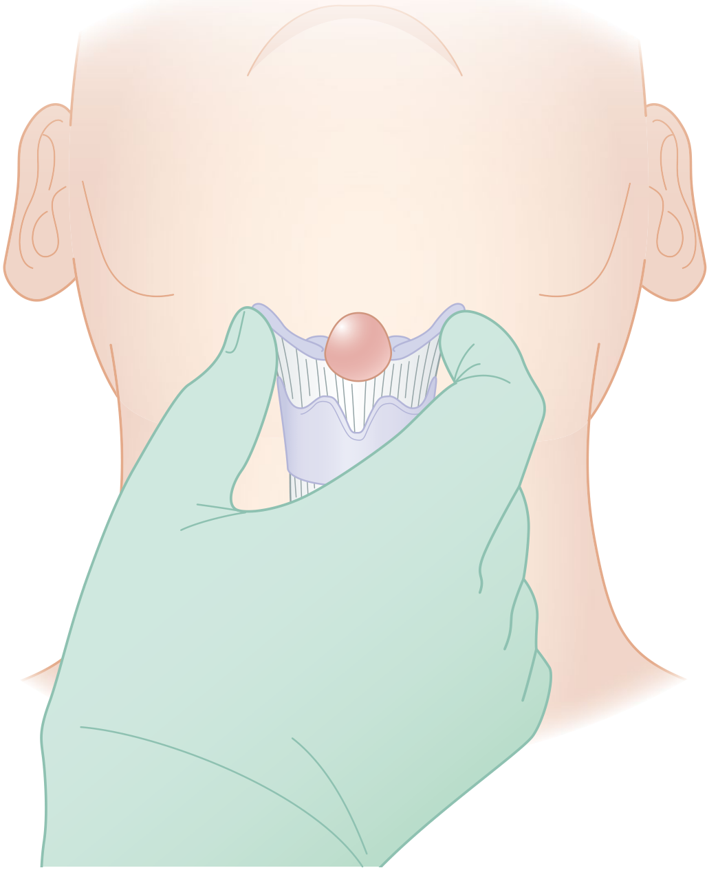
- Painless midline neck mass, most commonly just below the hyoid bone
- Classic sign: the cyst moves upward on swallowing AND on tongue protrusion (due to attachment to the hyoid and the tract running to the foramen cecum) - this distinguishes it from other midline masses
- Usually asymptomatic unless infected
- Infection causes pain, redness, and swelling; repeated infection may cause rupture through the skin, creating a thyroglossal fistula/sinus with mucoid discharge
- Rarely: dysphagia, dyspnea, or a discharging sinus on the anterior neck
- Bailey & Love's, p.828; Cummings Otolaryngology, p.3820
Pathology (Robbins)
- Cysts measure 1 to 4 cm
- Lined by stratified squamous epithelium near the base of tongue, or pseudostratified columnar epithelium at lower locations; transitional patterns also occur
- The fibrous cyst wall often contains lymphoid aggregates or thyroid remnants
- Malignant transformation of the lining epithelium is exceedingly rare
- Robbins & Cotran Pathologic Basis of Disease, p.694
Diagnosis
Clinical diagnosis based on the characteristic midline neck mass + movement with tongue protrusion.
Imaging
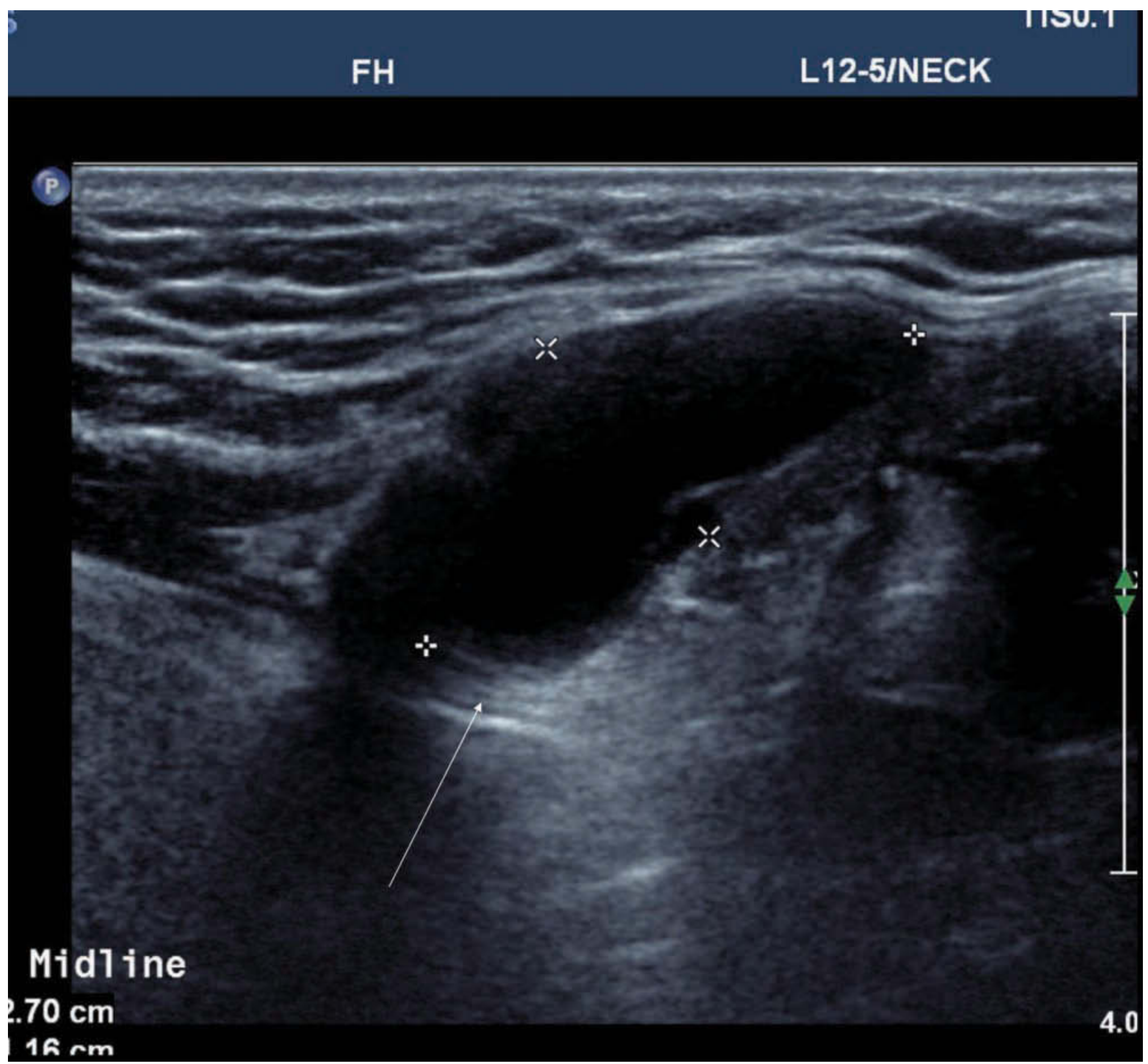
Ultrasound (first-line):
- Appearance varies: homogenous anechoic cyst with posterior wall enhancement (classic), pseudosolid mass (due to proteinaceous secretions), or heterogeneous (due to hemorrhage/infection)
- Key objectives: confirm the cyst, document its relationship to the hyoid bone, and confirm the presence of a normal thyroid gland in its usual pretracheal location
CT/MRI:
- Cysts are invariably high T2 signal on MRI; T1 signal varies with proteinaceous content
- Useful if infection or malignancy is suspected
Important caveat: In rare cases, the TDC may contain the only functioning thyroid tissue in the body. Confirmation of a normal thyroid gland before surgery is mandatory. If no normal thyroid is found, thyroid scintigraphy or further investigation is required before excision. - Cummings Otolaryngology, p.3820
FNA: Can be used to rule out malignancy or differentiate from dermoid cyst (mucus-filled vs keratin-filled).
Malignant Potential
- ~1% incidence of carcinoma within TDC - classically papillary thyroid carcinoma (PTC)
- A recent systematic review (2025) focused specifically on TDC carcinomas - [Thimsen et al., Virchows Arch, 2025 (PMID 40347268)]
- Presence of malignancy in TDC is associated with a 6.6% risk of microcarcinoma in the normal-appearing thyroid gland
- Thyroidectomy is not automatically required for malignant TDC, but lifelong surveillance of the thyroid is essential
- Cummings Otolaryngology, p.3820; Scott-Brown's, p.627
Treatment: The Sistrunk Procedure
Definitive treatment is surgical excision via the Sistrunk procedure (first described in 1928).
What is removed:
- The cyst itself
- The central portion of the hyoid bone (body of hyoid)
- The suprahyoid tract - a core of genioglossus muscle extending superiorly to the foramen cecum at the tongue base
Why it works: The thyroglossal duct has intimate anatomic relation to the hyoid bone and sends small side branches. Simple excision of just the cyst leaves remnant tract, causing recurrence.
| Procedure | Recurrence Rate |
|---|---|
| Simple cyst excision alone | ~46% |
| Modified Sistrunk procedure | ~5.8% |
| Incomplete resection (any technique) | up to 40-50% |
- Cummings Otolaryngology, p.3820; Sabiston, p.1343
If infected: Treat with antibiotics first (and drainage if needed). Defer definitive excision until acute inflammation has resolved.
Summary Table
| Feature | Detail |
|---|---|
| Origin | Foramen cecum → thyroglossal duct remnant |
| Most common site | Infrahyoid midline |
| Classic sign | Moves up with swallowing + tongue protrusion |
| Lining epithelium | Squamous (upper) / columnar (lower) |
| First-line imaging | Ultrasound |
| Malignancy risk | ~1% (usually papillary thyroid carcinoma) |
| Treatment | Sistrunk procedure |
| Recurrence without Sistrunk | ~46% |
Recent evidence note: A 2025 systematic review (PMID 40347268) on TDC carcinomas and a 2025 review on diagnostic utility of thyroid scan and ultrasound (PMID 40757114) are consistent with the textbook content above - no major changes to current guidelines are indicated by these recent publications.
This is a shared conversation. Sign in to Orris to start your own chat.