
Metastasis in a short note 5 to 7 marks harsh mohan
metastatic cascade tumor spread invasion extravasation diagram
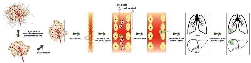
This pathophysiology diagram illustrates the sequential steps of the cancer metastatic cascade. The process begins with a primary tumor mass and progresses through the degradation of the extracellular matrix and basement membrane, followed by local invasion into surrounding tissues. The diagram depicts intravasation, where cancer cells enter the circulatory system. Within the vasculature, the image distinguishes between cell death and cell survival pathways. Surving circulating tumor cells are shown undergoing extravasation to exit the blood vessel. The final stages include the settlement of these cells in distant organs, specifically visualized in the lungs and liver. The diagram concludes by showing the proliferation of these cells into secondary metastatic tumors within those organs. This educational visual provides a comprehensive overview of oncology concepts related to tumor progression, hematogenous spread, and organ-specific metastasis for medical students and oncology professionals.
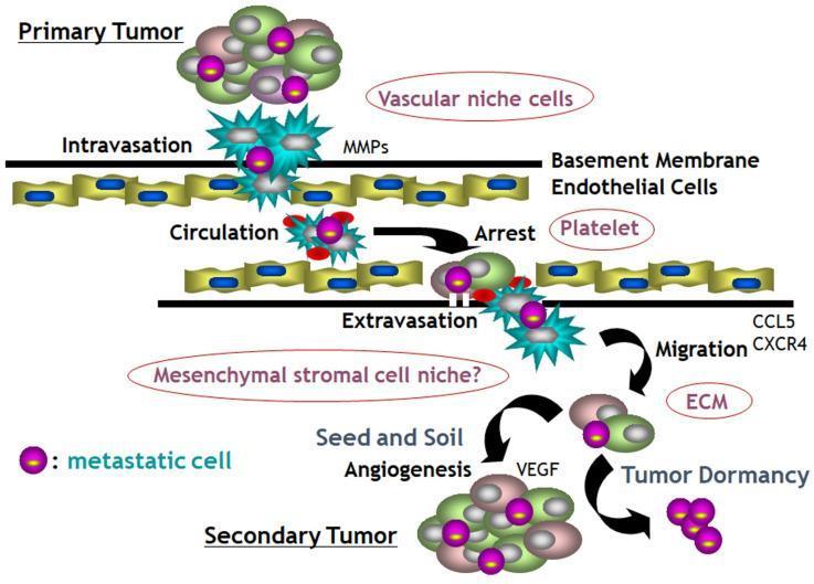
A pathophysiology diagram illustrating the invasion-metastasis cascade in cancer biology. The process begins at the top with a 'Primary Tumor' containing heterogeneous cells, including specific 'metastatic cells' (marked with purple nuclei). The cascade proceeds through 'Intravasation,' where tumor cells penetrate the 'Basement Membrane' and 'Endothelial Cells' facilitated by Matrix Metalloproteinases (MMPs) and vascular niche cells. Within the 'Circulation' phase, metastatic cells interact with 'Platelets' to form clusters before reaching 'Arrest' at a distant site. This is followed by 'Extravasation,' where cells exit the vasculature and undergo 'Migration' into the 'Mesenchymal stromal cell niche,' influenced by the extracellular matrix (ECM) and chemokines like CCL5 and CXCR4. The final stages include 'Seed and Soil Angiogenesis' driven by Vascular Endothelial Growth Factor (VEGF), which can lead to either 'Tumor Dormancy' or the successful colonization and formation of a 'Secondary Tumor.' The diagram effectively summarizes the sequential cellular interactions and molecular signaling involved in malignant progression.
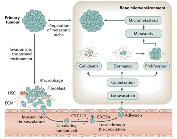
This pathophysiology diagram illustrates the multi-step process of cancer cell metastasis from a primary tumor to the bone microenvironment. The metastatic cascade begins with the 'Primary tumour' and the 'Preparation of metastatic niche'. It progresses to 'Invasion into the stromal environment', where cancer cells interact with the extracellular matrix (ECM), macrophages, fibroblasts, and hematopoietic stem cells (HSCs). This is followed by 'Invasion into the vasculature'. Once inside a blood vessel, a 'Circulating tumour cell' undergoes 'Travel through the circulation', mediated by the CXCL12/CXCR4 signaling pathway. The diagram shows the subsequent stages of 'Adhesion' to the vessel wall and 'Extravasation' into the 'Bone microenvironment'. Within this new niche, 'Colonization' occurs, leading to three possible outcomes for the tumor cells: 'Cell death', 'Dormancy', or 'Proliferation'. Proliferation eventually leads to 'Metastasis' and the formation of 'Micrometastasis'. The illustration effectively demonstrates the complex interactions between tumor cells and the systemic environment during disease progression.
Metastasis - Short Note (5-7 Marks)
Definition
- ~30% of patients with newly diagnosed solid tumors present with clinically evident metastases
- An additional ~20% have occult (microscopic) metastases at diagnosis
- Metastasis is a property exclusive to malignant tumors; benign tumors do not metastasize
Pathways of Metastatic Spread
1. Lymphatic Spread
- Most common route for carcinomas (epithelial tumors)
- Tumor cells enter regional lymphatics and colonize draining lymph nodes
- "Skip metastases" can occur when cells bypass proximal nodes to reach distal ones
- Sentinel lymph node - the first regional lymph node to receive lymph from a primary tumor; its biopsy guides staging and treatment
- Note: lymph node enlargement near a tumor does not always indicate metastasis - reactive hyperplasia (lymphadenitis) from tumor antigens can also cause it
2. Hematogenous Spread
- More common with sarcomas (mesenchymal tumors) but also occurs in carcinomas
- Tumor cells penetrate thin-walled veins (rather than thick-walled arteries)
- Cells arrest in the first capillary bed encountered:
- Portal blood drains to the liver - hence liver is a frequent metastatic site
- Caval blood drains to the lungs - hence lungs are frequently involved
- Cancers near the vertebral column (thyroid, prostate) can use the paravertebral venous plexus and spread to the spine
- Organ tropism examples:
- Prostatic carcinoma -> bone
- Bronchogenic carcinoma -> adrenals, brain
- Neuroblastoma -> liver, bones
- Uveal melanoma -> liver
3. Seeding of Body Cavities (Transcoelomic Spread)
- Occurs when tumors invade a body cavity (peritoneal, pleural, pericardial, subarachnoid)
- Classic example: ovarian carcinoma seeds the peritoneal surfaces widely without necessarily invading underlying tissue
- CNS tumors (medulloblastoma, ependymoma) can seed via cerebrospinal fluid along meningeal surfaces
The Metastatic Cascade
Phase 1 - Invasion of the Extracellular Matrix (ECM)
-
Loosening of intercellular connections: E-cadherins act as intercellular glue and transmit growth-inhibitory signals via beta-catenin. In many cancers, E-cadherin function is lost due to:
- Mutational inactivation
- Activation of beta-catenin genes
- Upregulation of SNAIL and TWIST transcription factors (which repress E-cadherin expression)
- This process is called Epithelial-to-Mesenchymal Transition (EMT)
-
Local degradation of basement membrane and ECM: Tumor cells (or stromal cells they recruit) secrete proteolytic enzymes:
- Matrix metalloproteinases (MMPs) - especially MMP-9 (gelatinase), which cleaves type IV collagen in basement membranes
- Cathepsin D
- Urokinase plasminogen activator
- MMPs also release ECM-sequestered growth factors (e.g., VEGF), promoting angiogenesis and tumor growth
- Importantly, MMP inhibitors are simultaneously reduced, tipping the balance toward degradation
-
Changes in attachment to ECM proteins: Normal cells use integrins to bind laminin and collagen; loss leads to apoptosis (anoikis). Tumor cells become resistant to anoikis. Cleavage of collagen IV and laminin by MMP-2/9 generates novel integrin-binding sites that stimulate tumor cell migration.
-
Locomotion: Tumor cells migrate through the degraded matrix. This is directed by:
- HGF/SCF (Hepatocyte Growth Factor/Scatter Factor) - elevated at invasive tumor edges
- Cleavage products of ECM (collagen, laminin)
- Insulin-like growth factors 1 and 2
Phase 2 - Vascular Dissemination and Homing
- Millions of tumor cells are shed daily into circulation from even small tumors
- Most circulating tumor cells die; only rare cells survive
- Cells form emboli by aggregating with platelets (protection from immune destruction)
- At distant sites: tumor cells adhere to endothelium, degrade the vascular basement membrane, and extravasate
- Formation of micrometastases -> angiogenesis -> growth into macroscopic tumors
Organ Tropism ("Seed and Soil" Hypothesis)
- First proposed by Stephen Paget (1889): tumor cells (seed) can only grow in compatible microenvironments (soil)
- Molecular basis: chemokine receptors on tumor cells match chemokine gradients in target organs
- Example: Breast cancer cells express CXCR4 (chemokine receptor); its ligand CXCL12 (SDF-1) is highly expressed in bone marrow, lungs, and liver - explaining common breast cancer metastatic sites
Exceptions and Important Points
| Feature | Detail |
|---|---|
| Basal cell carcinoma of skin | Locally invasive but rarely metastasizes |
| CNS primary tumors | Locally invasive, rarely metastasize outside CNS |
| Leukemias/Lymphomas | Always assumed disseminated; derived from cells that normally circulate |
| Local invasion vs. metastasis | These are separable properties - a tumor can be locally invasive without metastasizing |
Summary Diagram (Metastatic Cascade)
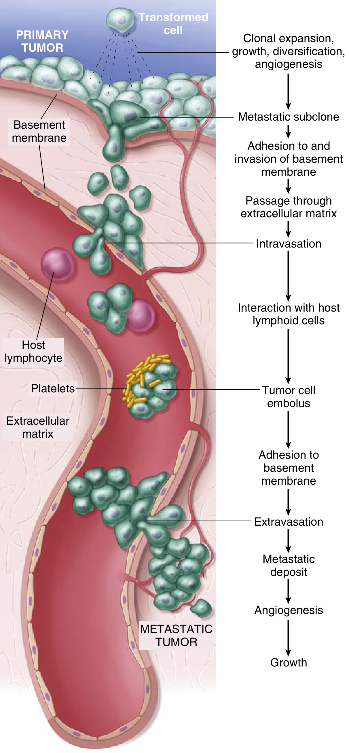
- Clonal expansion, angiogenesis in primary tumor
- EMT, loosening of E-cadherin junctions
- ECM degradation (MMPs)
- Invasion through basement membrane
- Intravasation into blood/lymphatics
- Survival in circulation (embolus formation with platelets)
- Arrest at distant capillary bed
- Extravasation
- Formation of micrometastases
- Angiogenesis and growth into macrometastatic tumor