
Kallman syndrome
Kallmann syndrome diagnosis treatment
Kallmann syndrome anosmia hypogonadotropic hypogonadism GnRH migration
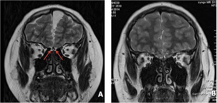
This diagnostic image displays two coronal T2-weighted brain MRI scans, labeled (A) and (B), focusing on the anterior cranial fossa of two different patients. In image A, red arrows specifically point to the olfactory region above the ethmoid bone's cribriform plate. Both images demonstrate bilateral dysplasia and hypoplasia of the olfactory bulbs, olfactory tracts, and the corresponding olfactory sulci. This anatomical area normally contains well-defined, rounded olfactory bulbs sitting within the olfactory sulci of the frontal lobes. However, in these patients, the sulci are shallow or poorly formed, and the olfactory structures are visibly diminished or absent. This radiological finding is a hallmark diagnostic feature of Kallmann syndrome, reflecting a failure in the development and migration of the olfactory nerves and gonadotropin-releasing hormone (GnRH) neurons. The images illustrate the neuroanatomical basis for clinical anosmia or hyposmia in the context of idiopathic hypogonadotropic hypogonadism.
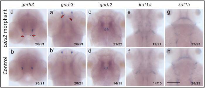
This diagnostic image set displays whole-mount in situ hybridization (WISH) of Danio rerio (zebrafish) embryos at 56 hours post-fertilization (hpf), used as a disease model for Kallmann syndrome (KS). The panels compare gene expression patterns between 'cars2 morphants' (knockdown group) and control embryos across four genetic markers: gnrh3, gnrh2, kal1a, and kal1b. In the gnrh3 panels (a, a', b, b'), red arrows highlight the ectopic migration of GnRH3 neurons in the cars2 morphants compared to the restricted, normal distribution in the control embryos. Conversely, the expression patterns for gnrh2 (c, d), kal1a (e, f), and kal1b (g, h) appear consistent between the morphant and control groups. These images demonstrate the specific role of the mitochondrial cysteinyl-tRNA synthetase (CARS2) in the developmental migration of GnRH neurons, a key pathophysiology in human hypogonadotropic hypogonadism. The visual evidence supports the educational concept of gene-environment or synergistic genetic interactions in neurodevelopmental migration disorders.
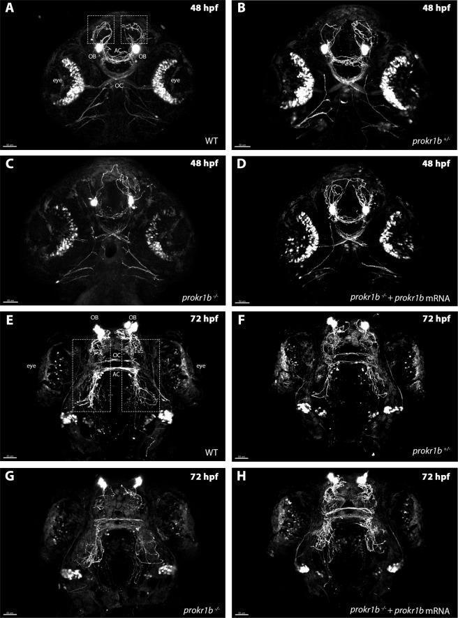
This set of fluorescence microscopy images illustrates the development of the GnRH3 neuron fiber network in zebrafish embryos (tg(gnrh3:EGFP)) at 48 hours post-fertilization (hpf) and 72 hpf across different genotypes. Panels A and E show wild-type (WT) embryos with highly organized fiber distribution throughout the olfactory bulbs (OB), optic chiasm (OC), and anterior commissure (AC). Panels B and F demonstrate that prokr1b heterozygous mutants retain a similar organized architecture. In contrast, prokr1b homozygous mutants (Panels C and G) exhibit significant axon misrouting and disorganization, particularly at the rostral region and the optic chiasm. Panels D and H demonstrate a successful phenotypic rescue in homozygous mutants following the injection of wild-type prokr1b mRNA, restoring the structural integrity of the GnRH3 network. This comparative visualization highlights the critical role of the prokr1b gene in guiding the migration and pathfinding of GnRH neurons, which is medically relevant for understanding the molecular basis of Kallmann syndrome and related hypogonadotropic hypogonadism disorders.
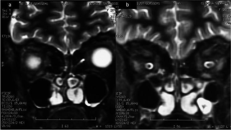
Two coronal magnetic resonance imaging (MRI) scans of the brain (labeled a and b) focusing on the anterior cranial fossa and olfactory region. The images demonstrate aplasia of the olfactory bulbs and tracts, a hallmark radiological finding in Kallmann syndrome. Image (a) shows the inferior frontal lobes with altered signal intensity and a lack of distinct olfactory bulb morphology within the olfactory sulci. Image (b) further confirms the bilateral absence of these structures. The nasal cavities and ethmoid sinus regions are visible inferiorly with normal aeration (dark signal). The imaging demonstrates the key diagnostic feature for patients presenting with congenital hypogonadotropic hypogonadism and anosmia/hyposmia. This comparison illustrates the developmental abnormalities of the olfactory system associated with genetic neuroendocrine disorders.
Kallmann Syndrome
Definition
Epidemiology
- Incidence: approximately 1 in 10,000 males and 1 in 50,000 females (Campbell Walsh Urology; Berek & Novak's Gynecology)
- Males are far more commonly affected
- Sporadic cases are more common than inherited forms
Pathophysiology and Embryology
- Anosmia - from failure of olfactory bulb/tract development (arhinencephaly)
- Hypogonadism - from failure of GnRH neurons to reach the hypothalamus, resulting in absent pulsatile GnRH, low LH and FSH, and consequently low sex steroids
Genetics
| Mode | Key Gene | Details |
|---|---|---|
| X-linked recessive | KAL1 (Xp22.3) | Encodes anosmin-1, a neurotropic growth factor guiding GnRH neuron migration. Accounts for ~15% of all cases and most X-linked cases |
| Autosomal dominant | FGFR1 (KAL2), others | Multiple genes involved |
| Autosomal recessive | GNRHR, PROK2, PROKR2, others | ~10% have mutations in GNRHR or KAL1 |
Clinical Features
Males (more severely affected)
- Delayed/absent puberty - no spontaneous pubertal development
- Eunuchoid body habitus - long limbs, poor muscle mass, lack of secondary sexual characteristics
- Micropenis, cryptorchidism - often present from infancy
- Infertility - azoospermia or severe oligospermia
- Small testes
- Anosmia/hyposmia - patients often unaware
Females
- Primary amenorrhea - the rule
- Sexual infantilism with possible partial breast development
- Small ovaries with follicles rarely developing beyond the primordial stage
- Very low but usually measurable gonadotropin levels (Berek & Novak's Gynecology)
Associated Features (classic triad in men: anosmia + hypogonadism + color blindness)
- Color blindness
- Cleft lip/palate
- Cerebellar ataxia
- Nerve deafness
- Renal agenesis
- Bimanual synkinesia (mirror movements)
- Optic atrophy
- Abnormalities of thirst and vasopressin release
- Dental agenesis
- Neurologic abnormalities
Diagnosis
- Low testosterone (males) / low estradiol (females)
- Low LH
- Low FSH
- Normal prolactin (distinguishes from hyperprolactinemia)
- Normal or low GnRH on stimulation testing
Treatment
Sex Hormone Replacement (puberty induction / maintenance)
- Males: Testosterone replacement (intramuscular or transdermal) to induce virilization, increase bone density, and improve wellbeing
- Females: Combined estrogen and progestin replacement to induce breast development, menses, and protect bone
Fertility Induction
| Agent | Role | Dose |
|---|---|---|
| hCG (LH substitute) | Stimulates Leydig cell testosterone production | 1,500-5,000 IU IM/SC 2-3x weekly; max 10,000 IU/wk, titrated to serum testosterone |
| hMG or rFSH (FSH substitute) | Stimulates Sertoli cells and spermatogenesis | 75 IU SC 2-3x weekly |
- Treatment with hCG alone may initiate spermatogenesis; hMG/rFSH added if needed
- 80% of men achieve sperm in the ejaculate after 12-18 months of gonadotropin therapy
- Men previously on exogenous androgen therapy may require 1-2 years before sperm appears
- Alternatively, pulsatile GnRH via subcutaneous pump can be used to stimulate endogenous gonadotropin secretion (Smith & Tanagho Urology; Campbell Walsh Urology)
Key Distinguishing Points
| Feature | Kallmann Syndrome | IHH (normosmic) |
|---|---|---|
| Anosmia/hyposmia | Present | Absent |
| GnRH deficiency | Yes | Yes |
| Olfactory bulb hypoplasia on MRI | Yes | No |
| Genetics | KAL1, FGFR1, others | GNRHR, GPR54, others |