Here is your complete NEET PG Hematology - From Scratch integrated teaching session. This covers all 19 subjects integrated (Physiology, Biochemistry, Pathology, Medicine, Pharmacology, Forensics, Pediatrics, Obs/Gynae, Microbiology, Surgery, etc.) with high-yield MCQ focus.
🩸 HEMATOLOGY FOR NEET PG - COMPLETE FROM SCRATCH
MODULE 1: HEMATOPOIESIS (Foundation)
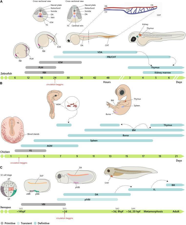
1.1 Sites of Hematopoiesis - THE MOST MCQ-TESTED TOPIC
| Age | Primary Site | Mnemonic |
|---|
| 0-2 weeks embryo | Yolk sac | Young Yolk |
| 2nd month | AGM region (Aorta-Gonad-Mesonephros) | |
| 3rd-7th month fetus | Liver (chief site) + Spleen | Liver Loves 3rd month |
| 4th month onwards | Bone marrow (starts) | |
| Birth onwards | Bone marrow only | |
| After puberty | Axial skeleton only (vertebrae, sternum, ribs, skull, pelvis) | |
MCQ ALERT: In adults, only ~50% of marrow space is hematopoietically active (red marrow). Yellow marrow = fat, inactive.
Pathology integration: When bone marrow fails (aplastic anemia, leukemia), the body reverts to liver and spleen = Extramedullary hematopoiesis (EMH). This causes hepatosplenomegaly.
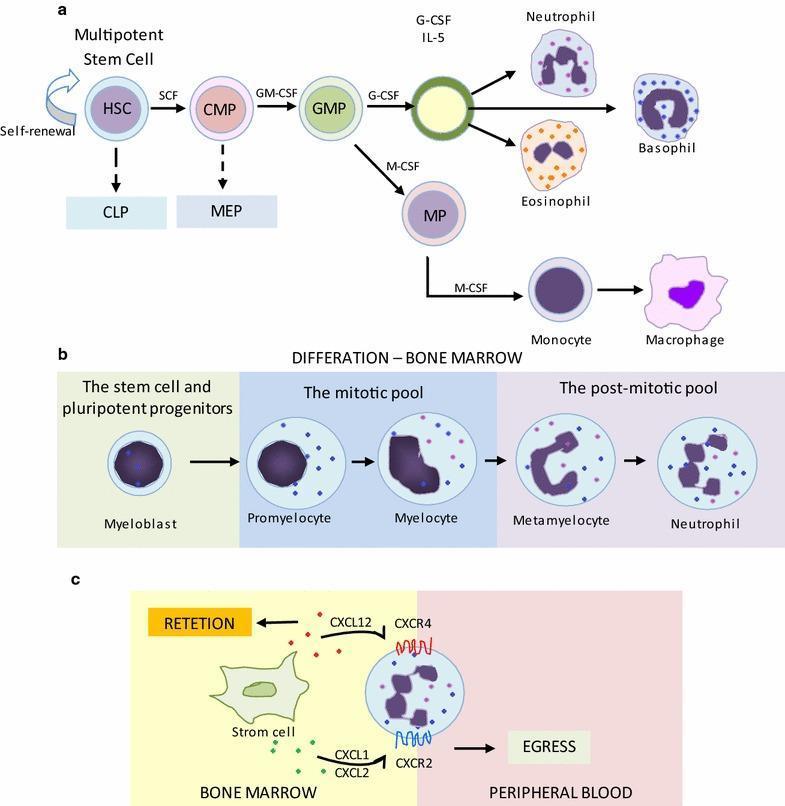
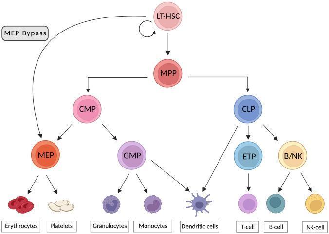
1.2 Hematopoietic Cell Hierarchy
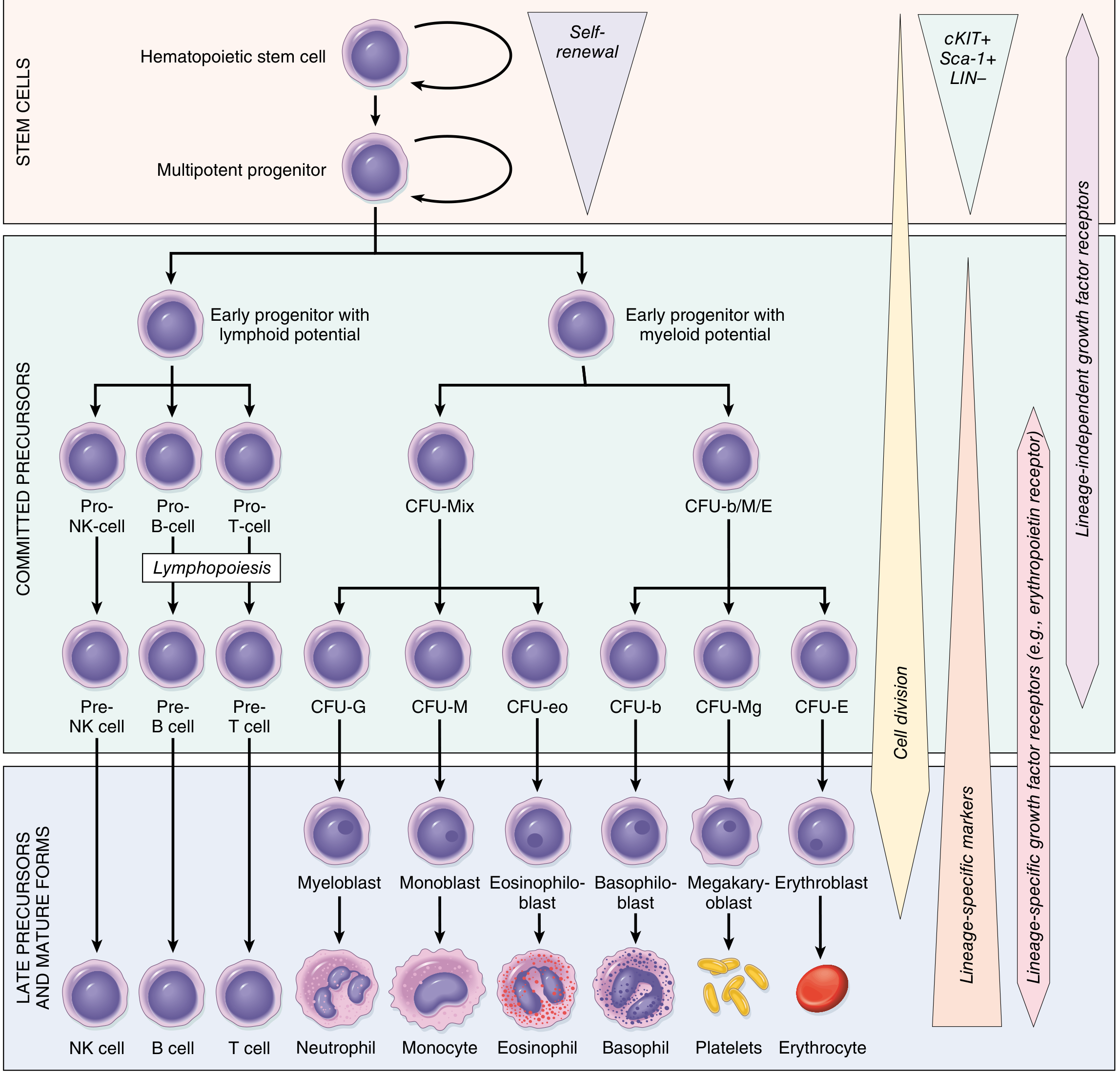
Here is the actual diagram from Robbins Pathology showing the full cell lineage:
Key hierarchy (NEET-critical):
HSC (Hematopoietic Stem Cell)
  ├── Common Lymphoid Progenitor (CLP)
  │     ├── Pro-B → Pre-B → B cell
  │     ├── Pro-T → Pre-T → T cell
  │     └── Pro-NK → NK cell
  └── Common Myeloid Progenitor (CMP)
        ├── CFU-GEMM (Mix)
        │     ├── CFU-E → Erythroblast → Erythrocyte
        │     ├── CFU-Meg → Megakaryoblast → Platelets
        │     ├── CFU-G → Myeloblast → Neutrophil
        │     ├── CFU-M → Monoblast → Monocyte
        │     ├── CFU-eo → Eosinophiloblast → Eosinophil
        │     └── CFU-b → Basophiloblast → Basophil
MCQ: HSC markers = CD34+, CD117 (c-KIT)+, Lin- (lineage negative). Used for stem cell transplantation identification.
Pharma integration: Growth factors used clinically:
- EPO (Erythropoietin) - stimulates CFU-E → used in renal anemia (Darbepoetin = long-acting EPO)
- G-CSF (Filgrastim) - stimulates neutrophil production → used post-chemotherapy
- GM-CSF (Sargramostim) - stimulates myeloid line broadly
- TPO (Thrombopoietin/Eltrombopag) - stimulates megakaryocytes → used in ITP
MODULE 2: NORMAL BLOOD VALUES (Physiology + Biochem)
2.1 Critical Normal Values - MEMORIZE THESE
| Parameter | Normal Value | MCQ Trick |
|---|
| Hemoglobin Men | 13.5-17.5 g/dL | <13 = anemia in men |
| Hemoglobin Women | 12-16 g/dL | <12 = anemia in women |
| Hb in pregnancy | <11 g/dL = anemia (WHO) | Lower due to dilution |
| Hb Newborn | 18-22 g/dL (highest!) | Due to HbF |
| MCV | 80-100 fL | Memory: 80-100 = normal |
| MCH | 27-32 pg | |
| MCHC | 32-36 g/dL | Most sensitive index of IDA |
| RBC count | M: 4.5-5.5 million; F: 3.8-4.8 million | |
| Reticulocytes | 0.5-2.5% | Elevated = active hemolysis/blood loss |
| WBC | 4000-11000/mm³ | |
| Platelets | 1.5-4 lakh/mm³ (150,000-400,000) | |
| ESR (Westergren) | M: <15 mm/hr; F: <20 mm/hr | |
| Hematocrit (PCV) | M: 40-52%; F: 37-47% | PCV = Hb × 3 (approx) |
MCQ: Wintrobe's indices (MCV, MCH, MCHC) are calculated from Hb, RBC count, and PCV. These are the indices used to classify anemia.
MODULE 3: HEMOGLOBIN (Biochemistry Integration)
3.1 Hemoglobin Structure
- Structure: Tetramer - 2 alpha + 2 non-alpha chains + 4 heme groups
- Each heme = Protoporphyrin IX + Fe²⁺
- Fe²⁺ (ferrous) carries O₂; Fe³⁺ (ferric) = methemoglobin (can't carry O₂)
3.2 Types of Normal Hemoglobin
| Type | Chains | % in Adult | Key Point |
|---|
| HbA | α₂β₂ | 96-98% | Main adult Hb |
| HbA₂ | α₂δ₂ | 2-3% | Increased in β-thalassemia trait |
| HbF | α₂γ₂ | <1% adult; 70% at birth | Higher O₂ affinity than HbA |
| HbGower | ζ₂ε₂ | Embryonic only | First embryonic Hb |
MCQ: HbF has higher O₂ affinity than HbA because 2,3-DPG binds less to γ chains. This is why HbF shifts the ODC to the LEFT.
3.3 Hemoglobin Switching
Embryo:  HbGower → HbPortland
Fetus:   HbF (peaks at 8-10 weeks, declines near birth)
Adult:   HbA takes over by 6 months of age
MCQ: At birth, 70% is HbF. By 6 months, HbA predominates. This is why sickle cell disease and β-thal manifest after 6 months of age (when HbF declines).
3.4 Oxygen Dissociation Curve (ODC) - HIGH YIELD
RIGHT shift (↓ O₂ affinity, ↑ O₂ delivery to tissues):
- ↑ CO₂, ↑ Temperature, ↑ 2,3-DPG, ↑ H⁺ (↓ pH) = Bohr effect
- Sickle cell disease (HbS polymerizes when deoxygenated)
LEFT shift (↑ O₂ affinity, ↓ O₂ release):
- CO poisoning (HbCO), Methemoglobin, HbF, Alkalosis, ↓ 2,3-DPG
MCQ: Stored blood has ↓ 2,3-DPG → LEFT shift → poor O₂ delivery. Packed cells transfused for major surgery need to be fresh for this reason.
MODULE 4: CLASSIFICATION OF ANEMIA - THE MASTER FRAMEWORK
4.1 Two-Way Classification (NEET standard)
BY MCV (Wintrobe's morphologic classification):
| Type | MCV | Causes | Mnemonic |
|---|
| Microcytic | <80 fL | Iron deficiency, Thalassemia, ACD, Sideroblastic | TAILS (Thalassemia, Anemia of chronic disease, Iron deficiency, Lead poisoning, Sideroblastic) |
| Normocytic | 80-100 fL | Aplastic anemia, Hemolysis, Renal failure, Acute blood loss | HARM (Hemolysis, Aplastic, Renal, Marrow infiltration) |
| Macrocytic | >100 fL | B12/Folate deficiency, Liver disease, Hypothyroidism, Alcohol, Drugs | Oval macrocytes = megaloblastic; Round macrocytes = non-megaloblastic |
BY MECHANISM (Pathology-based):
-
Decreased production (hypoproliferative - low reticulocytes)
- Iron/B12/Folate deficiency
- Aplastic anemia
- Anemia of chronic disease
- Bone marrow infiltration (myelophthisis)
-
Increased destruction (Hemolysis) (high reticulocytes)
- Intravascular: G6PD, PNH, mechanical valves
- Extravascular: Spherocytosis, Sickle cell, Thalassemia
-
Blood loss (high reticulocytes if chronic, normal if acute)
MCQ KEY: The reticulocyte count is the single most important test to distinguish production problem (low retics) from destruction/loss (high retics).
MODULE 5: IRON DEFICIENCY ANEMIA (IDA) - Most Tested
5.1 Epidemiology
- Most common nutritional deficiency worldwide (Medicine, Pathology)
- Most common cause of anemia globally
- Affects ~10% in high-resource countries, 25-50% in low-resource countries (Robbins)
5.2 Iron Metabolism (Biochemistry Integration)
Total body iron: 2.5 g (women) / 3.5 g (men)
- 80% in Hb, myoglobin, iron enzymes (cytochromes, catalase)
- 15-20% in storage form (ferritin + hemosiderin) in liver, spleen, bone marrow macrophages
Iron absorption:
- Duodenum (primary site of absorption)
- Fe³⁺ reduced to Fe²⁺ by duodenal cytochrome B (Dcytb/CYBRD1)
- Fe²⁺ enters enterocyte via DMT-1 (Divalent Metal Transporter-1)
- Exits basolateral side via Ferroportin
- Re-oxidized by Hephaestin/Ceruloplasmin to Fe³⁺ → binds Transferrin in plasma
Hepcidin - The MASTER REGULATOR:
- Made by liver
- Degrades ferroportin → reduces iron absorption and macrophage iron release
- ↑ Hepcidin in inflammation (IL-6 induces it) → Anemia of Chronic Disease
- ↓ Hepcidin in iron deficiency and hemochromatosis
MCQ: Transferrin is normally 33% saturated. Serum iron averages 120 μg/dL (men), 100 μg/dL (women). Total iron-binding capacity (TIBC) = 300-350 μg/dL normally.
5.3 Stages of Iron Deficiency
| Stage | Ferritin | Serum Iron | TIBC | Transferrin Sat | Hb | MCV |
|---|
| Stage 1: Storage depletion | ↓ | Normal | Normal | Normal | Normal | Normal |
| Stage 2: Iron-deficient erythropoiesis | ↓ | ↓ | ↑ | ↓ (<20%) | Normal | Normal |
| Stage 3: IDA | ↓ | ↓ | ↑ | ↓ | ↓ | ↓ |
MCQ: Ferritin is the earliest to fall. It is the best indicator of iron stores. But ferritin is also an acute phase reactant (rises in inflammation, liver disease, malignancy) - so can be falsely normal in IDA + infection.
5.4 IDA - Clinical Features (Medicine + Surgery Integration)
Symptoms of anemia: fatigue, pallor, dyspnea on exertion, palpitations
Iron-specific features:
- Koilonychia (spoon-shaped nails) - Pathognomonic
- Pica (craving for clay, ice = pagophagia, starch = amylophagia)
- Angular cheilitis, glossitis (smooth tongue)
- Plummer-Vinson (Paterson-Brown-Kelly) syndrome = IDA + dysphagia (postcricoid web) + atrophic glossitis → precancerous for hypopharyngeal carcinoma
5.5 Lab Findings in IDA
| Lab Test | Finding in IDA |
|---|
| Hb | ↓ |
| MCV | ↓ (<80 fL) |
| MCH | ↓ |
| MCHC | ↓ (hypochromia) - most sensitive index |
| Serum ferritin | ↓ (earliest, most sensitive) |
| Serum iron | ↓ |
| TIBC | ↑ (body tries to absorb more) |
| Transferrin saturation | ↓ (<15%) |
| Serum transferrin receptor (sTfR) | ↑ (most specific for IDA, unaffected by inflammation) |
| Blood film | Microcytic, hypochromic, pencil cells, target cells |
| Reticulocytes | Normal/↓ (production problem) |
| Bone marrow | Absent iron stores (Prussian blue stain negative) |
MCQ: In anemia of chronic disease (ACD): ferritin is NORMAL/↑, serum iron ↓, TIBC ↓ (unlike IDA where TIBC ↑). sTfR/log ferritin ratio differentiates IDA from ACD.
5.6 Treatment (Pharmacology Integration)
-
Oral iron: Ferrous sulfate 200 mg TDS (contains 60 mg elemental iron per tablet)
- Best absorbed on empty stomach (but GI side effects)
- Ascorbic acid (Vit C) increases absorption (reduces Fe³⁺ to Fe²⁺)
- Tetracyclines, antacids, PPIs, milk, phytates, tannins decrease absorption
- Duration: 3-6 months (continue 3 months after Hb normalizes to replenish stores)
- Reticulocyte count rises at 7-10 days (proof of treatment response)
- Hb rises by 1-2 g/dL per month
-
IV iron indications: Malabsorption, intolerance, inflammatory bowel disease, CKD on hemodialysis, need for rapid correction
MCQ: Cause of failure to respond to oral iron: 1) Non-compliance, 2) Ongoing blood loss, 3) Malabsorption, 4) Wrong diagnosis, 5) Concurrent folate/B12 deficiency
MODULE 6: MEGALOBLASTIC ANEMIA (Vitamin B12 and Folate)
6.1 Key Differences - B12 vs Folate
| Feature | B12 Deficiency | Folate Deficiency |
|---|
| Neural tube defects | No | YES (periconceptional folate prevents NTDs) |
| Neurological features | YES - SACD (Subacute Combined Degeneration) | No neuro features |
| Serum methylmalonic acid | ↑ | Normal |
| Serum homocysteine | ↑ | ↑ |
| Serum folate | Normal or ↑ | ↓ |
| RBC folate | ↓ (trapped as methylTHF) | ↓ |
| Schilling test | Diagnoses B12 malabsorption | Not applicable |
MCQ: The methyl-folate trap: B12 is needed to convert methylTHF → THF. Without B12, folate gets trapped as methylTHF → can't be used for DNA synthesis. This is why B12 deficiency causes megaloblastic anemia even with normal folate intake.
6.2 B12 (Cobalamin) - High-Yield Facts
Sources: Animal products only (meat, fish, eggs, dairy) - vegans at risk
Absorption pathway:
- Food → released by acid + pepsin
- Binds R-factor (haptocorrin) in stomach
- In duodenum: pancreatic enzymes cleave R-factor → B12 binds Intrinsic Factor (IF) (made by gastric parietal cells)
- IF-B12 complex absorbed at terminal ileum (via cubilin receptor)
- Transported by Transcobalamin II (TC-II) in plasma
Causes of B12 deficiency:
- Pernicious anemia (most common in developed world) = autoimmune gastritis → ↓ parietal cells → ↓ IF → ↓ B12 absorption. Anti-IF antibody = most specific (50% sensitive). Anti-parietal cell antibody = 90% sensitive but less specific
- Strict veganism (no animal products)
- Gastrectomy/gastric bypass
- Terminal ileum resection (Crohn's disease)
- Fish tapeworm (Diphyllobothrium latum) - competes for B12
- Metformin (reduces B12 absorption - MCQ in pharmacology!)
MCQ: Body stores of B12 last 3-5 years (liver stores large amounts). Body stores of folate last only 3-4 months.
6.3 Neurological Features of B12 Deficiency - SACD
Subacute Combined Degeneration of spinal cord:
- Affects posterior columns (vibration, proprioception lost first)
- AND lateral corticospinal tracts (upper motor neuron signs)
- Presents as: peripheral neuropathy + spastic paraparesis + ataxia
MCQ: SACD can occur WITHOUT macrocytic anemia (Hb can be normal). Giving folate to B12-deficient patient: corrects anemia BUT worsens neurological damage (the methyl-folate trap resolves, masking B12 deficiency). This is a classic NEET trap!
6.4 Blood Film in Megaloblastic Anemia
- Oval macrocytes (not round)
- Hypersegmented neutrophils (≥5 lobes in >5% neutrophils, or any cell with ≥6 lobes) - EARLIEST feature, appears before anemia
- Pancytopenia in severe cases
- Ineffective erythropoiesis (most RBC destroyed in marrow before reaching blood)
- Bone marrow: giant metamyelocytes, megaloblasts
MODULE 7: HEMOLYTIC ANEMIAS
7.1 Classification of Hemolysis
By site:
| Intravascular | Extravascular |
|---|
| Within blood vessels | In spleen, liver macrophages |
| Free Hb → hemoglobinuria (dark urine) | Bilirubin ↑ (indirect/unconjugated) |
| Haptoglobin ↓↓ | Haptoglobin ↓ (less severe) |
| Hemosiderinuria | Splenomegaly |
| Schistocytes on film | Spherocytes on film |
| PNH, G6PD (triggered), TTP/HUS, mechanical | Hereditary spherocytosis, warm AIHA |
Lab hallmarks of hemolysis (any type):
- ↑ LDH (released from RBCs)
- ↑ Indirect bilirubin → jaundice
- ↓ Haptoglobin (binds free Hb, then cleared by liver)
- ↑ Reticulocyte count (bone marrow response)
- Blood film: polychromasia (blue-staining young RBCs)
7.2 Hereditary Spherocytosis (HS) - Pediatrics Integration
- Most common inherited RBC membrane disorder (in Northern Europeans)
- Autosomal dominant (75%) or recessive
- Defect in spectrin (most common), ankyrin, band 3, or protein 4.2
- These proteins anchor lipid bilayer to cytoskeleton → loss of membrane area → sphere shape
Diagnosis:
- Osmotic fragility test (increased = spherocytes lyse in lower salt concentration) - traditional
- EMA (Eosin-5-maleimide) binding test - newer, more specific (↓ binding in HS)
- Coombs test negative (differentiates from AIHA which also has spherocytes)
- Blood film: spherocytes (small, hyperchromic, no central pallor)
Complications:
- Aplastic crisis - triggered by Parvovirus B19 (infects erythroblasts) - SUDDEN Hb drop
- Megaloblastic crisis - folate depletion due to high RBC turnover
- Hemolytic crisis - with infections
- Pigment gallstones (bilirubin gallstones) from chronic hemolysis
Treatment: Splenectomy (reduces hemolysis; give vaccinations pre-splenectomy)
MCQ: Pre-splenectomy vaccines: Pneumococcal (PCV/PPSV23), Meningococcal, Haemophilus influenzae type b. Post-splenectomy: lifelong penicillin prophylaxis in children.
7.3 G6PD Deficiency - High Yield
- X-linked recessive (males affected; females can be carriers)
- Most common enzyme deficiency worldwide (protects against malaria)
- Defect: G6PD enzyme (hexose monophosphate shunt) → ↓ NADPH → ↓ reduced glutathione → oxidative stress → Heinz body formation → RBC destruction
Triggers: Drugs (Primaquine, Dapsone, Nitrofurantoin, Sulfonamides), Fava beans, Infections, Naphthalene (mothballs)
Blood film: Bite cells (splenic macrophages bite out Heinz bodies) + Blister cells + Heinz bodies on supravital stain (crystal violet)
Diagnosis: G6PD enzyme assay (must do when patient is NOT in crisis - level falsely normal in crisis as deficient cells have already hemolyzed)
MCQ: G6PD deficiency is the most common cause of neonatal jaundice in many parts of India and Africa. Ascorbic acid in large doses can also trigger hemolysis.
7.4 Sickle Cell Disease (SCD) - Integrated High-Yield
Mutation: HbS = Glu → Val at position 6 of β-globin chain (GAG → GTG). Chromosome 11.
Pathophysiology: HbS polymerizes when deoxygenated → rigid sickle shape → vaso-occlusion + hemolysis
HbSS (sickle cell disease): ~100% HbS, severe
HbSC: moderate disease
HbS trait (HbAS): ~40% HbS, generally asymptomatic (protective against malaria)
Clinical features (Vaso-occlusive = Painful crisis):
- Precipitated by: cold, infection, dehydration, hypoxia, stress
- Dactylitis = hand-foot syndrome (first manifestation in infants, due to infarction of small bones)
- Avascular necrosis of femoral head (most common AVN site)
- Acute chest syndrome = commonest cause of death in SCD
- Splenic sequestration crisis = rapid pooling of blood in spleen (toddlers, medical emergency)
- Autosplenectomy by age 5 (repeated infarcts) → functional asplenia → susceptibility to encapsulated organisms (S. pneumoniae, H. influenzae, Salmonella typhi)
- Salmonella = MC cause of osteomyelitis in SCD (not Staph aureus as in normal patients)
- Priapism - painful, prolonged erection
- Proliferative retinopathy, CVA (stroke) in children
- Renal papillary necrosis → hematuria
Lab: Target cells, sickle cells, Howell-Jolly bodies (asplenia), elevated retics, ↑ bilirubin, ↑ LDH
Diagnosis:
- Hb electrophoresis (gold standard)
- Sickling test (sodium metabisulfite) - screening
- Sickledex/Solubility test - can't differentiate trait from disease
Treatment:
- Hydroxyurea - ↑ HbF production → dilutes HbS → fewer crises. Also ↑ NO production, reduces adhesion. (Pharma: myelosuppressive, need CBC monitoring)
- Folic acid supplementation
- Pneumococcal, meningococcal vaccines
- Bone marrow transplant = only curative option
- Voxelotor (new): prevents HbS polymerization
- Crizanlizumab: anti-P-selectin monoclonal antibody, reduces vaso-occlusion
- Painful crisis: IV fluids, analgesia (opioids), oxygen
MCQ: Prenatal diagnosis of SCD: chorionic villus sampling (CVS) at 10-12 weeks or amniocentesis at 15-20 weeks using PCR/DNA analysis.
MODULE 8: THALASSEMIA - Complete Coverage
8.1 α-Thalassemia
- α-globin genes on chromosome 16 (4 copies: αα/αα)
- Most common cause: gene deletion (unlike β-thal which is point mutations)
| Deleted genes | Condition | Clinical Features |
|---|
| 1 (αα/α-) | Silent carrier | Asymptomatic |
| 2 (αα/-- or α-/α-) | α-thal trait | Mild microcytic anemia, normal HbA2 |
| 3 (α-/--) | HbH disease | Moderate hemolytic anemia, HbH (β₄) = 4 beta chains unstable |
| 4 (--/--) | Hydrops fetalis (Hb Barts) | FATAL in utero; Hb Barts (γ₄) = no oxygen delivery |
MCQ: HbH (β₄ tetramers) precipitates in RBCs → Heinz body-like inclusions detected with brilliant cresyl blue stain. Hb Barts disease: baby born with severe hydrops, massive hepatosplenomegaly → incompatible with life.
8.2 β-Thalassemia
- β-globin on chromosome 11
- Caused by point mutations (>200 known), leading to ↓ (β+) or absent (β⁰) β-chain synthesis
- Excess α chains precipitate → destroy RBC precursors in marrow = ineffective erythropoiesis (hallmark)
| Condition | Genotype | Features |
|---|
| β-thal minor/trait | β/β+ or β/β⁰ | Mild microcytic anemia; HbA₂ elevated (>3.5%) - DIAGNOSTIC; HbF mildly elevated |
| β-thal intermedia | β+/β+ or β+/β⁰ | Moderate anemia; transfusion-independent; splenomegaly |
| β-thal major (Cooley's anemia) | β⁰/β⁰ | Severe transfusion-dependent anemia from 6 months of age |
β-thal major clinical features (Pathology + Pediatrics):
- Presents at 6 months (when HbF declines)
- Chipmunk facies / Frontal bossing (skull X-ray: hair-on-end appearance) due to marrow expansion
- Maxillary overgrowth (classic NEET image)
- Massive hepatosplenomegaly (EMH)
- Growth retardation, endocrinopathies (iron deposition)
- Gallstones (chronic hemolysis)
- Secondary hemochromatosis from transfusions
Lab:
- Severe anemia (Hb may be 3-7 g/dL)
- Target cells, nucleated RBCs, tear-drop cells, basophilic stippling
- Elevated HbF (major fraction in β⁰/β⁰)
- HbA absent (in β⁰/β⁰), HbA₂ elevated
Treatment:
- Regular blood transfusions every 3-4 weeks to maintain Hb >9-10 g/dL
- Deferasirox (oral, first-line iron chelation), Deferoxamine (IV/SC), Deferiprone (oral, crosses blood-brain barrier)
- Splenectomy if transfusion requirement > 200-250 mL/kg/year
- BMT = curative (best before development of iron overload)
- Gene therapy (Betibeglogene - recently approved by FDA)
MCQ: Diagnosis of β-thal trait: HbA₂ >3.5% on HPLC/Hb electrophoresis. This is how you differentiate β-thal trait from IDA (both microcytic, but HbA₂ is LOW-NORMAL in IDA).
8.3 Thalassemia vs IDA - The Classic Differentiator
| Feature | IDA | β-Thal Trait |
|---|
| MCV | ↓ | ↓ |
| Serum ferritin | ↓ | Normal/↑ |
| Serum iron | ↓ | Normal |
| TIBC | ↑ | Normal |
| HbA₂ | Normal | ↑ (>3.5%) |
| RBC count | ↓ | Normal or ↑ |
| Mentzer index (MCV/RBC) | >13 = IDA | <13 = Thalassemia |
MCQ: Mentzer Index = MCV ÷ RBC count. <13 = thalassemia; >13 = IDA. Simple screening tool when Hb electrophoresis unavailable.
MODULE 9: APLASTIC ANEMIA
Definition: Pancytopenia with hypocellular bone marrow (<25% cellularity) with fatty replacement
Causes:
- Idiopathic (most common, autoimmune T-cell destruction of HSCs)
- Drugs: Chloramphenicol (most common drug cause, dose-independent, idiosyncratic), NSAIDs, Gold, Carbamazepine, PTU
- Viral: Hepatitis (seronegative, ~10%), EBV, CMV, Parvovirus B19
- Radiation, Chemicals (Benzene - occupational exposure)
- PNH (clonal, related)
- Fanconi anemia (inherited, chromosome breaks, short stature, thumb anomalies)
Investigations:
- CBC: pancytopenia, absolute reticulocyte count <20,000/μL (severely reduced)
- Blood film: normocytic normochromic anemia
- Bone marrow trephine biopsy (gold standard): hypocellular, fat replaced, no abnormal cells
- Flow cytometry for PNH clone
Severity (Camitta criteria):
- Severe AA: BM cellularity <25% + 2 of 3: neutrophils <500/μL, platelets <20,000/μL, reticulocytes <20,000/μL
- Very severe AA: same + neutrophils <200/μL
Treatment:
- <40 years + HLA-matched sibling: Allogeneic BMT (curative)
- >40 years or no matched donor: Immunosuppression = ATG (Anti-thymocyte globulin) + Cyclosporin + Eltrombopag
- Supportive: transfusions, G-CSF, infection prophylaxis
MCQ: Chloramphenicol causes two types of bone marrow toxicity: (1) Dose-dependent, reversible suppression (all patients at high doses); (2) Idiosyncratic aplastic anemia (rare, dose-independent, irreversible, fatal).
MODULE 10: POLYCYTHEMIA VERA (PV) - Myeloproliferative Neoplasm
Definition: ↑ RBC mass (primary) due to JAK2 mutation
JAK2 V617F mutation: present in >95% of PV cases (also in 50% ET, 50% MF)
Clinical: Ruddy (plethoric) face, aquagenic pruritus (itching after hot bath - pathognomonic!), splenomegaly, Budd-Chiari syndrome, thrombosis, headache, hypertension
WHO Diagnostic Criteria (2022 revised):
Major:
- Hb >16.5 g/dL (men) / >16 g/dL (women) OR Hct >49%/48%
- BM biopsy: panmyelosis + pleomorphic mature megakaryocytes
- JAK2 V617F or exon 12 mutation
Minor:
- Subnormal serum EPO level
Diagnosis: 3 major OR 2 major + 1 minor
Treatment:
- Low-risk (<60y, no thrombosis): Phlebotomy + low-dose aspirin
- High-risk: Hydroxyurea (cytoreductive)
- Ruxolitinib (JAK1/2 inhibitor) for resistant/intolerant cases
MCQ: Aquagenic pruritus occurs due to mast cell and basophil degranulation triggered by water. EPO levels are LOW in PV (primary polycythemia) - this distinguishes it from secondary polycythemia where EPO is high.
MODULE 11: LEUKEMIAS - Overview
11.1 Quick Classification
| Type | Key Age | Chromosome | Marker |
|---|
| CML | Adults 30-60 | t(9;22) Philadelphia chromosome → BCR-ABL | CD34+, BCR-ABL |
| AML | Adults | Various; t(15;17) in M3 (APML) | Auer rods, MPO+ |
| ALL | Children (2-10 yr peak) | t(12;21) = best prognosis; t(9;22) = worst | TdT+, CD10+ (CALLA) |
| CLL | Elderly (>60 yr) | del(13q) most common | CD5+, CD19+, CD23+ |
MCQ: CML and Philadelphia chromosome = t(9;22) → ABL gene (chr 9) fused to BCR (chr 22) → BCR-ABL fusion protein with constitutive tyrosine kinase activity. Treatment: Imatinib (Gleevec) - first TKI, revolutionized CML treatment.
MCQ: Auer rods (pink needle-like inclusions in myeloblasts) are pathognomonic of AML. Most important in AML-M3 (APML) - presents with DIC. Treatment: ATRA (All-trans retinoic acid) + Arsenic trioxide.
MCQ: CLL = most common adult leukemia in the Western world. Smudge cells (Basket cells) on blood film are pathognomonic. CD5+ B cells (normally a T-cell marker, aberrantly expressed in CLL).
11.2 ALL - Most Tested Childhood Leukemia
- Peak age: 2-10 years
- Most common childhood malignancy
- TdT+ (Terminal deoxynucleotidyl transferase) - marker of immature lymphoid cells
- CD10 (CALLA = Common ALL Antigen) - most favorable marker
- t(12;21) → ETV6-RUNX1 = best prognosis
- t(9;22) (Ph+) = worst prognosis in ALL
- CNS involvement common → LP needed at diagnosis
Treatment: Induction (Vincristine + Prednisolone + L-Asparaginase + Anthracycline) → Consolidation → Maintenance (2 years)
MCQ: L-Asparaginase mechanism: leukemic cells lack asparagine synthetase → depend on exogenous asparagine → L-Asp depletes serum asparagine → selective leukemic cell kill. Side effects: pancreatitis, coagulopathy, hepatotoxicity.
MODULE 12: LYMPHOMAS - High Yield
12.1 Hodgkin's Lymphoma (HL)
Reed-Sternberg (RS) cells = hallmark, binucleated owl-eye cells, CD15+, CD30+
Classification (WHO/Modified Ann Arbor):
| Type | RS Cells | Prognosis |
|---|
| Nodular sclerosis | Lacunar cells | Most common (70%), young women, mediastinal |
| Mixed cellularity | Classic RS | 2nd most common; EBV associated |
| Lymphocyte predominant | Popcorn cells (L&H cells); CD20+ | Best prognosis |
| Lymphocyte depleted | Many RS, few lymphocytes | Worst prognosis |
Staging (Ann Arbor):
- I: Single node group
- II: ≥2 node groups, same side of diaphragm
- III: Both sides of diaphragm
- IV: Extranodal involvement (bone marrow, liver)
- B symptoms: Fever >38°C, night sweats, weight loss >10% in 6 months (worse prognosis)
Treatment: ABVD (Adriamycin/Doxorubicin + Bleomycin + Vinblastine + Dacarbazine)
MCQ: HL spreads contiguously (node to node in orderly fashion). NHL spreads non-contiguously. This is why Ann Arbor staging predicts prognosis better in HL than NHL.
12.2 Non-Hodgkin's Lymphoma (NHL) - Key Types
| Type | Translocation | Key Feature |
|---|
| Burkitt's lymphoma | t(8;14) → c-MYC | "Starry sky" pattern; EBV associated; jaw mass in Africa; most aggressive |
| Follicular lymphoma | t(14;18) → BCL-2 overexpression | Most common indolent NHL; CD10+, BCL-2+ |
| Diffuse Large B-Cell (DLBCL) | t(3;14) or various | Most common NHL overall; aggressive but potentially curable |
| Mantle cell lymphoma | t(11;14) → Cyclin D1 | CD5+, CD23-, Cyclin D1+; aggressive, poor prognosis |
| MALT lymphoma | t(11;18) | H. pylori associated gastric lymphoma; treat H. pylori first |
MCQ: Burkitt's lymphoma has the highest proliferation rate of any human tumor (Ki-67 = 100%). c-MYC translocation. Treated with intensive short-duration chemotherapy (not ABVD). EBV association: 100% in endemic (African) form, 30% in sporadic.
MODULE 13: MULTIPLE MYELOMA
Clonal plasma cell disorder producing a monoclonal protein (M-protein)
CRAB criteria:
- C = hyperCalcemia
- R = Renal failure (Bence Jones protein - free light chains damage tubules)
- A = Anemia
- B = lytic Bone lesions (punched-out on X-ray)
Also: Recurrent infections (hypogammaglobulinemia), hyperviscosity
Investigations:
- SPEP: M-spike (monoclonal band, usually IgG >IgA)
- Bence Jones proteinuria (free light chains in urine)
- X-ray: Punched-out lytic lesions (skull = "Rain drop skull")
- Bone marrow: >10% plasma cells (clonal)
- Serum free light chain assay
- Rouleaux formation on blood film (RBCs stack like coins - due to high protein)
Diagnosis (IMWG 2014): ≥10% clonal plasma cells in BM + any CRAB criterion OR biomarker (BM >60%, sFLC ratio >100, >1 focal lesion on MRI)
Treatment: Bortezomib (proteasome inhibitor) + Lenalidomide + Dexamethasone (VRD) → autologous SCT in eligible patients
MCQ: Waldenstrom's macroglobulinemia = IgM monoclonal gammopathy (lymphoplasmacytic lymphoma). Presents with hyperviscosity (visual disturbance, headache), NOT lytic bone lesions. Treatment: Ibrutinib, Rituximab.
MODULE 14: COAGULATION - Foundation
14.1 Coagulation Cascade (Simplified NEET Version)
Intrinsic pathway:       XII → XI → IX → VIII → X
                                     ↑
Extrinsic pathway:    Tissue factor + VII → X
                                              ↓
Common pathway:              X + V → Prothrombin (II) → Thrombin → Fibrinogen → Fibrin
Tests:
- PT (Prothrombin time) = tests EXTRINSIC + COMMON pathway (Factors VII, X, V, II, fibrinogen)
- aPTT/APTT = tests INTRINSIC + COMMON pathway (Factors XII, XI, IX, VIII, X, V, II, fibrinogen)
- PT prolonged = liver disease, warfarin, Vitamin K deficiency, Factor VII deficiency
- aPTT prolonged = heparin (UFH), hemophilia A (VIII↓), hemophilia B (IX↓), lupus anticoagulant
MCQ - Vitamin K dependent factors: II, VII, IX, X, Protein C, Protein S (mnemonic: "1972" = factors 1,9,7,2 → but K-dependent = 2,7,9,10 + C and S). Warfarin inhibits Vitamin K epoxide reductase → these factors aren't carboxylated → inactive.
14.2 Hemophilia A and B
| Feature | Hemophilia A | Hemophilia B (Christmas Disease) |
|---|
| Factor | VIII deficiency | IX deficiency |
| Inheritance | X-linked recessive | X-linked recessive |
| aPTT | Prolonged | Prolonged |
| PT | Normal | Normal |
| Bleeding time | Normal | Normal |
| Treatment | Factor VIII concentrate / DDAVP (mild) | Factor IX concentrate |
MCQ: DDAVP (Desmopressin) releases stored vWF and Factor VIII from endothelial cells → useful in mild hemophilia A and vWD type 1. NOT useful in hemophilia B or severe hemophilia A.
14.3 Von Willebrand Disease (vWD)
- Most common inherited bleeding disorder (autosomal dominant)
- vWF: bridges platelet to collagen (primary hemostasis) AND acts as carrier for Factor VIII
| Test | Result in vWD |
|---|
| Bleeding time | Prolonged (platelet plug defect) |
| aPTT | Prolonged (in severe vWD - low Factor VIII) |
| PT | Normal |
| Platelet count | Normal |
| Ristocetin cofactor assay | ↓ (diagnostic) |
MCQ: Ristocetin agglutination test is key for vWD diagnosis. Ristocetin induces vWF-dependent platelet aggregation. Absent in vWD type 1,2,3. Also absent in Bernard-Soulier syndrome (deficient GP Ib = vWF receptor on platelets). Normal in Glanzmann's thrombasthenia (GPIIb/IIIa defect - can't aggregate).
MODULE 15: THROMBOCYTOPENIA - DIC AND ITP
15.1 ITP (Immune Thrombocytopenic Purpura)
- Auto-antibodies (IgG) against GPIIb/IIIa on platelets → splenic macrophage destruction
- Acute ITP: Children, post-viral (6-8 weeks), self-limiting
- Chronic ITP: Adults (>6 months), H. pylori association
Features: Petechiae, purpura, ecchymoses, mucosal bleeding; NO hepatosplenomegaly (unlike leukemia/hypersplenism)
Investigations: Isolated thrombocytopenia, ↑ megakaryocytes in BM, normal PT/aPTT
Treatment:
- Platelet >30,000 + no bleeding: Observe
- 1st line: Prednisolone, IVIG (for urgent rise), anti-D (in Rh+ patients)
- 2nd line: Splenectomy, TPO receptor agonists (Eltrombopag, Romiplostim)
- Refractory: Rituximab, Fostamatinib
15.2 DIC (Disseminated Intravascular Coagulation)
- Simultaneously: widespread clotting + consumption coagulopathy → bleeding
- Causes: Sepsis (most common), APML (M3 AML), amniotic fluid embolism, abruption, obstetric catastrophes, snakebite, transfusion reactions, burns
Lab findings:
- PT prolonged, aPTT prolonged
- Platelet count ↓ (consumed)
- Fibrinogen ↓ (consumed)
- D-dimer ↑↑ (fibrin degradation products) - most sensitive
- Blood film: Schistocytes (microangiopathic hemolysis)
MCQ: APML (AML-M3) has high incidence of DIC - the granules of malignant promyelocytes release tissue factor. Treatment is ATRA + arsenic (ATO) - rapid induction helps prevent DIC death.
MODULE 16: INTEGRATED MCQ BOMBS (Cross-Subject)
16.1 NEET PG One-liners to Memorize
| Fact | Subject |
|---|
| Most common anemia in pregnancy = Iron deficiency | Obs-Gynae |
| Folic acid for NTD prevention: 400 mcg/day periconceptionally | Obs-Gynae |
| In fetus, hematopoiesis: Liver is the main site at 3rd month | Physiology |
| Extramedullary hematopoiesis = seen on X-ray as paraspinal mass in thalassemia | Radiology |
| Ferritin = acute phase reactant (↑ in inflammation; may mask IDA) | Biochemistry |
| Folate body stores last 3-4 months (B12 lasts 3-5 years) | Biochemistry |
| Heinz bodies = denatured Hb precipitate (G6PD, unstable Hb) - seen on supravital stain | Pathology |
| Howell-Jolly bodies = nuclear remnants in RBCs = asplenia/post-splenectomy | Pathology |
| Cabot rings = remnants of mitotic spindle in RBCs = megaloblastic anemia | Pathology |
| Basophilic stippling = aggregated ribosomes = lead poisoning, thalassemia, sideroblastic anemia | Pathology |
| Pencil/cigar cells = IDA | Pathology |
| Schistocytes = TTP, HUS, DIC, mechanical heart valves, MAHA | Pathology |
| Tear-drop cells (Dacrocytes) = Myelofibrosis (leukoerythroblastic picture) | Pathology |
| Target cells = HALT: HbC disease, Asplenia/post-splenectomy, Liver disease, Thalassemia | Pathology |
| Leukoerythroblastic blood film = nucleated RBCs + immature WBCs → bone marrow infiltration | Pathology |
| ESR raised in pregnancy (physiological) due to ↑ fibrinogen | Physiology |
| Wiskott-Aldrich syndrome = X-linked; eczema + thrombocytopenia + infections | Pediatrics |
| Fanconi anemia = autosomal recessive; aplastic anemia + radial ray defects + short stature; ↑ chromosomal breaks with MMC/DEB test | Pediatrics |
| Paroxysmal Cold Hemoglobinuria = Donath-Landsteiner Ab (IgG, biphasic) | Medicine |
| PNH = CD55, CD59 deficiency → complement-mediated intravascular hemolysis; thrombosis; Budd-Chiari | Medicine |
| Coombs positive hemolysis = AIHA (warm = IgG, cold = IgM) | Medicine |
| Ham's test (acidified serum lysis) = PNH (replaced by flow cytometry for CD55/CD59) | Medicine |
| Philadelphia chromosome = CML (>95%), ALL (25%), occasionally AML | Pathology |
16.2 Classic NEET PG Blood Film Images
| Blood Film Finding | Diagnosis |
|---|
| Hypersegmented neutrophils | Megaloblastic anemia (earliest sign) |
| Smudge/basket cells | CLL |
| Auer rods in blasts | AML |
| Owl-eye Reed-Sternberg cells | Hodgkin's lymphoma |
| Rouleaux formation | Multiple myeloma |
| Spherocytes + direct Coombs + | AIHA |
| Spherocytes + direct Coombs - | Hereditary spherocytosis |
| Target cells + ↑ HbA₂ | β-thalassemia |
| Pencil cells + ↑ TIBC + ↓ ferritin | IDA |
| Schistocytes + low platelets | TTP/HUS/DIC |
| Tear-drop cells + nucleated RBCs | Myelofibrosis |
MODULE 17: SUBJECT INTEGRATION SUMMARY
This is how the 19 NEET PG subjects appear in Hematology:
| Subject | Key Hematology Connection |
|---|
| Physiology | Normal values, ODC, erythropoiesis regulation, EPO |
| Biochemistry | Iron metabolism, Hb structure, B12/folate pathways, hepcidin |
| Pathology | Blood film, BM biopsy, leukemias, lymphomas, coagulation disorders |
| Medicine (Internal) | Anemia diagnosis/treatment, CML, CLL, MM, bleeding disorders |
| Pediatrics | SCD, thalassemia, ALL, HS, neonatal jaundice, aplastic anemia |
| Obstetrics | Anemia in pregnancy, folate for NTD, HELLP syndrome (thrombocytopenia) |
| Surgery | Coagulation before surgery, DIC, blood transfusion, splenectomy |
| Pharmacology | Hydroxyurea, Imatinib, ATRA, chelation therapy, hematinics, anticoagulants |
| Microbiology | Malaria (RBC destruction), EBV (lymphoma), Parvovirus B19 (aplastic crisis), Plasmodium falciparum (MAHA) |
| Forensic Medicine | CO poisoning (cherry red HbCO), lead poisoning (basophilic stippling, sideroblastic anemia) |
| SPM/Community | Prevalence of IDA, national programs (WIFS - weekly iron-folic acid), G6PD screening neonates |
| Radiology | Hair-on-end skull (thalassemia), punched-out lesions (myeloma), paraspinal mass (EMH) |
| Ophthalmology | Proliferative retinopathy (SCD), hyperviscosity fundus (Waldenstrom's, PV) |
| Genetics | Chromosomal translocations in leukemia, prenatal diagnosis, G-banding |
| Immunology | Lymphocyte development, lymphoma immunophenotyping, AIHA mechanisms |
| Orthopedics | AVN femoral head (SCD), bone infarcts, Salmonella osteomyelitis |
| Nephrology | Anemia of CKD (↓ EPO), TTP-HUS (thrombotic microangiopathy), myeloma kidney |
| Endocrinology | Pernicious anemia (thyroid autoimmunity association), iron overload endocrinopathy in thal |
| Dermatology | Koilonychia (IDA), petechiae/purpura (platelet disorders), jaundice (hemolysis) |
QUICK REVISION TABLE - MOST TESTED MCQ NUMBERS
| What | Number |
|---|
| Hb threshold for anemia: Men | <13 g/dL |
| Hb threshold for anemia: Women | <12 g/dL |
| Hb in pregnancy (WHO anemia) | <11 g/dL |
| Normal MCV | 80-100 fL |
| Normal MCHC | 32-36 g/dL |
| Normal platelets | 1.5-4 lakh/mm³ |
| Serum ferritin (IDA = low) | Normal: 12-300 ng/mL (men); 12-150 (women) |
| Transferrin saturation (IDA) | <15% (normal: ~33%) |
| Normal TIBC | 300-350 μg/dL |
| HbA₂ in β-thal trait | >3.5% |
| Mentzer index: IDA | >13 |
| Mentzer index: Thalassemia | <13 |
| Reticulocyte rise after iron therapy | 7-10 days |
| Hb rise after iron therapy | 1-2 g/dL per month |
| Body iron: women/men | 2.5 g / 3.5 g |
| B12 body stores duration | 3-5 years |
| Folate body stores duration | 3-4 months |
| Age for peak ALL | 2-10 years |
| Philadelphia chromosome in CML | >95% |
This session covers Module 1-17. Each module integrates the relevant subjects from across NEET PG.
What to do next:
- Tell me which module to go DEEPER into (e.g., "explain TTP vs HUS vs DIC in detail")
- Ask for a specific MCQ practice set from any module
- Ask for the next topic batch: Transfusion medicine, Bone marrow transplant, Myeloproliferative neoplasms, Myelodysplastic syndrome