
Melioidosis, Glanders, Lyme Disease & Skin Disease caused by NTM (L)
Reading File
Now I'll search for all four topics in parallel.
Finding Sources
Finding Sources
Finding Sources
Finding Sources
Finding Sources
Reading File
Reading File
Reading File
Finding Sources
Reading File
Reading File
Finding Sources
Reading File
Now let me get the NTM skin disease content from Andrews' and Harrison's:
Finding Sources
Reading File
Finding Sources
Reading File
Finding Sources
Reading File
Reading File
Loading Image
Loading Image
Reading File
Reading File
Reading File
Loading Image
Reading File
Loading Image
I now have comprehensive content for all four topics. Let me compile the full answer.
Melioidosis, Glanders, Lyme Disease & Skin Disease Caused by NTM
1. GLANDERS
Causative organism: Burkholderia mallei (formerly Pseudomonas mallei) - a non-motile, strictly aerobic, facultative intracellular, Gram-negative bacillus. Historically also known as equinia, farcy, and malleus.
Epidemiology & Risk:
- Primarily a disease of horses, mules, and donkeys
- Human infection occurs in those handling infected animals and laboratory personnel
- Not reported naturally in the US since 1934; still endemic in East Asia, South America, Eastern Europe, North Africa, and the Middle East
- Both B. mallei and B. pseudomallei are considered bioterrorism threats
Pathogenesis: Tissue damage results from exotoxins including pyocyanin (interferes with electron transfer), lecithinase (causes cell lysis), collagenase, lipase, and hemolysin.
Clinical Forms (4):
| Form | Features |
|---|---|
| Septicemic (Acute) | Malaise, chills, fever, diarrhea, joint pain; disseminated pustular eruption, cellulitis, lymphangitis, erythroderma, cyanosis, jaundice; multi-organ failure; mortality ~50% (>90% without treatment) |
| Localized | Nodule/pustule/vesicle surrounded by hemorrhagic edema at inoculation site after 1-5 days; superficial sloughing produces ulcer with gray-brown base; can involve nasal mucosa, causing epistaxis, mucoid discharge, or septal/palatal perforation |
| Pulmonary | After inhalation; catarrhal symptoms, then epistaxis/mucoid nasal discharge (characteristic) |
| Chronic | Malaise, myalgias, fevers; painful subcutaneous/intramuscular abscesses; "farcy buds" - multiple nodules along draining lymphatics; "farcy pipes" - firm cords from lymphatic involvement |
Skin Lesion Pattern:
- Inflammatory papule/vesicle at inoculation site
- Rapidly progresses: nodular → pustular → ulcerative
- Irregular excavation with undermined edges, purulent and sanguineous exudate
- "Farcy buds" develop along lymphatics in days to weeks, then break down
Diagnosis:
- Culture: Isolation of B. mallei from skin ulcers or nasal discharge is definitive
- Serum agglutination to confirm
- Also: serological tests, PCR-based assays, indirect hemagglutination assay
Treatment:
| Disease Extent | Regimen |
|---|---|
| Localized (skin) | Amoxicillin-clavulanate, doxycycline, or TMP-SMX for 60-150 days |
| Severe/septic | IV ceftazidime (parenteral) |
| Other options | Ciprofloxacin, streptomycin, gentamicin |
| Infected lab worker (reported cure) | Imipenem + doxycycline in combination |
Note: Bovine farcy (a related but distinct condition) is caused by Mycobacterium farcinogenes and M. senegalense in sub-Saharan Africa, presenting as suppurative granulomatous inflammation of skin and lymphatics.
- Andrews' Diseases of the Skin, p. 326; Dermatology 2-Volume Set 5e
2. MELIOIDOSIS (Whitmore Disease)
Causative organism: Burkholderia pseudomallei - a motile, obligately aerobic, intracellular, non-spore-forming Gram-negative bacillus found in soil and water.
- Name derived from Greek: melis (donkey distemper) + eidos (resembles) - i.e., "resembles glanders"
Epidemiology:
- Endemic in Southeast Asia and northern Australia; also Africa, Middle East, Central and South America
- B. pseudomallei identified along Gulf Coast of the US; contaminated imported products (e.g., aromatherapy spray from India) have caused US outbreaks
- Cases peak during rainy season (aerosolization of bacteria)
- Transmission: direct contact with contaminated soil/water (through abraded/lacerated skin), ingestion, inhalation, or sexual intercourse
Risk factors for severe disease: Diabetes mellitus (highest risk), alcohol use, chronic lung disease, chronic kidney disease, chronic renal failure
Clinical Forms:
| Form | Features |
|---|---|
| Acute localized | Cellulitis, subcutaneous abscesses, granulomatous lesions |
| Acute pulmonary | Pneumonia; often confused with TB in subacute form |
| Acute septicemic | Multiple miliary abscesses in viscera; rapid death |
| Cutaneous manifestations | Cellulitis, subcutaneous abscesses, granulomatous lesions, ecthyma gangrenosum, purpura, pustules, Sweet syndrome, urticaria |
| Chronic | Abscesses and granulomas in multiple sites |
| Uncommon complications | Severe urticaria, necrotizing fasciitis |
Key clinical features: Similar to glanders, disseminated fungal infections, and tuberculosis. Can remain latent for decades and recrudesce.
Diagnosis:
- Bacterial culture - gold standard, but sensitivity only ~60%
- Multiplex PCR - more sensitive and species-specific
- Complement-fixing and agglutinating antibodies appear 4-6 weeks after infection (serologic testing alone inadequate in endemic regions)
Treatment:
| Phase | Regimen |
|---|---|
| Intensive phase (acute/septicemic) | IV ceftazidime or carbapenem (meropenem/imipenem) for at least 10-14 days |
| Eradication phase | Oral TMP-SMX or amoxicillin/clavulanate for ≥3 months (up to 3-6 months) |
| Chronic cutaneous only | Oral treatment alone usually effective |
Resistance: B. pseudomallei shows intrinsic resistance to quinolones, macrolides, and aminoglycosides.
Prognosis: Sepsis mortality 50-90%; with intensive supportive care lowers to ~20%. Relapse rate ~10%.
- Andrews' Diseases of the Skin, p. 326; Dermatology 2-Volume Set 5e, p. 1541
3. LYME DISEASE
Causative organisms: Borrelia burgdorferi sensu lato species complex; transmitted by Ixodidae (hard ticks).
- B. burgdorferi sensu stricto - United States
- B. garinii - European Lyme neuroborreliosis
- B. afzelii - acrodermatitis chronica atrophicans, lymphocytoma (Europe)
- Borrelia lonestari - STARI (Southern Tick-Associated Rash Illness) via Lone Star tick (Amblyomma americanum)
Skin Manifestations
Stage 1 - Early Localized: Erythema Migrans (EM)
- ~50% of patients recall tick bite (leaves small red macule/papule)
- Sites: legs, groin, axilla (adults: lower extremity; children: trunk more common)
- 3-32 days (median 7) after bite: gradual expansion of redness
- Advancing border: slightly raised, warm, red to bluish-red, no scale
- Center may clear (bull's-eye/annular appearance) or remain red, indurated, vesicular, or necrotic
- US pattern: usually homogeneous central redness
- European pattern: large annular variety most common
- Median diameter 15 cm (range 3-68 cm)
- Burning sensation in ~50%; rarely pruritic; localized alopecia may develop
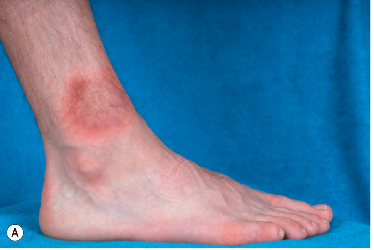
Fig. 14.50A - Erythema migrans: early expanding erythema
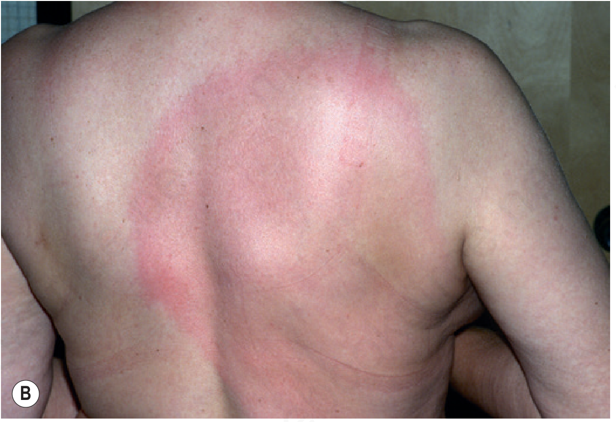
Fig. 14.50B - Erythema migrans: large annular pattern
Stage 2 - Early Disseminated (European features):
- Borrelia-induced lymphocytoma: B-cell proliferations presenting as red, indurated papules and plaques, most commonly on areola or earlobe; occurs from time of EM until 10 months later
Stage 3 - Late/Chronic (European, B. afzelii):
- Acrodermatitis chronica atrophicans (ACA): late cutaneous sequela
- Also: morphea, atrophoderma of Pasini and Pierini, anetoderma, lichen sclerosus et atrophicus
- Some morphea-type lesions: interstitial granulomatous dermatitis with histiocytic pseudorosettes
Systemic Manifestations
| System | Features |
|---|---|
| Musculoskeletal | Chronic arthritis of knees in ~10% of untreated US patients; half progress to severe disability |
| Cardiac | Fluctuating AV block or complete heart block (3 days - 6 weeks); dilated cardiomyopathy (Europe) |
| Neurologic | Stiff neck, headache, meningitis, Bell palsy, optic neuritis, radiculopathy, cranial/peripheral neuropathies; Bannworth syndrome in Europe (radicular pains + lymphocytic meningitis + cranial nerve paralysis) |
| Transplacental | Rare; infant deaths reported |
Histopathology of EM
- Superficial and deep perivascular and interstitial mixed-cell infiltrate
- Lymphocytes, plasma cells, eosinophils (especially at center)
- Warthin-Starry stain may reveal spirochetes in upper dermis
Diagnosis
| Test | Detail |
|---|---|
| Clinical | EM recognition is most sensitive for early infection |
| Serologic conversion | 27% at <7 days symptoms; 41% at 7-14 days; 88% at >2 weeks |
| ELISA (screening) | 89% sensitive, 72% specific |
| Western blot (confirmatory) | IgM: 2 of 3 bands positive; IgG: 5 of 10 bands positive |
| False positives | Syphilis, pinta, yaws, leptospirosis, relapsing fever, EBV, autoantibody diseases |
| VDRL | Negative in B. burgdorferi infection |
| PCR | Specific but not sensitive; not widely available |
Treatment
| Population | Drug | Dose/Duration |
|---|---|---|
| Adults (EM/early) | Doxycycline | 100 mg BID × 3 weeks |
| Alternative (adults) | Amoxicillin | 500 mg TID |
| Children / pregnant | Amoxicillin | 500 mg TID |
| Pregnant women | Penicillin | preferred alternative |
| Cardiac (complete heart block) | IV ceftriaxone | 2 g/day |
| Late neurologic/arthritis | IV ceftriaxone | 2 g/day × 30 days |
| ACA / late skin | Doxycycline or penicillin | 3-4 weeks |
- Andrews' Diseases of the Skin, pp. 334-336
4. SKIN DISEASES CAUSED BY NONTUBERCULOUS MYCOBACTERIA (NTM)
NTM are acid-fast mycobacteria that do not cause tuberculosis. >190 species are now known. Human skin disease is increasing due to: cosmetic procedures, expanding immunocompromised populations, improved identification techniques.
Classification by growth rate:
- Rapidly growing mycobacteria (RGM): subcultured within 1 week - M. fortuitum, M. chelonae, M. abscessus
- Slowly growing mycobacteria:
- Photochromogens (pigment only with light): M. marinum, M. kansasii
- Scotochromogens (pigment regardless of light): M. gordonae, M. scrofulaceum
- Nonchromogens (no pigment): MAC organisms, M. ulcerans
Diagnostic approach: Culture at both high and low temperatures on special media; AFB stain; PCR from fresh or paraffin-fixed tissue; molecular identification.
A. Mycobacterium marinum - Fish Tank Granuloma / Swimming Pool Granuloma
Source: Fresh and salt water; kills aquarium fish. Grows optimally at 30°C (photochromogen).
Exposure: Home aquariums, fishermen, fish sellers, aquaculture. A preceding injury with water exposure is usually present. Males 60% of cases.
Skin lesions:
- Start ~3 weeks after exposure as indolent papule/nodule on hands, knees, elbows, or feet
- Keratotic or warty surface
- Sporotrichoid pattern (ascending nodules up the arm) is very common
- Immunosuppressed: ulcers and abscesses
- Deep structures: tenosynovitis, bursitis, arthritis, osteitis; tendon sheath involvement limiting range of motion
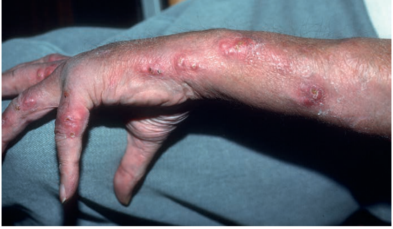
Fig. 16.27 - M. marinum infection in a sporotrichoid pattern (Andrews' Diseases of the Skin)
Treatment: Combination of macrolide + ethambutol + rifamycin. Therapy continued 1-2 months after clinical resolution; longer for tendon/bone involvement. Also active: sulfonamides, TMP-SMX, doxycycline, minocycline, clarithromycin.
B. Mycobacterium ulcerans - Buruli Ulcer
Geography: West and Central Africa, Australia, Papua New Guinea, Southeast Asia.
Transmission: Contact with contaminated soil or water (abraded skin); in Australia, mosquito bites are associated.
Skin lesions:
- Starts as painless nodule, plaque, or edema (preulcerative)
- Progresses to massive, undermined ulcer - destroys skin, subcutaneous tissue, and fascia
- Histotoxin mycolactone causes tissue necrosis and immune suppression
Diagnosis: AFB smear (up to 80% positive), culture, PCR, histology; at least one test positive in 94% when all three done simultaneously.
Treatment: Daily observed therapy for 8 weeks with streptomycin + rifampin; ciprofloxacin may substitute for streptomycin. Healing is slow (half healed by 24 weeks). Surgical excision with delayed grafting for those refusing/intolerant to antibiotics.
Complications: Severe scarring, contracture deformity, amputation; periocular involvement may require enucleation; metastatic skin lesions; rarely bone lesions (75% distant from primary ulcer).
C. Rapidly Growing Mycobacteria (RGM): M. fortuitum, M. chelonae, M. abscessus
Exposure sources:
- Trauma, surgery, injections in immunocompetent patients
- Cosmetic procedures: laser resurfacing, laser hair removal, dermal fillers, botulinum toxin, mesotherapy, liposuction, Mohs surgery, acupuncture, tattoos, skin piercing
- Nail salon whirlpool footbaths (M. fortuitum leg abscess outbreaks)
- Renal transplant patients: tender nodular leg lesions
Skin lesions:
- Small erythematous papules, many spontaneously heal
- Others progress to large, fluctuant, painful abscesses that can ulcerate
- Sporotrichoid or disseminated disease in immunocompromised patients
- Deep extension into underlying bone possible
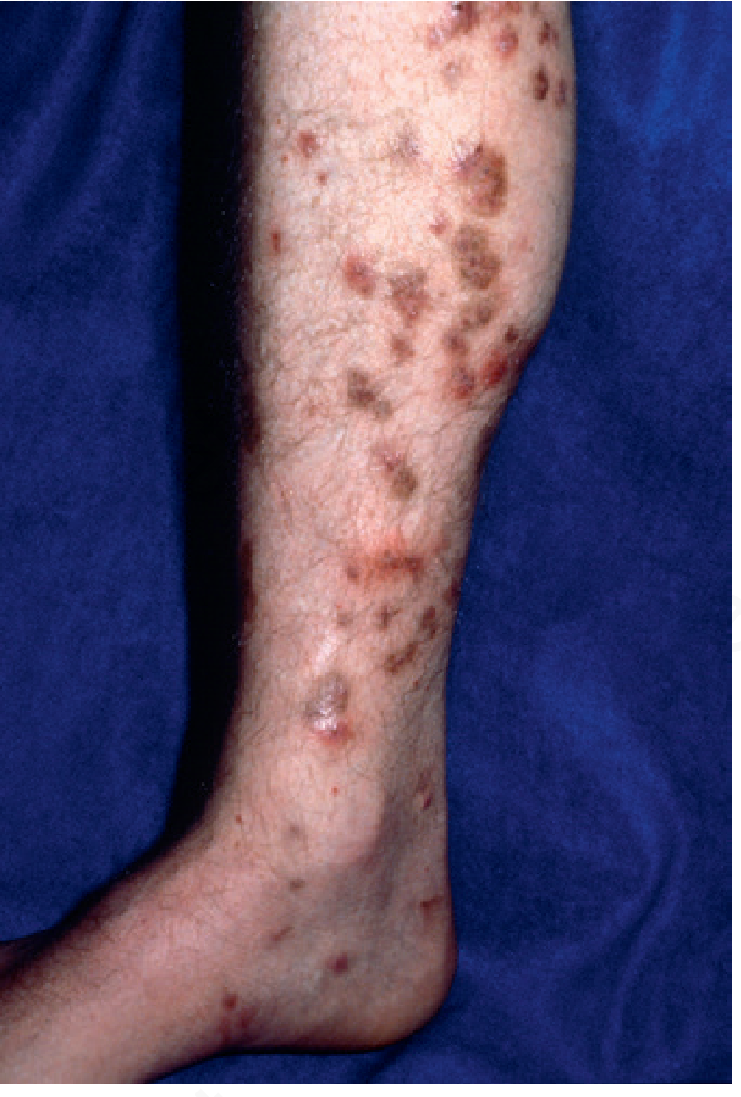
Fig. 16.13 - Mycobacterium fortuitum infection (Andrews' Diseases of the Skin)
Treatment:
| Organism | Treatment |
|---|---|
| M. chelonae / M. abscessus (skin) | Clarithromycin 500 mg BID × ≥6 months |
| M. chelonae / M. abscessus (severe/immunosuppressed) | Moxifloxacin + surgical debridement |
| M. fortuitum | Amikacin + cefoxitin + probenecid (6 weeks) then doxycycline or TMP-SMX up to 1 year; combination recommended |
| Pulmonary M. abscessus | Macrolide + IV amikacin, carbapenem, cefoxitin, or tigecycline (extremely difficult to cure) |
D. Mycobacterium haemophilum
Hosts: Primarily immunosuppressed patients (HIV, organ transplant, biologic agents, leukemia/lymphoma).
Growth requirement: Grows preferentially at 30-32°C (acral sites predominate - cooler body areas).
Skin lesions: Papules, plaques (cellulitis-like), dermal/subcutaneous nodules; break down to form painful draining ulcers. Also: septic arthritis, osteomyelitis, pulmonary nodules.
Special populations: Also reported in immunocompetent patients after acupuncture, permanent eyebrow makeup, and tattoos.
Culture: Requires special media (must notify the lab if suspected - not isolated on routine media).
Treatment: Ciprofloxacin + clarithromycin + rifabutin (all three in combination) for 1 year; adjunctive surgery may be needed.
E. Mycobacterium avium-intracellulare (MAC)
Skin involvement: Uncommon except in AIDS with disseminated infection (hematogenous spread).
- Presents as nodules, ulcers, pustules, or cellulitis-like appearance
- Rare cases of inoculation-type lesions in immunocompetent hosts
Treatment (disseminated): Clarithromycin or azithromycin + ethambutol + rifabutin (at least 3 drugs). Ensure adequate antiretroviral therapy in HIV patients.
F. Mycobacterium kansasii
Skin infection: Rare; usually after minor trauma; 75% in immunosuppressed persons.
Lesions: Papules, nodules, pustules, cellulitis, or sporotrichoid pattern.
Treatment: Isoniazid + rifampin + ethambutol until 12 months after culture clearance (15 months for HIV patients); surgical excision adjunctively.
Summary Table: NTM Skin Infections
| Organism | Setting | Characteristic Lesion | Key Treatment |
|---|---|---|---|
| M. marinum | Aquarium/water exposure | Papule/nodule → sporotrichoid ascending nodules | Macrolide + ethambutol + rifamycin |
| M. ulcerans | Africa, Australia | Massive undermined ulcer (Buruli ulcer) | Streptomycin + rifampin × 8 weeks |
| M. fortuitum | Trauma, nail salon, cosmetics | Subcutaneous abscess, cellulitis | Amikacin + cefoxitin → doxycycline/TMP-SMX |
| M. chelonae/abscessus | Surgery, cosmetics, trauma | Subcutaneous abscesses, erythematous papules | Clarithromycin ± moxifloxacin |
| M. haemophilum | Immunosuppressed, acral | Papules, nodules, draining ulcers at acral sites | Ciprofloxacin + clarithromycin + rifabutin |
| M. kansasii | Minor trauma, immunosuppressed | Papules, nodules, sporotrichoid | INH + rifampin + ethambutol |
| MAC | AIDS, disseminated | Nodules, ulcers, cellulitis-like | Clarithromycin/azithromycin + ethambutol + rifabutin |
- Andrews' Diseases of the Skin, pp. 388-395; Harrison's Principles of Internal Medicine 22E, pp. 1461-1470
This is a shared conversation. Sign in to Orris to start your own chat.