
maximum fluid removal in pleural effusion
Reading File
The classic teaching is to limit drainage to 1000-1500 mL in a single session to avoid re-expansion pulmonary edema. Now let me check the textbooks for the full picture.
Searching sources...
Finding Sources
Finding Sources
Searching PubMed
thoracentesis maximum fluid removal pleural effusion safety
Good - Tintinalli's confirms the 1.0-1.5 L limit. Let me read that section in full and search for more detail on the mechanism and exceptions.
Reading File
Finding Sources
Searching PubMed
thoracentesis large volume pleural effusion reexpansion pulmonary edema
Excellent - found a 2024 RCT and a 2024 paper specifically on REPE after large-volume thoracentesis. Let me get those details and read the key textbook sections in parallel.
Reading Article
PMID: 38858252
Reading Article
PMID: 39029784
Reading File
Reading File
I now have comprehensive information from multiple textbooks and recent literature. Let me also quickly check Harrison's for the pleural manometry/trapped lung angle.
Reading File
I now have everything needed for a thorough, well-sourced answer.
Maximum Fluid Removal in Pleural Effusion (Thoracentesis)
The Traditional Limit
The long-standing teaching is to remove no more than 1,000-1,500 mL of pleural fluid in a single thoracentesis session. This limit exists to prevent re-expansion pulmonary edema (REPE), a rare but potentially life-threatening complication.
- Tintinalli's Emergency Medicine: "Therapeutic thoracentesis with drainage of 1.0 to 1.5 L of fluid is indicated if the patient has dyspnea at rest. Acute drainage of larger volumes is associated with reexpansion pulmonary edema, so large-volume drainage is to be avoided."
- Fishman's Pulmonary Diseases and Disorders: "Traditional teaching recommends that drainage should be limited to 1 to 1.5 L to avoid the risk of re-expansion pulmonary edema."
The Modern Challenge to This Limit
Recent evidence increasingly challenges the strict 1,500 mL ceiling:
- A 2024 retrospective study of 1,376 thoracenteses (PMID: 38858252) found that only 6 patients (0.7%) developed REPE overall, and there was no statistically significant difference in REPE incidence between those with ≥1,500 mL removed vs. <1,500 mL (p = 0.599). The authors concluded large-volume thoracentesis may safely improve symptoms while reducing the need for repeat procedures.
- Fishman's itself notes: "Evidence suggesting that the volume of fluid removed is not correlated with re-expansion pulmonary edema challenges this assertion."
When to Stop Drainage - Symptom-Based Endpoint
Rather than a fixed volume cutoff, current best practice uses symptoms as the primary stopping point:
| Stop drainage immediately if: |
|---|
| Chest pain or tightness develops |
| Persistent cough (non-productive) |
| Dyspnea worsens (rather than improves) |
| Pleural pressure falls excessively (if manometry used: <-20 cmH₂O) |
Special Consideration: Trapped Lung
In trapped lung (lung encased by fibrous visceral peel), continued drainage without re-expansion causes rapidly falling pleural pressure. Signs include:
- Air appearing on imaging after drainage (pneumothorax ex vacuo)
- Persistent chest tightness despite small volumes removed
- Fluid reaccumulating rapidly after drainage
Pleural manometry can guide safe drainage in this setting - Harrison's notes that measuring pleural pressure during thoracentesis helps confirm trapped lung. Drainage should stop if the pressure drops below approximately -20 cmH₂O.
Summary Table
| Parameter | Value |
|---|---|
| Traditional single-session limit | 1,000-1,500 mL |
| Modern approach | Symptom-guided; can exceed 1,500 mL if tolerated |
| REPE incidence (overall) | ~0.7-1% |
| REPE association with large volume | Not statistically confirmed in recent data |
| Manometry stop point | Pleural pressure < -20 cmH₂O |
| Indwelling catheter drainage (chronic) | ~500-1,000 mL per session, every 1-2 days |
Key Takeaway
The 1,000-1,500 mL limit remains a reasonable practical guideline, especially in routine settings without manometry. However, it is not absolute - well-tolerated, symptom-guided large-volume thoracentesis appears safe. The driver of REPE is likely rapid re-expansion against a trapped or non-compliant lung, not simply total volume. Always stop for symptoms regardless of how much has been drained.
Sources:
- Tintinalli's Emergency Medicine (Comprehensive Study Guide), p. 473
- Fishman's Pulmonary Diseases and Disorders, Management section
- Harrison's Principles of Internal Medicine 22E, p. 2313
- Scott et al. (2024) - REPE and large-volume thoracentesis, Cardiovasc Intervent Radiol
- Shojaee et al. (2024) - Gravity vs. wall suction RCT, Chest
What are the signs and symptoms of re-expansion pulmonary edema?
Re-expansion pulmonary edema (REPE) typically presents with sudden onset cough, progressive dyspnea, chest tightness, and frothy/pink sputum within minutes to hours after lung re-expansion. It can range from mild (radiographic only) to severe (hypoxemia, hypotension).
Searching sources...
Finding Sources
Finding Sources
Murray & Nadel's has a dedicated section. Let me read it along with broader paragraph searches.
Reading File
Finding Sources
Searching PubMed
reexpansion pulmonary edema clinical features pathophysiology
Good. Fischer's Mastery of Surgery also has relevant content. Let me read that and search for the Barash anesthesia and Fishman's sections for more clinical detail.
Reading File
Reading File
Searching PubMed
reexpansion pulmonary edema management treatment outcome
I have a rich picture from multiple textbooks and recent literature. Let me fetch the CXR image of REPE from Fischer's for visual reference and get the 2024 review abstract.
Loading Image
Reading Article
PMID: 38731196
I now have everything needed for a thorough answer. Here it is:
Re-Expansion Pulmonary Edema (REPE) - Signs and Symptoms
What It Is
REPE is unilateral pulmonary edema that develops after rapid re-inflation of a lung that has been collapsed - either from pneumothorax or pleural effusion drainage. It is caused by mechanical stress leading to increased vascular permeability (reperfusion/ventilation injury), not hydrostatic pressure overload.
Clinical Presentation
Symptoms typically appear within minutes to 24 hours after drainage:
Respiratory symptoms (most common):
- Persistent cough - often the earliest sign, non-productive, begins during or shortly after drainage
- Dyspnea - worsening breathlessness despite successful fluid removal (paradoxical deterioration)
- Chest tightness / pleuritic chest pain
- Frothy or pink-tinged sputum (in severe cases)
Systemic signs (severe cases):
- Hypoxemia - declining O₂ saturation, cyanosis
- Hypotension - due to fluid shift into the lung interstitium
- Tachycardia
- Diaphoresis
Spectrum of severity:
| Grade | Features |
|---|---|
| Mild (most common) | Radiographic infiltrates only, asymptomatic or minimal cough |
| Moderate | Cough, dyspnea, hypoxemia requiring supplemental O₂ |
| Severe (<1% of cases) | Respiratory failure, hypotension, may require intubation/ICU |
As Murray & Nadel's states: "Typically, patients with RPE will report cough or chest tightness. In extreme cases, patients can be hypoxemic and hypotensive."
Imaging Findings
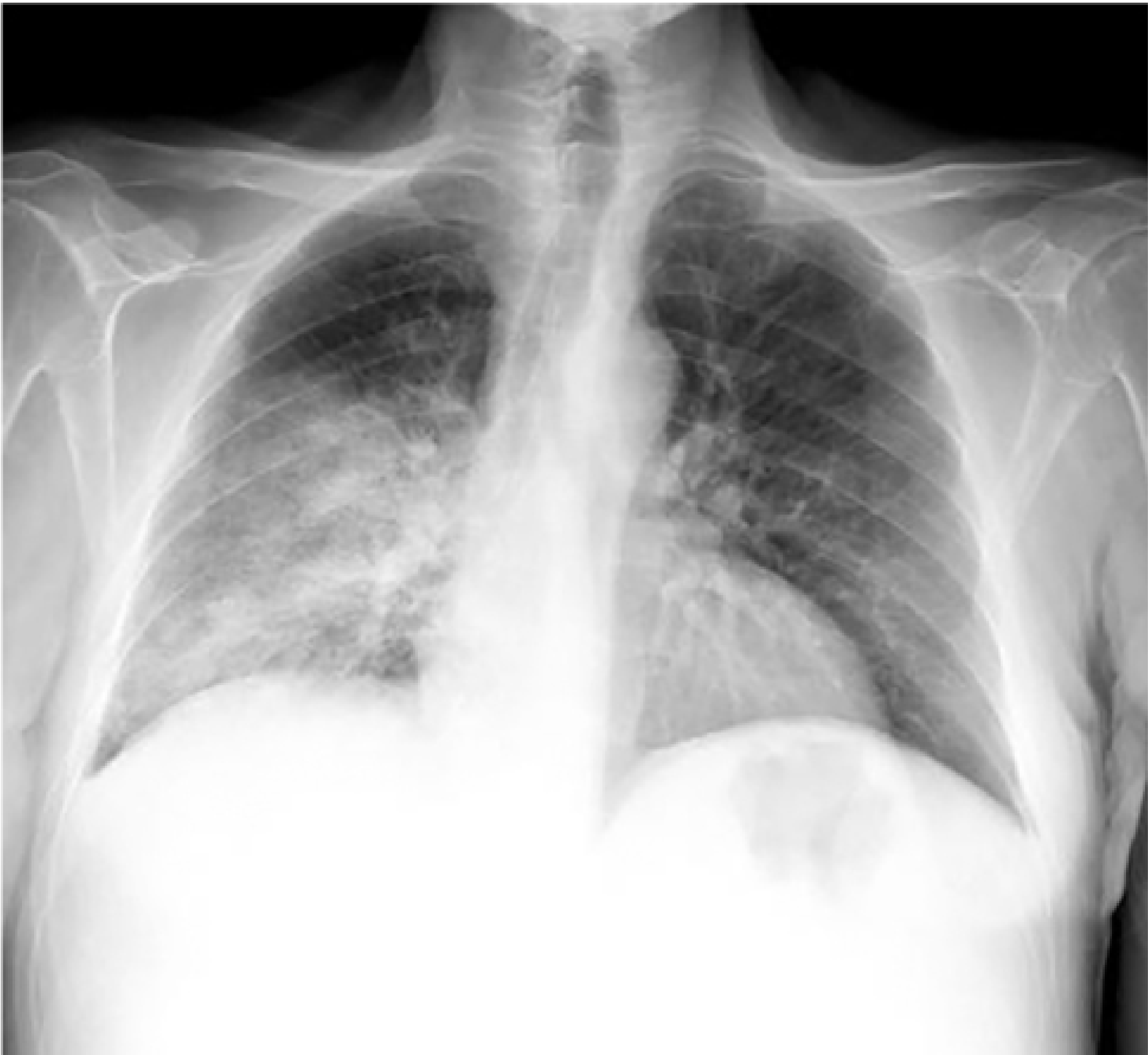
On chest X-ray: new fluffy infiltrates and ground glass opacification on the ipsilateral (re-expanded) side - classically unilateral, distinguishing it from cardiogenic pulmonary edema which is bilateral.
Risk Factors
According to Fischer's Mastery of Surgery:
- Elderly age
- Effusion / collapse present for >72 hours (chronic collapse is the biggest risk factor)
- Large volume effusion >3 L
- Drainage of >1.5-1.8 L during the procedure
- Rapid re-expansion (vs. gradual)
Incidence and Mortality
- Overall incidence after thoracentesis: <1% (though likely underreported as mild cases are missed)
- Most cases (~16-27% in some series) are asymptomatic, detected only on imaging
- Reported mortality: up to 20% in severe cases, though this likely reflects the underlying comorbid burden of patients requiring drainage - Fischer's Mastery of Surgery
- A 2024 review (PMID 38731196) notes clinical manifestations range from "almost asymptomatic to a rapidly fatal condition"
Treatment
Treatment is supportive:
- Supplemental oxygen (first-line for mild-moderate)
- High-flow nasal oxygen (HFNO) or CPAP for moderate-severe hypoxemia
- Diuretics (limited evidence; used adjunctively)
- Intubation and mechanical ventilation with PEEP for severe respiratory failure
- There is no specific antidote - management mirrors standard ARDS/pulmonary edema protocols
Sources:
- Murray & Nadel's Textbook of Respiratory Medicine - Reexpansion Pulmonary Edema section
- Fischer's Mastery of Surgery 8th ed., p. 2439-2440
- Barash's Clinical Anesthesia 9e - Cardiovascular Complications
- Cusumano et al. (2024) - REPE case series and literature review, J Clin Med
This is a shared conversation. Sign in to Orris to start your own chat.