
Orris we are starting from first to learn the legal med, I say the chapter name we ll go one by one chapter, for every chapter you have to teach each and everything in the chapter you have to teach one by one topic you shouldn't leave out any topic any info understood and teach me like I am 5,at the end give all mcqs from book back for each chapter which you will be teaching for me with explanation clearly remember I am 5
📚 Welcome to Legal Medicine Class!
🏆 Your Textbooks (We Have These!)
Book 1: "The Essentials of Forensic Medicine and Toxicology" - 36th Edition (2026)
Book 2: "Parikh's Textbook of Medical Jurisprudence, Forensic Medicine and Toxicology"
📋 Chapter List - Book 1 (Essentials of Forensic Medicine, 36th Ed)
- History of Development of Forensic Medicine
- Acts and Amendments
- Principles of Ethics (Autonomy, Beneficence, Nonmaleficence, Justice)
- Legal Competencies
- Inquest
- Offence
- Punishments
- Courts of Law
- (+ more topics in this block)
Which book and which chapter shall we start with?
Which book do you want to study from? · Which chapter do you want to START with?
Shall we start
Start chapter medicolegal aspects of autopsy
The Essentials of Forensic Medicine & Toxicology, 36th Edition
🧒 IMAGINE THIS...
📌 TOPIC 1: WHAT IS AN AUTOPSY?
- Why? Because the evidence of death may be hiding in more than one organ.
- A poor or incomplete autopsy is WORSE than no autopsy at all - because it misleads the court and causes injustice!
- The famous saying: "What the mind does not know, the eyes cannot see, and hands cannot do" - meaning: the doctor must be knowledgeable or they'll miss clues.
📌 TOPIC 2: TYPES OF AUTOPSY
| Type | Who orders it? | Purpose |
|---|---|---|
| Medicolegal/Forensic Autopsy | Police or Magistrate | Find cause of death in suspicious/criminal cases |
| Clinical/Pathological Autopsy | Hospital/family consent | Find why a patient died (for medical knowledge) |
| Exhumation Autopsy | Court/Magistrate | Re-examine a body that was already buried |
📌 TOPIC 3: WHO PERFORMS THE AUTOPSY?
- The doctor must personally remove all organs. The attendant only helps (like preparing the body, handing instruments).
- Must be done by an authorized government doctor.
- Private medical institutions CANNOT do autopsies without government permission.
📌 TOPIC 4: WHERE IS AUTOPSY DONE? (Mortuary/Postmortem Room)
- 🌞 Well-lit - preferably with natural daylight (because colour changes like bruise colours, jaundice, lividity CANNOT be seen properly in artificial light!)
- 🌬️ Well-ventilated - so smells and infections don't spread
- 🧹 Clean and easy to disinfect
- Has a proper autopsy table (usually stainless steel with drainage)
- Has instruments: scalpels, scissors, bone saw, forceps, brain knife, etc.
📌 TOPIC 5: WHEN SHOULD AUTOPSY BE DONE?
- As soon as possible after receiving the official requisition (order from police/magistrate).
- Best time = Daylight, because:
- Color of bruises, jaundice, lividity cannot be seen in artificial light
- If body arrives late at night → do preliminary examination that night, then complete autopsy next morning early
- There is NO law preventing nighttime autopsy, BUT decomposed bodies, murder cases, and suspected foul play cases should NOT be done at night.
📌 TOPIC 6: AUTHORIZATION (PERMISSION)
- Police (in most cases)
- Magistrate (in special cases like dowry death, rape/death in custody)
📌 TOPIC 7: PRE-AUTOPSY REVIEW (Before You Cut Anything!)
- Read the Inquest Report (Panchanama) - find out the apparent cause of death
- Review case sheets/accident register if the person was treated in hospital before death
- Decide what special investigations are needed (toxicology, microbiology, radiology, etc.)
- Mistaking a surgical incision for a wound
- Not recognizing resuscitation injuries (CPR can fracture ribs!)
- Not knowing if a wound was sutured during surgery
- Drainage tube wounds looking like stab wounds
📌 TOPIC 8: SCENE OF CRIME / EXAMINATION AT SCENE
- The body is in a remote location and transport is difficult
- Evidence would be lost during transport
- Immediate examination is essential for some special reason
📌 TOPIC 9: EXTERNAL EXAMINATION (Outside the Body First!)
🥼 Step 1: Clothes and Ornaments
- Listed, examined, and described in detail (type, color, size, tears, disarrangement suggesting struggle)
- Removed gently - to preserve trace evidence like hair, fibers, glass, paint, sand
- Cut ONLY if they cannot be removed normally, and cuts should be made away from bullet holes
- Placed in separate clean plastic bags - do not mix items
- Wet clothes should be hung up to dry - NOT heat dried (heat destroys evidence, and wet folded clothes grow fungus!)
- Stained and unstained areas should NOT come in contact with each other
- All clothing handed over to police in sealed packets
🔍 Step 2: Nail Scrapings and Trace Evidence
- Nail scrapings are taken from EACH finger separately (10 separate envelopes!)
- A matchstick or folded filter paper is run under each nail
- Each envelope is labeled with the finger number and sealed
- Why? Because the person may have scratched their attacker - and the attacker's skin cells, blood, hair may be under the nails!
🔍 Step 3: External Body Examination
- Identity - age, sex, build, nutrition, complexion
- Postmortem changes - lividity (PM staining), rigor mortis, decomposition
- Injuries - describe all wounds, their nature, size, location (measured from bony landmarks)
- Natural disease signs - jaundice, anemia, etc.
📌 TOPIC 10: INCISIONS (HOW THE BODY IS OPENED)
1. "I" - Shaped Incision (Straight cut)
- One straight vertical cut from the top of the chest (suprasternal notch) down to the pubic area
- The incision goes around the umbilicus (belly button) - because the belly button has tough fibrous tissue that's hard to stitch up later!
2. "Y" - Shaped Incision
- Starts near the shoulder (acromial process) on both sides
- The two lines meet and go down below the breast to the xiphoid (bottom tip of breastbone)
- From xiphoid, one vertical line goes down to the pubic area
- Looks like the letter Y!
3. Modified "Y" - Shaped Incision
- Similar to Y, but the arms of the Y extend over the clavicle (collar bone) and upward behind the ear
- Used when neck examination is also needed
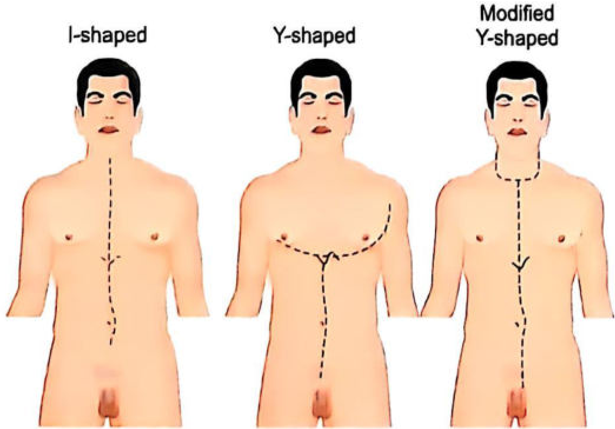
📌 TOPIC 11: INTERNAL EXAMINATION (Opening the Body Cavities)
Opening the Abdomen:
- Doctor stands on the RIGHT side (if right-handed)
- Cuts the abdominal muscles, opens peritoneum
- Before disturbing anything, OBSERVE and NOTE:
- Any blood, pus, or fluid in the cavity?
- Any perforation (hole) in any organ?
- Position and condition of all organs
Opening the Chest (Thorax):
- Ribs are cut with a rib-cutter (costotome)
- The breastplate (sternum + ribs) is lifted off like a lid
- Examine for blood or fluid in chest cavity, condition of lungs, heart
📌 TOPIC 12: SPECIAL TEST - AIR EMBOLISM
- Float test: If the right ventricle contains air, it will float in water
- Pericardial water test: Fill the pericardial sac with water, then puncture the right heart - bubbles will appear if air is present
- Syringe test: Insert a wide-bore needle into right ventricle - air will bubble through water in the syringe
- Inferior vena cava test: Puncture it under water, look for bubbles
- Chest X-ray
- Pyrogallol Test: A chemical test - if air (oxygen) is present, a mixture turns brown. A second syringe acts as a control. This test distinguishes air from decomposition gas!
📌 TOPIC 13: POSTMORTEM CLOTS vs. ANTEMORTEM THROMBUS
Postmortem Clots (Formed after death - NOT significant):
- Two types:
- Red currant jelly clot - blood clotted quickly, dark red, soft, lumpy, slippery
- Chicken fat clot - blood settled (red cells sank), leaving pale yellow fibrin layer on top
- Features: Moist, smooth, shiny, rubbery, NOT firmly attached to vessel wall, NO striae of Zahn (fine white fibrin lines)
- When pulled from vessels: forms a cast of the vessel branches
Antemortem Thrombus (Formed before death - significant!):
- Features: Firm, attached to vessel wall, dull, dry, granular, HAS striae of Zahn
- Significance: Can indicate heart attack, pulmonary embolism, etc.
Agonal Thrombus (Formed during the dying process):
- Person dying slowly with circulatory failure
- Firm, stringy, tough, pale-yellow
- Usually forms on RIGHT side of heart
- Can extend like a tree-like cast into pulmonary artery
Postmortem Blood Fluidity:
- Shortly after death, blood is usually FLUID
- Why? Because fibrinolysins released from vascular endothelium break down clots
- In most deaths from asphyxia, blood is fluid and incoagulable
📌 TOPIC 14: EXAMINATION OF SPECIFIC ORGANS
🫁 Lungs:
- Open pulmonary artery - look for thrombi, emboli, atherosclerosis
- Examine for: consolidation, oedema, emphysema, Tardieu spots (tiny hemorrhages from asphyxia!), emboli, tumour
- Antemortem embolus: May be coiled, when straightened out looks like a cast of the originating vessel. Does NOT fit the lung vessel perfectly. Massive pulmonary emboli commonly block the RIGHT side and are always fatal!
❤️ Heart:
- Remove with great vessels attached (at least 1 cm beyond pericardial fold)
- Open with inverted T-shaped incision
- Measure: height, width, circumference 1 cm below AV sulcus
- Look for: fibrosis, recent infarcts, valve problems
🩸 Aorta:
- Cut on its posterior surface, full length from iliac vessels to aortic valve
- Look for aortitis, plaque formation blocking coronary arteries
🧠 Brain (Examination of Head):
- Cut from behind one ear (mastoid process), over the top of the head (vertex), to behind the other ear
- Scalp is reflected forward (to superciliary ridges) and backward (below occipital protuberance)
- Any bruising in the scalp is noted
- Sawed open with oscillating saw
- Dura mater examined for hemorrhage
- Brain is removed by cutting cranial nerves and brainstem
- Flattening of convolutions, obliteration of sulci
- Herniation of temporal poles through tentorial hiatus
- In severe cases: lateral ventricles reduced to mere slits, white matter appears glistening
- Brain may weigh up to 1750 g
- Cerebellar tonsils pushed through foramen magnum ("coning")
🦷 Neck Organs:
- The tongue, hyoid bone, larynx, trachea are examined together as a block
- Very important in strangulation cases (hyoid bone fracture!)
🟤 Liver:
- Look for fatty change, cirrhosis, congestion
- Sectioned on the right side of the body
🫘 Kidneys:
- Capsule stripped to examine cortical surface
- Cut longitudinally to see cortex, medulla, pelvis
🍖 Spleen:
- Weighed, examined for size, softness, infarcts
🔵 Female Genitalia (Important in sexual offence/abortion cases!):
- Tubes, ovaries, and uterus freed and removed from pelvis
- Uterus opened from external OS to fundus
- If fetus present: age of fetus determined
- Uterus and appendages preserved in criminal abortion cases
🔵 Male Genitalia:
- Prostate: vertical cross-sections made
- Testes: pulled out, cut longitudinally, check for orchitis, carcinoma
🦴 Vertebral Column:
- Atlanto-occipital joint checked for fracture-dislocation
- Cervical spine examined
- Pelvis squeezed from side to side - any mobility indicates fracture
📌 TOPIC 15: LABORATORY INVESTIGATIONS AT AUTOPSY
Histopathology:
- Tissue pieces: 20 mm × 12 mm × 5 mm size
- Fixed in 10% neutral formalin or 95% alcohol
- Amount of preservative = 6-10 times the volume of tissue
Bacteriology:
- All specimens collected under STERILE conditions
- Blood for culture obtained BEFORE organs are disturbed
- After opening pericardial sac, right ventricle surface is seared with a hot spatula, then aspirated
Toxicology - Viscera to Preserve:
| Organ | Amount | Preservative | What it detects |
|---|---|---|---|
| Stomach + contents | All | None (sealed) | Ingested poisons |
| Small intestine + contents | All | None | Poisons |
| Liver | 500g | Saturated salt (NaCl) | Most poisons |
| Kidney | One whole | Saturated NaCl | Poisons |
| Blood | 100 mL | Sodium fluoride | Alcohol, CO, drugs |
| Urine | All available | Sodium fluoride | Drugs, alcohol |
| Spleen | 500g | Saturated NaCl | General |
| Brain | 500g | Saturated NaCl | Volatile poisons |
| Vitreous humor | Both eyes | None | Alcohol, glucose, electrolytes |
| Hair | 20-30 strands with roots | None | Arsenic, chronic poisoning |
| Nails | All nails removed entirely | None | Arsenic, chronic poisoning |
| Bone | 10 cm femur shaft | None | Arsenic, antimony, lead, mercury |
| Fat | 10g from abdominal wall | None | Pesticides, insecticides |
| Lung/Air | Sealed nylon bag | None | Volatile poisons (HCN, alcohol, chloroform) |
📌 TOPIC 16: PHOTOGRAPHY AND VIDEO AT AUTOPSY
- To provide a permanent record of findings
- To supplement PM report findings - prevent suppression of information
- To allow independent review of PM report later
- 20-25 coloured photographs covering whole body
- Some photos taken BEFORE removing clothes
- Doctor's voice should be recorded during video (narrating findings)
- Photos must include: PM number, date, and a scale for dimensions in the frame
- Face (front, right lateral, left lateral, back of head)
- Front and back of body
- Upper and lower extremities (front and back)
- Each injury (numbered properly)
- Internal findings
- Palms and soles (with incision to show deep seated injuries)
- For firearm injuries: distance from heel AND midline for each injury
- Both hands wrapped in white paper bags
- Body covered in special body bags for transport
- PM report + video + photos sent to commission within 2 months of incident
- If viscera sent for examination, PM report is sent WITHOUT waiting for viscera report (viscera report sent later)
📌 TOPIC 17: AUTOPSY IN INFECTIOUS CASES (AIDS, CHOLERA, etc.)
- Cholera
- Rabies
- Plague
- Anthrax
- Gas gangrene
- Smallpox
- Full protective gear (gloves, gown, mask, eye protection, boots)
- Minimize number of personnel in room
- No unnecessary instruments or equipment
- Dispose of materials properly
- Disinfect everything
📌 TOPIC 18: AUTOPSY IN RADIOACTIVE CASES
- Use a Geiger counter to measure radiation level of the body first
- Wear protective gear
- Work quickly and in shifts (minimize exposure time)
- Rinse instruments with running water, dispose into sewer
- Contaminated clothing: wash with soap and water, stored for radioactive decay before sending to laundry
- If body contains >300 millicuries after autopsy - it must be embalmed in hospital morgue
- Cardiac pacemakers must be recorded - some contain mercury or radioactive substances that can cause explosions in crematoria!
📌 TOPIC 19: OCCUPATIONAL HAZARDS IN MEDICOLEGAL AUTOPSIES
| Hazard | Example |
|---|---|
| Infection by puncture/cut | HIV, Hepatitis B from needle stick |
| Aerosol inhalation | Tuberculosis, Rabies, Anthrax, Plague |
| Eye contact | Localized or generalized infection |
| Noxious chemical exposure | Formaldehyde, pesticides, corrosives |
| Radiation exposure | From radioactive victims |
| Psychological harm | Constant exposure to mutilated/decomposed bodies → depression, personality disorders, alcohol abuse |
📌 TOPIC 20: EXAMINATION OF SKELETAL REMAINS (BONES)
Is it Bone or Not?
- Large pieces = easy to identify by gross appearance
- Small/burnt pieces = histological examination needed
Human or Animal?
- Usually easy when whole skeleton available
- Difficult with: fragments, burnt bones, small animal bones
- Tests:
- Precipitin test - if fresh bone with blood proteins still present
- DNA analysis - from marrow cells (if bones are fresh)
- Microscopy - Non-human bones have plexiform structures in cross-section; great apes cannot be distinguished from human bones by this method alone
- Chemical analysis of bone-ash
- Short-wave UV lamp - different individuals' bones fluoresce different colors (useful to separate mixed/commingled bones!)
How Many Individuals?
- Reconstruct the skeleton
- If no disproportion, no duplication of same bones, correct articulation, same age/sex/race = one individual
- If mixed: use short-wave UV lamp - bones of different individuals emit different fluorescent colors
Sex from Bones?
- Sex differences NOT present before puberty
- After puberty: examine pelvis (most reliable), skull, manubrium-gladiolus ratio, femur head diameter, humerus head diameter
- Scars of parturition = irregular dorsal border of pubic symphysis in women who have given birth (caused by trauma during childbearing)
Age from Bones?
- Teeth (most reliable in children)
- Ossification centers
- Epiphyseal union
- Length of long bones
- Pubic symphysis changes
Cause of Death from Bones?
- Usually cannot be determined unless:
- Fractures of skull, upper cervical vertebrae, hyoid bone (strangulation!), several ribs
- Marks of deep cuts in long bones
- Marks of burning
- Foreign bodies (knife blade, bullet)
- Disease (caries, necrosis)
- Metallic poisons (arsenic, antimony, lead, mercury) can be found in bones long after death
- Arsenic can be detected even in burnt bones!
- Diatoms in bone marrow = death from drowning!
📌 TOPIC 21: POSTMORTEM REPORT (PM REPORT)
- Name, age, sex of deceased
- Name of police station, case number
- Date, time, place of examination
- External examination findings
- Internal examination findings (organ by organ)
- Special investigations ordered
- Opinion on cause of death
- Based ONLY on personal findings - not on information from others
- Should be clear, concise, complete, legible
- Avoid technical terms where possible
- Include relevant NEGATIVE findings too (e.g., "no fractures found")
- Written in DUPLICATE using carbon paper
- First copy → investigating officer
- Second copy → retained by doctor
- Doctor signs/initials at bottom of EACH page
- Name and designation noted on last page
📌 TOPIC 22: MEDICAL CERTIFICATE OF CAUSE OF DEATH (MCCD)
- (a) Immediate Cause = the final disease/injury directly causing death
- (b) Intermediate Cause(s) = conditions linking underlying to immediate cause
- (c) Underlying Cause = the disease/injury that STARTED the whole chain (this gets the ICD-11 code!)
- Immediate: Septicemia
- Intermediate: Extensive burns (70% TBSA)
- Underlying: Assault by burning (dowry-related) → ICD-11 Code: PK80.1
📌 TOPIC 23: THANATOLOGY (Science of Death)
- Somatic/Systemic/Clinical death = irreversible stoppage of vital functions
- Molecular/Cellular death = death of individual cells (happens AFTER somatic death, different cells die at different times)
| Type | Mechanism | Examples |
|---|---|---|
| Anoxic anoxia | Not enough O₂ reaching blood | Smothering, choking, hanging, drowning, asphyxia |
| Anemic anoxia | Blood can't carry O₂ | Massive hemorrhage, CO poisoning, chlorates |
| Stagnant anoxia | Poor circulation | Heart failure, embolism, shock |
| Histotoxic anoxia | Cells can't USE O₂ | Cyanide poisoning |
📌 TOPIC 24: ASPHYXIA
- Asphyxia is a mode of dying, NOT a cause of death
- Normal arterial O₂: 90-100 mmHg (young adults), 60-85 mmHg (>60 years)
- Fatal asphyxia: O₂ falls to 20-40 mmHg
- Brain = only 1.4% of body weight BUT uses 20% of total body oxygen!
- Cerebral cortex stops functioning: 8-15 seconds after total ischemia
- Brainstem ganglia: 25-30 seconds
- Irreparable cortex damage: ~3 minutes
- Basal ganglia damage: 6-7 minutes
- Vagal center damage: ~9-10 minutes
- Thumb rule: "Breathing stops within 20 seconds of cardiac arrest; heart stops within 20 minutes of stopping of breathing"
🎯 FAMOUS CASES (The Book Includes These - Remember Them!)
Alavander Murder Case (1952):
- Murdered by Prabhakar Menon
- Headless trunk found in third-class compartment of Indo-Ceylon Express
- Head found at Roya Puram sea beach, Chennai
- Identity established by: fingerprints, circumcised penis, socks, waist thread, overriding canine teeth, pierced earlobes
Acid-Bath Murder Case (John George Haigh, 1949):
- Haigh dissolved a victim (Mrs. Durand Deacon) in sulphuric acid
- Identity established by: acrylic dentures (recognized by her dentist!), gallstones, bloodstained coat, jewelry
- This is why it's said: acid cannot destroy gallstones or dentures!
📝 MCQs FROM THE CHAPTER (With Full Explanations)
- A) Artificial light
- B) Natural daylight ✅
- C) UV light
- D) Night time
- A) Examining only the most affected organ
- B) Opening the chest cavity only
- C) Opening all body cavities and examining every organ ✅
- D) External examination only
- A) Carbon monoxide poisoning
- B) Air embolism ✅
- C) Fat embolism
- D) Cyanide poisoning
- A) Antemortem thrombus
- B) Agonal thrombus
- C) Postmortem clot ✅
- D) Fibrin thrombus from disease
- A) Postmortem clots
- B) Agonal thrombi
- C) Antemortem thrombus ✅
- D) Chicken fat clot
- A) Skull
- B) Femur
- C) Humerus
- D) Pelvis ✅
- A) Sacrum
- B) Dorsal border of pubic symphysis ✅
- C) Ischial tuberosity
- D) Acetabulum
- A) Only fresh bones
- B) Only intact bones
- C) Even in burnt bones ✅
- D) Only in bone marrow
- A) Arsenic poisoning
- B) Death by strangulation
- C) Death by drowning ✅
- D) Death by burning
- A) Midline vertical
- B) Coronal/Intermastoidal ✅
- C) Horizontal over forehead
- D) Circular around crown
- A) Opening the skull first
- B) Opening the abdomen
- C) Opening the heart ✅
- D) Opening the spine
- A) Formalin
- B) 95% Alcohol
- C) Saturated common salt (NaCl) solution ✅
- D) Distilled water
- A) -20°C
- B) 0°C
- C) 4°C ✅
- D) 10°C
- A) 5-10
- B) 10-15
- C) 20-25 ✅
- D) 50-100
- A) 1 month
- B) 2 months ✅
- C) 3 months
- D) 6 months
- A) 5%
- B) 10%
- C) 20% ✅
- D) 40%
- A) 1 minute
- B) 3 minutes ✅
- C) 6 minutes
- D) 10 minutes
- A) Fingerprints
- B) DNA analysis
- C) Dentures and gallstones ✅
- D) Footprints
- A) Haversian systems
- B) Plexiform structures ✅
- C) Lamellar bone only
- D) Osteoid seams
- A) Heat dried before packing
- B) Folded and packed wet
- C) Hung up to dry (air dried) ✅
- D) Discarded
🌟 QUICK MEMORY TRICKS
| Fact | Memory Aid |
|---|---|
| A poor autopsy is worse than no autopsy | "Half job = Injustice!" |
| Autopsy in daylight | "Colors need sunlight!" |
| Skull opened AFTER heart drained | "Drain first, then Brain!" |
| Chicken fat = postmortem clot | "After death, fat floats!" |
| Striae of Zahn = antemortem thrombus | "Zahn = Alive (Zel in German means living)" |
| Pyrogallol turns brown = air present | "Brown = Air around!" |
| Brain uses 20% O₂ | "Small but greedy!" |
| Irreparable brain damage = 3 minutes | "3 minutes to save the brain!" |
| Arsenic in burnt bones | "Arsenic is immortal!" |
| Diatoms in marrow = drowning | "Water bugs swim to the bone!" |
You didn't tell techniques in autopsy like Virchow or letulle didn't I say you shouldn't leave any topic right
⚠️ MISSED TOPIC: TECHNIQUES FOR REMOVAL OF ORGANS AT AUTOPSY
🧒 IMAGINE THIS FIRST...
- Take out each item ONE BY ONE
- Take out everything in big chunks all at once
- Take out sections together but study each section separately
🔬 THE 4 AUTOPSY DISSECTION TECHNIQUES
Key principle from the book: "Autopsy techniques differ very little between centres. The pathologist can depend on the method he masters best." They differ mainly in: (1) the ORDER organs are removed, (2) how they are SECTIONED, and (3) the METHOD of organ removal.
🥇 TECHNIQUE 1: VIRCHOW'S TECHNIQUE
- Organs are removed ONE BY ONE, individually
- Order of removal:
- Cranial cavity opened FIRST (brain removed)
- Spinal cord removed from the BACK
- Then thoracic (chest) organs
- Then cervical (neck) organs
- Then abdominal organs
- ✅ Each organ can be studied in great detail individually
- ✅ Most widely used technique in the world
- ❌ The anatomico-pathological relationships between organs are NOT preserved - so you cannot study how one organ relates to its neighbor in disease
🥈 TECHNIQUE 2: ROKITANSKY'S TECHNIQUE
- Involves in situ dissection (dissecting inside the body, in the original position) combined with en bloc removal in part
- The organs are partially examined while still IN the body, then removed as a block
- ✅ Preferred choice for highly infectious cases - HIV, Hepatitis B - because it LIMITS the spread of infection (less handling, less exposure)
- ❌ The organs cannot be studied in detail individually
🥉 TECHNIQUE 3: LETULLE'S TECHNIQUE
- Cervical + Thoracic + Abdominal + Pelvic organs are ALL removed EN MASSE (as one giant block) together
- Then the big mass is taken away from the body and dissected as organ blocks on the examination table
- ✅ All attachments between organs are left intact - you can study the connections!
- ✅ Best technique for routine inspection and preservation of connections between organ systems
- ✅ Body is ready for the undertaker in less than 30 minutes - without having to rush the dissection (dissection is done separately on the organ mass)
- ❌ The organ mass is often awkward/heavy to handle
4️⃣ TECHNIQUE 4: GHON'S TECHNIQUE
- Organs are removed as separate blocks by system:
- Block 1: Cervical + Thoracic + Abdominal organs together
- Block 2: Urogenital system (kidneys, bladder, genitals) separately
- Block 3: Neurological system (brain, spinal cord) separately
- Modern practice uses a modification where organs are removed en bloc (as one complete block)
📊 MASTER COMPARISON TABLE (The Most Important One for Exams!)
| Feature | Virchow | Rokitansky | Letulle | Ghon |
|---|---|---|---|---|
| Method | One by one | In situ + partial en bloc | All en masse | By system blocks |
| Order | Brain → Spine → Chest → Neck → Abdomen | In situ first | All together | Chest+Neck+Abdomen, Urogenital, Neural |
| Organ relationships preserved? | ❌ NO | Partial | ✅ YES (best!) | Partial |
| Individual organ detail? | ✅ BEST | ❌ Poor | Good (after removal) | Good |
| Best for infections (HIV, Hep B)? | No | ✅ YES | No | No |
| Body available fast? | No | Yes | ✅ YES (<30 min) | No |
| Organ mass awkward? | No | No | ❌ YES | No |
| Most widely used? | ✅ YES | No | No | No |
🎯 EXAM TRIGGER LINES (One-Liners to Remember)
| If exam says... | Answer is... |
|---|---|
| "Organs removed one by one" | Virchow |
| "En masse removal" | Letulle |
| "En bloc removal" / "most popular MCQ trap" | Letulle (en masse) |
| "Preferred in HIV / Hepatitis B" | Rokitansky |
| "In situ dissection combined with en bloc" | Rokitansky |
| "Anatomico-pathological relations NOT preserved" | Virchow |
| "All attachments left intact / connections preserved" | Letulle |
| "Body ready in <30 minutes" | Letulle |
| "Organs removed by system (urogenital separately, neural separately)" | Ghon |
| "Most widely used technique" | Virchow |
📝 THE BOOK'S OWN MCQ ON THIS TOPIC
- A. Virchow technique
- B. Rokitansky technique
- C. Ghon technique
- D. Letulle technique ✅
- A. R. Virchow's technique ✅
- B. C. Rokitansky's technique
- C. M. Letulle's technique
- D. A. Ghon's technique
🧒 Super Simple Summary (Like You're 5):
| Person | What they do |
|---|---|
| Virchow | 🍬 Takes out ONE candy at a time from the jar |
| Rokitansky | 🦠 Opens the jar carefully without touching (because it's dirty/infected!) |
| Letulle | 🛒 Takes out ALL candies in one big shopping bag, sorts them later |
| Ghon | 📦 Takes out candies in separate boxes by TYPE (chocolate in one box, gummy in another) |
Have legal med evals today no time let's be quick start chapter Mechanical Asphyxia and Asphyxiants DYSBARISM AND BAROTRAUMA please make it understandable and quick also remember the doc style and give book back of that chapter
⚡ MECHANICAL ASPHYXIA, ASPHYXIANTS, DYSBARISM & BAROTRAUMA
Essentials of Forensic Medicine & Toxicology, 36th Ed - QUICK EXAM PREP
🧒 SIMPLE IDEA FIRST
PART 1: MECHANICAL ASPHYXIA
📌 DEFINITION
📌 CLASSIFICATION (Professor Gorden)
| Type | What happens | Example |
|---|---|---|
| Anoxic anoxia | No O₂ in the air you breathe | High altitude |
| Stagnant anoxia | Blood flow decreases/blocked | Vascular blockage, heart failure |
| Anemic anoxia | Blood can't carry O₂ | Anemia, CO poisoning, methemoglobin |
| Histotoxic anoxia | Cells can't USE O₂ | Cyanide (blocks cytochrome oxidase) |
📌 CAUSES OF MECHANICAL ASPHYXIA
| Category | Examples |
|---|---|
| Nose & mouth blocked | Smothering, bagging |
| Airways blocked from within | Gagging, café coronary, drowning |
| Compression of neck | Hanging, strangulation, mugging, garroting, bansdola |
| Compression of chest | Traumatic asphyxia, burking |
📌 CARDINAL SIGNS OF ASPHYXIA (Common to ALL types)
- 🔵 Cyanosis - bluish discoloration of skin/lips
- 🔴 Petechial hemorrhages / Tardieu's spots - tiny pinpoint bleeds
- 🩸 Congestion of internal organs
- 💧 Fluidity of blood (doesn't clot)
- ❤️ Left heart empty; Right heart has dark fluid blood
PART 2: HANGING
📌 DEFINITION
"Hanging is always suicidal unless contrary is proved beyond reasonable doubt"
📌 CLASSIFICATION
- Complete hanging - body fully suspended, no part touching ground
- Partial hanging - toes/feet touch the ground, or sitting/kneeling posture. Even the weight of the head alone (5-6 kg) is enough to cause death!
- Typical hanging - knot at back of neck (occipital region), ligature runs symmetrically upward on both sides of neck above thyroid cartilage
- Atypical hanging - knot anywhere else (side or front of neck)
📌 LIGATURE MARK IN HANGING - KEY FEATURES
- The classic mark is oblique/slanting (NOT horizontal) - goes upward toward the knot
- Inverted V shape on the neck - the apex points toward the knot
- The area under the knot has NO mark (suspension peak/point) - because the rope is pulled AWAY from skin there
- Suspension peak = distinguishing feature of hanging vs. strangulation!
- Mark is: pale, yellowish-brown, parchment-like, dry, hard
- Usually above the level of the thyroid cartilage
- Running noose (weight causes it to tighten horizontally)
- Low suspension point
- Partial hanging with body leaning forward
📌 SYMPTOMS/STAGES OF HANGING
- Loss of power, flashes of light
- Unconsciousness (in seconds)
- Convulsions
- Death (from asphyxia, venous obstruction, carotid compression, or vagal inhibition)
📌 POSTMORTEM FINDINGS IN HANGING
- Face: congested, cyanotic, or pale (if carotids compressed → pale)
- Petechiae in conjunctiva
- Tongue: protruded, tip between teeth, dark
- Saliva: dribbling from corner of mouth (dried track - "le faci sympathicae")
- Ligature mark: oblique, pale/parchment, above thyroid cartilage
- Neck may show fracture of hyoid, thyroid cartilage (in judicial hanging)
- Fracture-dislocation of C2 (judicial hanging from long drop)
- Soft tissues of neck: ecchymoses, hemorrhage into neck muscles
- Fracture of hyoid bone - in throttling AND hanging (inner border)
- Carotid arteries: intimal tear sometimes
- Signs of asphyxia in internal organs
📌 CAUSE OF DEATH IN HANGING (4 mechanisms)
- Asphyxia (most common) - airway compressed
- Venous obstruction - jugular veins compressed → cerebral congestion
- Carotid artery compression → cerebral ischemia
- Vagal inhibition - reflex cardiac arrest (sudden death, especially in partial hanging)
- Fracture-dislocation of cervical spine (judicial hanging)
📌 SUICIDAL vs. HOMICIDAL vs. ACCIDENTAL HANGING
| Feature | Suicidal | Homicidal | Accidental |
|---|---|---|---|
| Common? | Very common | Very rare | Rare |
| Suspension point accessible? | Yes | May be staged | Yes |
| Signs of struggle? | No | May be present | No |
| Hands/feet tied? | Sometimes (to prevent mind change) | May be tied by assailant | No |
| Ligature mark | Oblique | May be horizontal (if strangled first) | Oblique |
PART 3: STRANGULATION
📌 TYPES
- Ligature applied to neck, constricting force is NOT body weight but manual tightening
- Ligature mark is horizontal (circumferential, goes all around the neck)
- Mark is below the thyroid cartilage
- NO suspension peak (unlike hanging)
- Almost always homicidal
- Using hands/fingers to compress the neck
- Shows: fingernail abrasions and fingertip bruises on neck
- Hyoid bone fracture more common than in hanging
- Almost always homicidal
- Mugging = forearm/elbow used to compress neck from behind (carotid sleeper hold)
- Bansdola = bamboo stick or rod across the neck and back - victim's body weight used
- Garroting = wire twisted around neck
📌 STRANGULATION vs. HANGING - EXAM TABLE
| Feature | Hanging | Ligature Strangulation |
|---|---|---|
| Ligature mark direction | Oblique/diagonal | Horizontal |
| Level | Above thyroid cartilage | Below thyroid cartilage |
| Suspension peak | Present | Absent |
| Mark complete? | Incomplete (gap at knot) | Complete (goes all around) |
| Usually | Suicidal | Homicidal |
| Hyoid fracture | Less common | More common |
| Manner | Self-suspension | External force |
PART 4: SUFFOCATION
📌 DEFINITION
📌 TYPES
- Breathing vitiated (oxygen-deficient) atmosphere
- O₂ in air normally = 21%; N₂ = 79%; CO₂ = 0.033%
- O₂ below 16% = dangerous
- O₂ at 5% = rapid loss of consciousness and death in minutes
- Causes: CO, CO₂, methane, H₂S, SO₂, chlorine, phosgene
- Examples: locked refrigerators (children), silos, sewers, unused wells, grain bins, tanks
- Blocking of nose and mouth by hand, pillow, plastic bag
- Face shows: bruises, petechiae, abrasions around mouth/nose
- Almost always homicidal in adults; accidental in infants
- Foreign body inside the airways (between pharynx and tracheal bifurcation)
- Common in: children (coins, marbles), elderly, drunk people, epileptics, psychiatric patients
- Café Coronary = food (usually meat) suddenly blocks airway during meal → collapse mimicking cardiac arrest
- Heimlich maneuver is the treatment
- Foreign material placed in the mouth/throat (cloth, tape) to silence victim
- If victim vomits, they can die of choking
📌 TRAUMATIC ASPHYXIA (Burking/Crush Asphyxia)
- Compression of chest and abdomen preventing breathing
- Seen in: crowd crushes, burial under rubble, industrial accidents
- Face/neck shows: intense bluish-red congestion and petechiae (like a mask)
- Called "Masque ecchymotique"
- Burking = simultaneous compression of chest + smothering (used by Burke and Hare, Scotland, 1820s)
PART 5: DROWNING
📌 DEFINITION
📌 TYPES
- Wet drowning - water enters lungs (most common, ~85%)
- Dry drowning (~10-15%) - laryngeal spasm prevents water entry; dies of asphyxia with NO water in lungs
- Secondary/Near drowning - survives initial submersion, dies later from complications
- Immersion syndrome (Hydrocution) - sudden cold water contact → vagal inhibition → instant death before drowning
📌 SEQUENCE OF EVENTS IN DROWNING
- Person falls into water → initial panic, breath-holding
- CO₂ rises → forced inspiration → inhales water
- Violent coughing, water enters alveoli
- Specific gravity of body rises → starts sinking
- Fear/anoxia → may vomit and aspirate gastric contents
- Struggle, unconsciousness, convulsions, death
- Body stays at bottom until intestinal bacteria produce gas → floats in 12-18 hours (summer) or 18-36 hours (winter) in India
📌 POSTMORTEM FINDINGS IN DROWNING
- 🫧 Fine white froth at mouth and nose - mushroom-shaped froth from air mixing with mucus
- 🦢 Goose skin / Cutis anserina - erection of hair follicles from cold water
- 👐 Washerwoman's hands - sodden, wrinkled, white skin (starts on fingertips in 2-4 hours)
- 🌿 Cadaveric spasm - weeds/grass firmly grasped in hands (proof person was alive when drowned!)
- Skin of hands/feet peels off like a glove or stocking in 2-4 days
- Body usually floats face down (spine uppermost); obese persons/women may float face up
- Aquatic animals (fish, crabs, eels) attack soft parts: eyelids, lips, nose, ears, genitals - lesions are circular/oval punched-out
- Voluminous, waterlogged, ballooned lungs - most important sign
- Froth in air passages
- Dark fluid blood (diluted by inhaled water)
- Right heart dilated with dark blood
- Water in stomach (70% of cases) - valuable if same type as drowning medium
- SEHRT's sign - micro-ruptures in gastric mucosa from overstretching
- Wydler's sign - gastric contents separate into 3 layers (solids bottom, liquid middle, foam top)
- UENO's sign - water in middle ear (forced through Eustachian tubes)
- Sveshnikov's sign - water in maxillary and sphenoid sinuses
- Temporal bone hemorrhages (also seen in hanging)
📌 RELIABLE SIGNS OF DROWNING (6 points to remember)
- Fine white froth at mouth and nose
- Weeds/sand firmly grasped in hands
- Weeds/sand/froth in lungs and air passages
- Voluminous water-logged lungs
- Water in stomach and intestines
- Diatoms in tissues (most specific!)
📌 DIATOM TEST (THE MOST IMPORTANT TEST!)
- Diatoms = microscopic single-celled algae with silica shells, found in natural water
- If person was alive when submerged → diatoms enter circulation → found in bone marrow, brain, kidney, liver
- Bone marrow = most reliable site (survives decomposition and putrefaction)
- Dry ashing method used to find diatoms
- If diatoms in body match diatoms in the drowning medium → confirms drowning at THAT location
- Diatoms not found if: death was due to vagal inhibition (no water inhaled), dry drowning, or body examined after putrefaction
📌 GETTLER TEST (Chloride test)
- Based on: when a person drowns in fresh water, blood in left heart becomes diluted → less salt (chloride)
- In sea water drowning: blood in right heart becomes more concentrated
- Normal blood chloride = 580 mg/100 mL
- In fresh water drowning: left heart chloride < right heart chloride (diluted)
- In sea water drowning: right heart chloride > left heart chloride (concentrated)
- Limitation: Not reliable in putrefied bodies
PART 6: SEXUAL ASPHYXIA
- Also called Autoerotic asphyxia / Asphyxiophilia
- Very rare - almost exclusively in males
- Partial asphyxia (carotid compression or partial airway obstruction) causes cerebral disturbances/hallucinations of erotic nature
- Person deliberately induces partial asphyxia during masturbation for enhanced sexual gratification
- Death occurs accidentally - when the safety mechanism fails
- Scene: elaborate setup - bondage equipment, women's clothing, pornographic material
- Manner of death: accidental (not suicidal!)
- Rule out suicide: suicide note absent, no motive, elaborate safety setup
PART 7: DYSBARISM AND BAROTRAUMA
📌 DEFINITIONS
- Dysbarism = ALL adverse effects of pressure changes
- Barotrauma = mechanical tissue damage caused by failure of a gas-filled body cavity to equalize its internal pressure with changes in ambient (surrounding) pressure
- Most commonly occurs under water
📌 MECHANISM (Think of a Coke Bottle!)
- Deep underwater = HIGH pressure → gas dissolves INTO liquid (like CO₂ dissolves in soda under pressure)
- Come up TOO FAST = pressure drops suddenly → gas comes OUT of solution as BUBBLES (like shaking the bottle then opening it!)
- Those bubbles in your blood and tissues = DECOMPRESSION SICKNESS
📌 TWO MAIN PROBLEMS
A. Nitrogen Narcosis ("Rapture of the Deep")
- At depths >30 meters: nitrogen dissolves progressively into plasma and tissue fluids under high pressure
- Causes narcosis = state resembling drunkenness
- Reversible on ascending
B. Decompression Sickness ("The Bends")
- Diver ascends TOO QUICKLY → dissolved nitrogen comes out of solution → gas bubbles form in:
- Blood (gas emboli → block vessels → infarction, especially CNS)
- Tissues
- Joint cavities (→ severe joint pain = "the bends")
- Additional effects:
- Platelet aggregation and DIC (disseminated intravascular coagulation)
- Subcutaneous emphysema
- Can also occur at HIGH ALTITUDE (aircraft depressurization - decrease from normal to low pressure)
📌 PHYSICAL DAMAGE (Pulmonary Barotrauma)
- When rapid decompression occurs, gas in body cavities expands as diver rises
- Pressure builds up in: paranasal sinuses, tooth pulp cavities, middle ear
- In lungs: alveolar walls rupture → interstitial emphysema → bullae on pleural surface → if bullae rupture = pneumothorax
- Air tracks to mediastinum, then up neck (surgical emphysema)
- Air enters lung capillaries and veins → systemic arterial circulation → impacts in arterioles/capillaries of myocardium, spinal cord, brain → micro-infarcts, hemorrhagic necrosis
- This pulmonary barotrauma can occur in as little as 2-3 meters of water!
📌 AUTOPSY FINDINGS IN DYSBARISM/BAROTRAUMA
- Lungs: oedema, patchy hemorrhages, torn alveoli, focal emphysema, subpleural bullae
- Pneumothorax
- Subcutaneous, mediastinal, retroperitoneal, subpleural emphysema
- Fat embolism (both in post-descent shock and decompression)
- Petechial hemorrhages in CNS and spinal cord
- Early infarcts (if victim survives a day or more)
- Microscopically: ring-shaped hemorrhages around affected vessels
📌 QUICK COMPARISON TABLE
| Feature | Decompression Sickness | Barotrauma |
|---|---|---|
| Mechanism | Gas bubbles from dissolved gas | Physical expansion of gas in cavities |
| Where | Blood, tissues, joints | Sinuses, teeth, ears, lungs |
| Key sign | "Bends" (joint pain), gas emboli | Pneumothorax, subcutaneous emphysema |
| Minimum depth | Deep diving (>30m for narcosis) | Only 2-3 meters! |
| Altitude | Also at high altitude | Also at altitude |
📝 BOOK MCQs WITH EXPLANATIONS
- A) Homicidal
- B) Accidental
- C) Suicidal ✅
- D) Due to sexual asphyxia
- A) The lowest point of the ligature mark
- B) The point where the mark is deepest
- C) The point where the rope leaves the skin, leaving no mark ✅
- D) The site of knot impression
- A) Horizontal, below the thyroid cartilage
- B) Oblique, above the thyroid cartilage ✅
- C) Horizontal, above the thyroid cartilage
- D) Oblique, below the thyroid cartilage
- A) 21%
- B) 19%
- C) 16% ✅
- D) 10%
- A) Cardiac arrest during coffee drinking
- B) Coronary artery spasm after a meal
- C) Sudden death from food bolus blocking airway, mimicking heart attack ✅
- D) Choking from café food only
- A) Hanging
- B) Drowning
- C) Traumatic asphyxia ✅
- D) Manual strangulation
- A) Gettler's chloride test
- B) Hydrostatic test
- C) SEHRT's sign
- D) Diatom test ✅
- A) Urine vs. blood
- B) CSF vs. blood
- C) Left heart blood vs. Right heart blood ✅
- D) Stomach contents vs. blood
- A) Postmortem injury
- B) Homicidal drowning
- C) Person was alive when they entered the water ✅
- D) Dry drowning
- A) Back of hand in 1 hour
- B) Whole palm in 1 hour
- C) Fingertips in 2-4 hours ✅
- D) Sole of feet in 2-4 hours
- A) 50% of drowning deaths
- B) 30% of drowning deaths
- C) 10-15% of drowning deaths ✅
- D) 1% of drowning deaths
- A) 10 meters
- B) 5 meters
- C) 30 meters
- D) 2-3 meters ✅
- A) Water enters the lungs
- B) O₂ becomes toxic at depth
- C) Dissolved nitrogen comes out of solution as bubbles on rapid ascent ✅
- D) CO₂ accumulates in blood
- A) 10 meters
- B) 20 meters
- C) 30 meters ✅
- D) 50 meters
- A) Water in maxillary sinuses
- B) Goose skin
- C) Water forced into middle ear through Eustachian tubes ✅
- D) Micro-ruptures in gastric mucosa
- A) 2-4 hours
- B) 6-8 hours
- C) 12-18 hours ✅
- D) 3-4 days
- A) Suicidal
- B) Homicidal
- C) Natural
- D) Accidental ✅
- A) UENO's sign
- B) SEHRT's sign
- C) Wydler's sign
- D) Sveshnikov's sign ✅
🎯 LAST MINUTE CHEAT SHEET
| Condition | Key word |
|---|---|
| Hanging | Oblique mark, ABOVE thyroid, suspension PEAK, suicidal |
| Ligature strangulation | HORIZONTAL mark, BELOW thyroid, NO peak, homicidal |
| Throttling | Fingernail marks, hyoid fracture, homicidal |
| Drowning | White froth, waterlogged lungs, diatoms, cadaveric spasm |
| Dry drowning | NO water in lungs, laryngospasm |
| Café coronary | Food bolus, accidental, mimics heart attack |
| Traumatic asphyxia | Chest crushed, masque ecchymotique |
| Decompression sickness | N₂ bubbles, rapid ascent, "bends" |
| Barotrauma | Gas expansion, pneumothorax, 2-3 m depth |
| Nitrogen narcosis | >30 m depth, like drunkenness |
| Sexual asphyxia | Accidental, male, erotic setting |
Good evening. Evals 6 for Legal Medicine tomorrow Saturday , August 8 at 5PM on the 4th floor paperless. Coverage: Topics of Groups 13 (Liabilties of Doctors and Hospitals) and 14 (Indian Courts and Jurisprudence) and my lectures on Medical Malpractice and Physicians in court Reference: Dr. Reddy’s book and the Groups’ presentations. Good luck!
- Group 13: Liabilities of Doctors and Hospitals
- Group 14: Indian Courts and Jurisprudence
- Medical Malpractice
- Physicians in Court
Good evening. Evals 6 for Legal Medicine tomorrow Saturday , August 8 at 5PM on the 4th floor paperless. Coverage: Topics of Groups 13 (Liabilties of Doctors and Hospitals) and 14 (Indian Courts and Jurisprudence) and my lectures on Medical Malpractice and Physicians in court Reference: Dr. Reddy’s book and the Groups’ presentations. Good luck! Have only 10 min
⚡ EVAL 6 RAPID PREP - LEGAL MEDICINE
Groups 13 & 14: Liabilities of Doctors/Hospitals + Indian Courts & Jurisprudence
Dr. Reddy's Essentials of Forensic Medicine, 36th Ed
🏛️ GROUP 14: INDIAN COURTS & JURISPRUDENCE
TYPES OF COURTS IN INDIA
Criminal Courts (Hierarchy - top to bottom):
| Court | Key Facts |
|---|---|
| Supreme Court | Highest tribunal, New Delhi. Purely appellate in criminal cases. Binding on ALL courts (Article 134) |
| High Court | State capital. Highest for the state (Article 214). Can try any offence. Judges appointed by President of India |
| Sessions Court | Established by state govt at district HQ. Can only try cases committed by a Magistrate. Death sentence passed must be confirmed by High Court |
| Magistrates' Court | 3 types: Chief Judicial, 1st Class, 2nd Class |
Powers of Magistrates (TABLE - HIGH YIELD!):
| Magistrate | Max Imprisonment | Max Fine |
|---|---|---|
| Chief Judicial Magistrate | Up to 7 years | Unlimited |
| 1st Class Judicial Magistrate | Up to 3 years | ₹10,000 |
| 2nd Class Judicial Magistrate | Up to 1 year | ₹5,000 |
Other Special Courts:
- Juvenile Court - For offenders below 18 years. Presided by 1st class woman magistrate + 2 social workers (at least 1 woman)
- Labor Courts - Industrial disputes
- Family Courts - Marriage and family disputes
- Supreme Court + High Courts = APPELLATE only - do not hold prima facie trials
Who Appoints Whom?
- Sessions judges + Magistrates → appointed by High Court
- High Court judges → appointed by President of India
INQUEST
1. Police Inquest (Section 194, BNSS / S.174 Cr.P.C.)
- Conducted by Sub-Inspector of Police or above
- Done when: person dies under suspicious/unnatural circumstances
- 2+ respectable persons (panchas) present
- Prepares Panchanama (inquest report)
- Body sent for PM with dead body challan + copy of inquest
- If NO foul play suspected → body handed to relatives
2. Magistrate's Inquest (Section 196, BNSS / S.176 Cr.P.C.)
- Dowry death
- Death of woman within 7 years of marriage (suicide, suspicious circumstances, relative's request, any doubt)
- Exhumation
- Any person dies/disappears or rape is alleged while in police custody
COURTS COMPETENCY - PHYSICIANS IN COURT
Summons / Subpoena
- A written order from court compelling a witness to appear
- Conduct money = fee paid to the witness to cover travel/expenses for attending court
- A doctor MUST appear when summoned as treating physician or eyewitness - he cannot refuse
- He MAY refuse if: he is not sufficiently qualified, cannot spare time, or fears a painful experience
Types of Witnesses:
| Type | Who |
|---|---|
| Common witness | Describes facts (e.g., describes wounds on body) |
| Expert witness | Gives opinion (e.g., wounds were antemortem, cause of death) |
| Hostile witness | Supposed to have interest in concealing truth (Section 227, BNS) |
- Describing wounds = common witness
- Opining cause of death = expert witness
Examination Types:
- Examination-in-Chief - Questioned by lawyer who called you (leading questions NOT allowed)
- Cross-examination - Questioned by opposing lawyer (leading questions ALLOWED)
- Re-examination - Clarification by your own lawyer (no NEW matters; leading questions NOT allowed)
- Judge's questions - Judge may ask anything, in any form, at any stage
Oath/Affirmation:
- Witnesses take oath (religious) or affirmation (non-religious, secular)
- Dying declaration: NO oath administered (dying person assumed to tell truth)
Perjury:
- Giving false evidence under oath = Perjury
- Punishment: Up to 7 years imprisonment + fine
DYING DECLARATION (Leterm Mortem = "Words before death")
- Section 26(1), BSA (S.32, I.E.A.)
- Written or oral statement of dying person about cause of his own death
- Doctor certifies person is conscious and mentally competent (compos mentis) before recording
- Ideally recorded by Executive Magistrate
- If no time for magistrate: doctor records in presence of 2 witnesses
- NO OATH administered (dying person assumed to speak truth)
- Can be recorded by village headman, police - but evidential value less
- Must be in patient's own words (not paraphrased)
PROCEDURE OF CRIMINAL TRIAL
- FIR (First Information Report)
- Police Investigation
- Charge Sheet filed in court
- Cognizance taken by Magistrate
- Committal to Sessions Court (for serious offences)
- Framing of Charges
- Prosecution evidence (examination-in-chief, cross-examination)
- Statement of accused (Section 313 BNSS)
- Defense evidence
- Final arguments
- Judgment
👨⚕️ GROUP 13: LIABILITIES OF DOCTORS & HOSPITALS
MEDICAL NEGLIGENCE - THE 3 ESSENTIAL ELEMENTS ("3 D's")
- Duty - Doctor owed a duty of care to the patient
- Dereliction (Breach of duty) - Doctor failed in that duty
- Damage - Patient suffered actual harm as a result
If even ONE element is absent → NO negligence
BOLAM TEST (Most Important Standard!)
"The standard of the ordinary skilled man exercising and professing to have that special skill. A man need not possess the highest expert skill; it is sufficient if he exercises the ordinary skill of an ordinary competent man exercising that particular art."
- Bolam v. Friern Hospital Management Committee
- A GP is NOT required to have the skills of a specialist
- A junior doctor can discharge duty by seeking help from a superior
- A doctor who claims superior skill is judged by that higher standard
- Locality rule: GP judged by standards of doctors with similar training in similar geographic location
TYPES OF NEGLIGENCE
1. Civil Negligence
- Standard: Breach of duty causing damage
- Tried by: Civil courts
- Punishment: Monetary compensation/damages
- Limitation: File within 2 years from date of negligence
- Contract breach: 3 years
2. Criminal Negligence
- Higher threshold - must be gross/culpable negligence (not mere error of judgment)
- Conditions for criminal negligence (any ONE of these):
- Indifference to obvious risk of injury to health
- Foresight of risk but continuation of same treatment
- Inattention/failure to avert risk
- Conscious disregard of health and safety of patient
- Tried by: Criminal courts
- Punishment: Imprisonment + fine
3. Professional Misconduct (Infamous Conduct)
- Violation of Code of Medical Ethics
- Tried by: State Medical Council
- Punishment: Erasure of name from register or warning
- No requirement for duty of care or damage to person
Negligence vs. Infamous Conduct (TABLE):
| Feature | Professional Negligence | Infamous Conduct |
|---|---|---|
| What | Absence of care/skill | Violation of Ethics Code |
| Duty of care needed | YES | NO |
| Damage needed | YES | NO |
| Tried by | Courts (civil/criminal) | State Medical Council |
| Punishment | Fine or Imprisonment | Erasure of name or warning |
| Appeal | Higher Court | State and Central Governments |
DUTIES OF A DOCTOR (Key for exam!)
- Must attend with reasonable care and skill
- Code of ethics: In emergency, no physician should refuse to treat
- No law compels a doctor to attend except in military necessity
- Must personally examine the patient
- Cannot rely on another doctor's notes without examining yourself
- Case: Newton vs Central Middlesex - 2nd doctor negligent for relying on card, not examining the knee
- Give legible prescriptions with full instructions
- Liable for harm from wrong prescription
- Tell patient about adverse reactions, exact doses, timing, and to stop drug if reaction occurs
- Must refer when case is beyond competence
- Case: Ball vs Howard - Surgeon went away without leaving instructions and refused consultation, held negligent
- Must maintain records of indoor patients for 3 years
- Must provide records within 72 hours of request
- Explain nature of operation and get consent
- Avoid operating on wrong patient/wrong limb
- Cannot delegate operative duty to another
- Must not experiment
- Must ensure all swabs/instruments are removed (count before and after!)
- Must use sterilized instruments
- Keep properly labeled in separate bottles
- Assist police in determining if poisoning was accidental/suicidal/homicidal
- Must report communicable diseases (smallpox, cholera, plague, typhoid, measles, diphtheria, yellow fever, food poisoning) to Public Health authorities
- Failure = criminal penalty AND civil negligence liability
- Warn patients of risk involved in treatment
- Case: Kankan vs Beharilal - Doctor negligent for NOT warning patient of risk of dangerous mixture
CONSENT
- Informed consent = patient given sufficient information about purpose, methods, risks, alternatives BEFORE agreeing
- Emergency = implied consent (patient cannot consent, doctor acts in best interest)
- No consent needed when: patient unconscious in emergency, identity not disclosed in publications
- Required for: operations that may result in sterility (BOTH husband and wife must consent)
- IVF/Artificial insemination: written informed consent of female patient AND spouse AND donor
VICARIOUS LIABILITY (Hospital Liability)
"Let the master answer" - Respondeat Superior principle
- Hospital/employer is vicariously liable for negligent acts of employees done in course of employment
- Doctor employed by hospital = hospital is liable for doctor's negligence
- Private nursing home run by physician = physician ultimately responsible for assistants' actions
RES IPSA LOQUITUR
"The thing speaks for itself"
- Negligence is self-evident from the facts - no detailed proof needed
- Examples:
- Swab/instrument left inside patient after surgery
- Wrong limb amputated
- Wrong patient operated
- The burden of proof shifts to the doctor to explain!
CONTRIBUTORY NEGLIGENCE
- Patient's own negligence contributed to the damage
- Last Clear Chance Doctrine: If doctor had last chance to avoid injury and failed, doctor cannot use patient's prior negligence as defense
- Avoidable Consequences Rule: If patient's negligence AFTER the doctor's negligence aggravated damage - doctor cannot claim contributory negligence
DEFENSES AGAINST MEDICAL NEGLIGENCE
- No duty owed to plaintiff
- Duty discharged according to prevailing standards
- Misadventure (unavoidable complication)
- Error of judgment (honest mistake, not negligence)
- Contributory negligence (patient's own fault)
- Res judicata (Section 337, BNSS) - already decided by court, cannot re-litigate
- Limitation - suit must be filed within 2 years; 3 years for contract breach
SERIOUS PROFESSIONAL MISCONDUCTS - "THE 6 A's" ⭐
- Adultery - arising from professional relationship
- Advertising - improper self-promotion
- Abortion - unlawful
- Association - with unqualified persons in professional matters
- Addiction - to drugs
- Alcohol - consumption at workplace
RIGHTS OF REGISTERED MEDICAL PRACTITIONERS
- Right to practice medicine
- Right to choose a patient
- Right to dispense medicines
- Right to possess and supply dangerous drugs to patients
- Right to add titles/descriptions to name
- Right to recovery of fees
- Right to appointment in public/local hospitals
- Right to issue medical certificates
- Right to give evidence as expert
RIGHTS/PRIVILEGES OF PATIENTS
- Choice - choose own doctor freely
- Access - regardless of age, sex, religion, economic status
- Dignity - treated with respect
- Privacy - during consultation and therapy
- Confidentiality - all information kept confidential
- Information - full disclosure of diagnosis, treatment, alternatives
- Safety - know risks and complications
- Refusal - right to refuse any treatment
- Second opinion - at any time
- Records - access to own records
- Complaint - right to complain and seek grievance redressal
- Compensation - for medical negligence
CONSUMER PROTECTION ACT (CPA) 2019
- Medical services = "services" under CPA
- Patient can file complaint as consumer
- Complaint fee (District level only):
- ≤₹5 Lakh: NIL
- ₹5-10 Lakh: ₹200
-
₹10 Lakh: ₹400
- Limitation period: Within 2 years (Section 69)
- Can appear with OR without a lawyer
- New in CPA 2019: E-commerce, unfair contracts, formal mediation, higher pecuniary limits
SUPREME COURT GUIDELINES ON MEDICAL NEGLIGENCE (11 Points - High Yield!)
- Negligence = breach of duty; a prudent and reasonable man would not do it
- Must be culpable or gross - not merely an error of judgment
- Doctor expected to bring reasonable degree of skill - neither highest nor lowest
- Doctor liable only if conduct fell below the reasonably competent practitioner standard
- Difference of opinion ≠ negligence
- Higher risk taken to save patient in grave situation ≠ automatically negligence
- Choosing one acceptable course over another ≠ negligence
- Doctor should not be made to practice "with a halter round his neck"
- Medical professionals should not be harassed or humiliated unnecessarily
- Criminal process should not be used as a tool to pressure doctors for compensation
- Doctors entitled to protection as long as they perform with reasonable skill and in patient's interest
MEDICAL INDEMNITY INSURANCE
- Contract where insurance company reimburses doctor against claims of professional negligence
- Objects:
- Protect professional reputation
- Pay legal costs
- Pay awarded damages
- Every practicing doctor should have it
DEFENSIVE MEDICINE
- Ordering every possible test to have "hard data" backup
- May lead to withholding of beneficial treatment to majority due to statistical risk to minority
- Doctor must NOT practice "defense by denial" (claiming patient is not his) = abandonment
IATROGENIC DISEASE
- Disease resulting from medical treatment, drug administration, or a procedure
- Doctor + hospital can be held liable
- Must be distinguished from natural disease progression
📝 BOOK MCQs WITH ANSWERS & EXPLANATIONS
- A) High Court
- B) Sessions Court
- C) Supreme Court ✅
- D) District Court
- A) Supreme Court
- B) High Court ✅
- C) District Magistrate
- D) President of India
- A) 1 year
- B) 3 years
- C) 5 years
- D) 7 years ✅
- A) All deaths
- B) Road traffic accident deaths
- C) Dowry death ✅
- D) Death due to natural causes
- A) 14 years
- B) 16 years
- C) 18 years ✅
- D) 21 years
- A) Duty and damage only
- B) Breach of duty only
- C) Duty + Breach of duty + Damage ✅
- D) Duty + Intent
- A) The highest level of medical skill
- B) The skill of the best doctor in the country
- C) The standard of the ordinary skilled doctor exercising that particular art ✅
- D) The skill of a specialist in every case
- A) The doctor has spoken
- B) Let the master answer
- C) The thing speaks for itself ✅
- D) No evidence needed
- A) The Supreme Court's ruling
- B) A doctor's right to refuse treatment
- C) Vicarious liability of hospital/employer for employee's negligence ✅
- D) Right of patient to second opinion
- A) Adultery
- B) Advertising
- C) Abortion (unlawful)
- D) Absenteeism ✅
- A) 1 year
- B) 3 years ✅
- C) 5 years
- D) 10 years
- A) 1 year
- B) 2 years ✅
- C) 3 years
- D) 5 years
- A) Ball vs Howard
- B) Maynard vs West Midlands
- C) Newton vs Central Middlesex ✅
- D) Kankan vs Beharilal
- A) It is a legal exemption
- B) The person is too ill
- C) The dying person is presumed to speak the truth ✅
- D) It is recorded by police, not court
- A) 1 year imprisonment
- B) 3 years imprisonment
- C) Up to 7 years imprisonment + fine ✅
- D) Only a fine
- A) Strictly prohibited
- B) Allowed only by the judge
- C) Allowed ✅
- D) Allowed only in civil cases
- A) ₹200
- B) ₹100
- C) ₹500
- D) NIL ✅
- A) Common witness
- B) Hostile witness
- C) Expert witness ✅
- D) Prosecution witness
- A) Newton vs Central Middlesex
- B) Maynard vs West Midlands
- C) Kankan vs Beharilal
- D) Ball vs Howard ✅
- A) Treating patients aggressively
- B) Practicing medicine only after getting legal clearance
- C) Ordering every test to have data backup to avoid litigation ✅
- D) Refusing all high-risk procedures
⚡ LAST-MINUTE CHEAT SHEET
| Key Rule | Remember |
|---|---|
| 3 D's of negligence | Duty + Dereliction + Damage |
| Bolam test | Ordinary skilled doctor = standard |
| Res ipsa loquitur | Thing speaks for itself (swab inside!) |
| Respondeat superior | Hospital liable for employee's acts |
| Perjury punishment | 7 years + fine |
| Record keeping | 3 years (produce in 72 hours) |
| Negligence suit limitation | 2 years |
| Dying declaration oath | No oath administered |
| 6 A's | Adultery, Advertising, Abortion, Association, Addiction, Alcohol |
| Juvenile court age | Under 18 |
| CJM max sentence | 7 years |
| Cross-examination | Leading questions ALLOWED |
| Exam-in-chief | Leading questions NOT allowed |
| Magistrate's inquest | Dowry death + Death in custody + Exhumation |
Good afternoon . Prefi Exam in Legal Medicine tomorrow Wednesday, August 12 at 7PM on the 4th floor paperless. Coverage: Topics of Groups 11, 12, 13 (Liabilties of Doctors and Hospitals) and 14 (Indian Courts and Jurisprudence) and my lectures on Medical Jurisprudence, Physician-patient relationship. Rights, Duties and Liabilities of Physicians, Hospitals and patients,Medical Malpractice and Physicians in court Reference: Dr. Reddy’s book and the Groups’ presentations. Good luck!
📚 PREFINAL EXAM STUDY GUIDE
Legal Medicine - Groups 11, 12, 13 & 14
Dr. Reddy's Essentials of Forensic Medicine, 36th Ed (2026)
🧠 GROUP 11: MEDICAL JURISPRUDENCE & MEDICAL ETHICS
What is Medical Jurisprudence?
HISTORY OF MEDICAL ETHICS - TIMELINE ⭐
| Year | Person/Event | Significance |
|---|---|---|
| 1750 BCE | Code of Hammurabi (Babylonia) | Earliest documented standards for medical practitioners |
| 1500 BCE | Sushruta Samhita | Father of Indian Surgery + Plastic Surgery; early rhinoplasty |
| 200 BCE | Charaka Samhita (Charaka's Oath) | Father of Indian Medicine |
| 5th c BCE | Hippocratic Oath | Father of Medicine and Medical Ethics; begins with Apollo + Asclepius |
| 1500 CE | University of Wittenberg (Germany) | First institution to administer oath to graduating medical students |
| 1803 | Thomas Percival (English physician) | Coined "medical ethics" + "medical jurisprudence"; wrote first modern code |
| 1847 | AMA Code of Ethics | Based on Percival's work |
| 1948 | Declaration of Geneva | World Medical Association; modernized Hippocratic Oath (amended 1968, 2017) |
| 2022 | NMC India | Introduced Charaka Oath for white coat ceremony (replaced Hippocratic Oath) |
Memory trick: Hammurabi → Sushruta → Charaka → Hippocrates → Wittenberg → Percival → AMA → Geneva → NMC
NATIONAL MEDICAL COMMISSION (NMC)
- Replaced the Medical Council of India (MCI)
- Conducts: NEET (entrance) and NEXT (exit exam)
- Has 4 Boards:
- UGMEB - Undergraduate Medical Education Board
- PGMEB - Postgraduate Medical Education Board
- MEAB - Medical Assessment and Rating Board
- EMRB - Ethics and Medical Registration Board
THE 4 PRINCIPLES OF BIOETHICS ⭐
| Principle | Meaning | Simple explanation |
|---|---|---|
| Autonomy | Patient's own right/will | The patient decides, not you. |
| Beneficence | "Do good" - fruitful result | Always act in the patient's best interest. |
| Nonmaleficence | "Do no harm" (Primum non nocere) | First - do NOT hurt them. This is primary! |
| Justice | Fair, equitable distribution | Treat all patients equally - no favoritism. |
TABLE: Beneficence vs Nonmaleficence
| Feature | Beneficence | Nonmaleficence |
|---|---|---|
| Meaning | Do good | Do no harm |
| Focus | Positive action for patient | Avoidance of harm |
| Priority | Secondary | Primary (Primum) |
| Example | Giving antibiotics for infection | Not giving a drug with dangerous side effects |
ACTS AND AMENDMENTS (Key Laws in Medicine)
| Act | Year | What it does |
|---|---|---|
| IPC / BNS | 1860 / 2023 | Indian Penal Code (now Bharatiya Nyaya Sanhita) |
| Mental Health Act | 2017 | Rights of mentally ill persons |
| MTP Act | 1971 (amended 2021) | Medical Termination of Pregnancy |
| POCSO Act | 2012 | Protection of children from sexual offences |
| PCPNDT Act | 1994 | Prohibits sex determination of fetus |
| Consumer Protection Act | 2019 | Patient as consumer |
| NMC Act | 2020 | Replaced MCI |
👨⚕️ GROUP 12: PHYSICIAN-PATIENT RELATIONSHIP, RIGHTS, DUTIES
TYPES OF PHYSICIAN-PATIENT RELATIONSHIP
(I) Therapeutic Relationship
- Doctor is free to accept OR refuse treatment - subject to emergency obligations
- 15 circumstances when a doctor MAY REFUSE treatment:
- Beyond practicing hours
- Not his specialty
- Beyond his competence/qualifications
- Doctor is unwell / family member ill
- Important family social function
- Doctor consumed alcohol
- Patient has been defaulting on payment
- Patient/relations are violent or abusive
- Malingerer (faking illness)
- Patient refuses consent / refuses to accept risk
- Patient demanding specific drugs (amphetamines, steroids for bodybuilding)
- Patient rejecting low-cost for high-cost alternatives
- At night on grounds of security, if patient not brought to him
- Unaccompanied minor or female patient
- Any new patient, if he is NOT the only doctor available
(II) Formal Relationship
- A third party refers the person (insurance, pre-employment, medicolegal cases, rape cases)
- Doctor is NOT obliged to give info to the patient about findings
- Must comply with directives of the referring party
- Exception: if serious clinical finding detected, inform family physician or third party to inform patient
RIGHTS OF REGISTERED MEDICAL PRACTITIONERS (9)
- Right to practice medicine
- Right to choose a patient
- Right to dispense medicines
- Right to possess and supply dangerous drugs to patients
- Right to add titles/degrees to his name
- Right to recovery of fees
- Right to appointment in public/local hospitals
- Right to issue medical certificates
- Right to give evidence as an expert witness
RIGHTS/PRIVILEGES OF PATIENTS (15) ⭐
- Choice - choose own doctor
- Access - healthcare regardless of age, sex, religion, economic status; including emergency services
- Dignity - compassion, respect, no discrimination
- Confidentiality - all information kept secret
- Privacy - treated privately during consultation and therapy
- Information - full disclosure of diagnosis, investigation, treatment plans, alternatives
- Safety - know risks, complications, expected results, facilities available
- Right to Know - day to day progress, diagnosis, prognosis
- Refusal - right to consent or refuse any/all measures
- Second Opinion - at any time
- Records - access to own records, demand summary
- Continuity - continuous care from physician/institution
- Comfort - treated comfortably during illness and follow-up
- Complaint - right to complain and seek grievance redressal
- Compensation - for medical injuries/negligence
DUTIES OF A PATIENT
- Furnish doctor with complete history (past illness, family history, facts of illness)
- Strictly follow doctor's instructions (diet, medicine, mode of life)
- Pay a reasonable fee to the doctor
DUTIES OF DOCTORS (All 14+ duties) ⭐
(I) Duty to Exercise Reasonable Degree of Skill and Knowledge
- Duty arises as soon as you examine someone or give telephone advice
- Doctor does NOT guarantee cure
- Not liable just because another, better doctor would have done it differently
- Case: Whiteford vs Hunter & Gleed (1950) - Surgeon diagnosed prostate cancer without cystoscopy. Court held: failure to use available diagnostic tools = negligence
(II) Duty to Exercise Due Care in Diagnosis
- Must examine personally; cannot rely on another doctor's notes without own examination
- Case: Newton vs Central Middlesex - 2nd doctor relied on card ("no fracture"), didn't examine knee himself. Patient complained of pain. Held NEGLIGENT.
(III) Duty to Prescribe Properly
- Legible prescriptions with full instructions
- Liable for harm from wrong prescription
(IV) Duty to Give Instructions
- Tell patient about adverse reactions, exact doses, timings, what to do if reaction occurs
- Case: Kankan vs Beharilal - Doctor prescribed dangerous mixture for ear trouble. Did not warn patient of risks. Patient's eardrum perforated. Held NEGLIGENT.
(V) Duty to Refer / Consult
- Must refer when case is beyond competence
- Case: Ball vs Howard - Surgeon operated for appendicitis, refused consultation when complications arose, went away without leaving instructions. Held NEGLIGENT.
(VI) Duty to Keep Records
- Maintain indoor patient records for 3 years from start of treatment
- Routine case records: 6 years after treatment completion, or 3 years after death of patient
- Medicolegally important records: 10 years
- Where litigation possible (minors): 25 years
- Public interest records → public library after 50 years; confidential identity released only after 100 years
- Must provide records within 72 hours of patient request
(VII) Duty Regarding Operations
- Explain nature of operation, get consent
- Avoid wrong patient / wrong limb / wrong site
- Cannot delegate operation to another
- Must NOT experiment
- Count all swabs/instruments before AND after (res ipsa loquitur if left inside!)
- Use sterilized instruments
(VIII) Duty to Inform Patients of Risks
- Tell patient of all known risks
- Inherent risk = known adverse effect of a drug or procedure
- Material risk = a specific inherent risk the patient would use to decide whether to accept treatment
- Case: Hot Water Bottle case - Patient burned between shoulders by hot water bottle left after operation. Doctor saw her, she complained of pain, he ignored it. Held NEGLIGENT.
(IX) Duty Regarding Poisons
- Keep properly labeled, separate containers
- Assist police in determining accidental/suicidal/homicidal poisoning
(X) Duty to Notify Communicable Diseases
- Must report communicable diseases to Public Health authorities
- Diseases: smallpox, cholera, plague, typhoid, measles, diphtheria, yellow fever, food poisoning
- Failure = criminal penalty + civil negligence
(XI) Duty of Professional Secrecy
- Must maintain confidentiality of all patient information
- Exceptions (when doctor MAY disclose):
- Patient's consent
- Notifiable diseases (duty to report)
- Suspected crime (gunshot, stab wounds, murder, rape - must inform police)
- Patient about to commit crime (warn potential victim - "Duty to Warn")
- Marriage with communicable disease - warn spouse or parents
- Court of law - if judge insists (cannot claim privilege in court)
- Self-defense (when patient sues doctor)
- Suspected child abuse
- Patient attempted suicide (not legally bound to report, but if dies, must inform police)
- Insurance (implied consent)
(XII) Privileged Communication
- A statement made bona fide to a concerned authority out of duty to protect community/state
- Must be made to a person having interest in it
- Doctor protected from civil liability for privileged communications
(XIII) Duty Toward Dangerously Ill Patients
- Give full and honest information to the patient and family
- Can disclose to relatives in patient's own interest
(XIV) Duty Toward Children and Incapable Adults
- Special precautions for children (hot water bottles, medications)
- Special precautions for mentally ill or physically disabled adults
PROFESSIONAL SECRECY - KEY RULES
- Doctor CANNOT disclose illness of:
- Government servant to employer without consent
- Employee of firm to employer without consent
- Domestic servant to master without consent
- Undertrial prisoner (has right of privacy); convicted prisoner - NO such right
- Publishing cases in journals: Must NOT reveal patient identity
- Insurance examination: Implied consent to disclose findings
- Dead body: Maintain secrecy if disclosure causes embarrassment to family
- Sex of unborn baby = NEVER disclose (PCPNDT Act)
⚖️ GROUP 13: LIABILITIES OF DOCTORS AND HOSPITALS
MEDICAL NEGLIGENCE = MALPRACTICE
Definition
"Absence of reasonable care and skill, or willful negligence of a medical practitioner in treatment of a patient, which causes bodily injury or death."
THE 4 D's OF NEGLIGENCE ⭐ (Exam Favorite!)
| D | Element | Explanation |
|---|---|---|
| Duty | Duty of care owed to patient | Doctor-patient relationship established |
| Dereliction | Breach of that duty | Doctor failed to meet standard of care |
| Direct causation | Breach caused the damage | Legal = proximate cause. Tests: "But For" test; Increased Risk test |
| Damage | Actual harm suffered | Physical, psychological, financial. Patient must show measurable injury |
If even ONE element is absent → NO negligence
What counts as "Damage/Injury"?
- Death
- Diminished chances of recovery
- Prolonged illness
- Physical harm / bodily impairment / disfigurement
- Increased suffering
- Loss of earning
- Loss of potency
- Pain and suffering (physical or mental) - includes fright, humiliation, grief
STANDARD OF CARE - BOLAM TEST ⭐
"The standard of the ordinary skilled man exercising and professing to have that special skill."
- Bolam v. Friern Hospital Management Committee, 1957
- A doctor need NOT have the highest expert skill
- A GP is NOT judged by specialist standard
- Locality rule: GP judged by standard of doctors with similar training in similar geographic location
- A doctor who claims superior skill is judged by that higher standard
- A junior doctor discharges duty by seeking help from a senior
- Doctor IS NOT liable just because another doctor would have done it better
INSTANCES / EXAMPLES OF MEDICAL NEGLIGENCE
- Refusal to admit patients requiring urgent hospitalization
- Failure to obtain informed consent before a procedure
- Failure to examine patient himself
- Prescribing wrong drug or dose
- Swab or instrument left inside after surgery (res ipsa loquitur!)
- Operating on wrong patient or wrong limb
- Failure to warn of drug side effects
- Failure to refer when beyond competence
- Drunk doctor operating on patient
- Doctor going on vacation without proper handover
TYPES OF NEGLIGENCE - COMPARISON TABLE ⭐
| Feature | Civil Negligence | Criminal Negligence | Professional (Ethical) Negligence |
|---|---|---|---|
| Standard | Breach of duty + damage | GROSS/Culpable breach | Violation of Code of Ethics |
| Tried by | Civil courts | Criminal courts | State Medical Council |
| Punishment | Monetary compensation | Imprisonment + fine | Erasure of name or warning |
| Duty of care needed | YES | YES | NOT necessary |
| Damage needed | YES | YES | NOT necessary |
| Limitation | 2 years (negligence), 3 years (contract) | No fixed limitation | - |
| Contributory negligence | IS a defense | NOT a defense | - |
For Criminal Negligence (must show):
- Indifference to obvious risk of injury to health
- Foresight of risk but continued same treatment
- Inattention / failure to avert risk
- Conscious disregard for patient's health and safety
LANDMARK CRIMINAL NEGLIGENCE CASES ⭐
| Case | Facts | Held |
|---|---|---|
| State vs Hakim (1947) | Hakim gave penicillin injection; patient died. Hakim was ignorant about penicillin injections | Ignorance about drug = CRIMINALLY NEGLIGENT |
| Kobiraj vs Empress | Quack cut internal piles with ordinary knife; patient died of hemorrhage. Said "I've done it before" | Not educated in surgery = CRIMINALLY NEGLIGENT |
| Desouza vs Emperor | Pharmacist prepared strychnine mixture instead of quinine (didn't read label). Gave to several patients, most died | Failure to read label = CRIMINALLY NEGLIGENT (Sec. 304-A) |
| German doctor smallpox case (1958) | Doctor returned from India without vaccination, resumed practice while showing smallpox symptoms. 18 patients infected, 2 died | Criminal negligence - 4 months imprisonment + fine |
| Drunk surgeon case | Doctor drunk, operated for eclampsia; woman died 2 days later | 1 year imprisonment - want of care due to intoxication |
| Jacob Mathew vs State of Punjab (2005) | SC: Simple lack of care, error in judgment, accident = NOT proof of negligence | Negligence must be GROSS |
Supreme Court Guidelines on Criminal Negligence:
- A private complaint against a doctor may NOT be entertained unless complainant has prima facie evidence from another competent doctor
- Investigating officer must obtain independent medical opinion preferably from a govt. doctor
- Doctor must NOT be arrested routinely unless arrest is necessary to further investigation
SPECIAL DOCTRINES ⭐
Res Ipsa Loquitur ("The thing speaks for itself")
- Negligence is self-evident - no detailed proof needed
- Burden of proof shifts to the doctor to explain!
- Examples: swab/instrument left inside patient, wrong limb amputated, wrong patient operated
- Patient need NOT prove standard of care AND causative act
- Variant: Doctrine of Common Knowledge (patient must prove causative act, but NOT standard of care - e.g., failure to give fluids in dehydration)
Contributory Negligence
- Patient's own fault contributed to damage
- IS a defense in civil negligence
- NOT a defense in criminal negligence
- Last Clear Chance Doctrine: If doctor had last chance to avoid injury and failed, patient's prior contributory negligence is irrelevant
- Avoidable Consequences Rule: If patient's negligence AFTER doctor's negligence aggravated the damage - doctor cannot claim this as defense
- Case: Maynard vs West Midlands (1984) - Patient told nurse (not surgeon) about swab in vagina. Held contributorily negligent.
Composite Negligence
- Two or more persons are negligent (without patient's fault)
- Patient can claim from ANY ONE of them
- That person can then claim contribution from others
Assumption of Risk (Volenti Non Fit Injuria)
- Doctor NOT responsible if patient COMPELLED a particular treatment even after being warned by doctor
- "You can't complain about a risk you voluntarily accepted"
Medical Mal Occurrence
- Bad outcome NOT due to negligence (biological variation)
- Examples: needle breaking during IM injection due to sudden muscle spasm; damage to recurrent laryngeal nerve during thyroidectomy
- These are inevitable accidents - not avoidable by reasonable precautions
VICARIOUS LIABILITY ⭐
"Respondeat Superior" - Let the master answer
- Hospital/employer is vicariously liable for negligent acts of employees committed during employment
- Corporate Negligence: Hospital has independent duty to investigate adequacy and review competence of staff physicians
- Private nursing home: Physician ultimately responsible for all staff
- A hospital doctor → hospital is liable
- An independent consultant → only consultant is liable
SERIOUS PROFESSIONAL MISCONDUCT - "THE 6 A's" ⭐
| A | Misconduct |
|---|---|
| Adultery | Improper conduct with patient (now decriminalized but still professional misconduct) |
| Advertising | Improper self-promotion |
| Abortion | Unlawful abortion |
| Association | With unqualified persons in professional matters |
| Addiction | To drugs |
| Alcohol | Consumption at workplace |
- Issuing false, misleading, or improper certificates (sickness benefit, insurance, passport, attendance in court)
- Contravening Drugs and Cosmetics Act
- Using Red Cross emblem illegally
- Conviction for offences involving moral turpitude
Erasure from Medical Register - 3 reasons:
- Death of practitioner
- Entry made in error or by fraud
- Penal erasure = due to serious professional misconduct ("professional death sentence")
CONSENT ⭐
Definition
Types:
- Implied consent - patient attends hospital, holds out arm for injection
- Informed Express consent - specifically stated (verbal or written)
- Full Disclosure: patient should be told everything (but doctor uses discretion with psychotic/extremely anxious patients)
- Therapeutic Privilege: Exception to full disclosure - can withhold information if disclosure would seriously harm a mentally fragile patient
When consent is implied (emergency):
- Comatose patient needing immediate treatment
- Mentally incompetent patient without legal guardian
- Intoxicated patient temporarily lacking capacity
- Doctor may extend a procedure beyond consent scope in a life-threatening emergency found during surgery (Extension Doctrine)
Special consent rules:
- Minors in hostel: Consent needed if above 12 years; below 12 years, headmaster/warden can consent
- Prisoner: Can be treated forcibly without consent in interest of society
- Operation for sterility: Consent of both husband and wife
- IVF / Artificial insemination: Written consent of patient + spouse + donor
- Anesthesia expressly prohibited by patient: If given anyway, doctor liable for all resulting harm even without negligence in administration
- Consent for crime (criminal abortion) = INVALID
- Consent is NOT a defense in professional negligence cases
Age of consent:
- 12 years for hostel inmates
- 18 years for adults (general)
DEFENSES AGAINST MEDICAL NEGLIGENCE (7) ⭐
- No duty owed to plaintiff
- Duty discharged according to prevailing standards (Bolam test satisfied)
- Misadventure - unavoidable complication
- Error of judgment - honest mistake, not gross negligence
- Contributory negligence - patient's own fault (civil only)
- Res judicata - already decided by court, cannot re-litigate the same question
- Limitation - civil negligence: must file within 2 years; breach of contract: 3 years
CONSUMER PROTECTION ACT 2019 ⭐
- Medical services = "services" under CPA
- Patient = consumer
- No need for lawyer (but can use one)
- District Commission filing fee:
- Claim ≤ ₹5 Lakh: NIL
- ₹5-10 Lakh: ₹200
-
₹10 Lakh: ₹400
- Limitation: 2 years from date of cause of action (Section 69)
- New in 2019: E-commerce complaints, unfair contracts, formal mediation
MEDICAL INDEMNITY INSURANCE
- Contract where insurance company reimburses doctor against claims of professional negligence
- Doctor pays premiums; company covers legal costs + awarded damages
- Objects:
- Protect professional reputation
- Pay legal costs
- Pay awarded damages
- Every practicing doctor should have it
IATROGENIC DISEASE
- Disease resulting from medical treatment, drug, or procedure
- Doctor + hospital can be held liable
- Examples:
- Penicillin/aspirin/tetracycline hypersensitivity
- Excessive antidote causing death
- Prolonged stilboestrol → breast cancer
- I-131 therapy → thyroid cancer
- Blood transfusion reactions (hemolytic, hemosiderosis, viral hepatitis)
- Burns from heating pads
- Barium enema → rupture of rectum
MALINGERING
- Conscious, planned feigning of disease for gain
- Reasons: avoid military duty, avoid prison work, claim compensation, attract sympathy
- Diseases feigned: epilepsy, blindness, deafness, paralysis, burns
- Detection: observation without patient's knowledge, complete examination, sometimes anesthesia
- Examples:
- Injuring nasopharynx → mimics hematemesis
- Eating carrots → carotenemia mimics jaundice
- Digitalis overdose → mimics heart condition
THERAPEUTIC PRIVILEGE
- Exception to full disclosure in consent
- Doctor may withhold disturbing information from:
- Frankly psychotic patient
- Extremely psychoneurotic patient who may refuse treatment despite little real risk
- Doctor must carefully document the rationale in records
🏛️ GROUP 14: INDIAN COURTS & JURISPRUDENCE
COURTS OF LAW - HIERARCHY
Criminal Courts (Top to Bottom):
| Court | Location | Key Powers |
|---|---|---|
| Supreme Court | New Delhi | Highest tribunal; purely appellate in criminal cases; decisions binding on ALL courts (Article 134) |
| High Court | State capital | Highest for the state (Article 214); can try any offence; judges appointed by President of India |
| Sessions Court | District HQ | Can only try cases committed by Magistrate; can pass death sentence but must be confirmed by High Court |
| Chief Judicial Magistrate | - | Max 7 years imprisonment + unlimited fine |
| 1st Class Magistrate | - | Max 3 years + ₹10,000 fine |
| 2nd Class Magistrate | - | Max 1 year + ₹5,000 fine |
Who Appoints Whom:
- Sessions judges + Magistrates → appointed by High Court
- High Court judges → appointed by President of India
Special Courts:
- Juvenile Court - offenders under 18 years; presided by 1st class woman magistrate + 2 social workers (at least 1 woman)
- Family Courts - marriage and family disputes
- Labor Courts - industrial disputes
OFFENCES AND PUNISHMENTS
Types of Offences:
- Cognizable - police can arrest without warrant (murder, rape, robbery)
- Non-cognizable - police CANNOT arrest without warrant (minor offences)
- Bailable - bail as of right
- Non-bailable - bail at court's discretion
- Compoundable - can be settled between parties
- Non-compoundable - cannot be settled privately (serious crimes)
Types of Punishments (BNS):
- Death
- Imprisonment for life
- Imprisonment (rigorous or simple)
- Forfeiture of property
- Fine
INQUEST ⭐
1. Police Inquest (Section 194, BNSS)
- Conducted by Sub-Inspector of Police or above
- When: unnatural/suspicious death
- 2+ respectable persons (panchas) present
- Prepares Panchanama (inquest report)
- Body sent for PM with dead body challan
- If no foul play → body handed to relatives
2. Magistrate's Inquest (Section 196, BNSS) ⭐
- Dowry death
- Death of woman within 7 years of marriage (under suspicious circumstances)
- Exhumation
- Death/disappearance or rape alleged while in police custody
PHYSICIANS IN COURT
Summons
- Written order compelling witness to appear
- Conduct money = fee paid to cover witness's travel/expenses
- Doctor MUST appear when summoned as treating physician or eyewitness
- May refuse only if: not qualified enough, cannot spare time, fears painful experience
Types of Witnesses ⭐
| Type | Role |
|---|---|
| Common witness | Describes facts (what the wounds looked like) |
| Expert witness | Gives opinions (wounds were antemortem; cause of death was asphyxia) |
| Hostile witness | Expected to conceal truth (Section 227 BNS) |
A doctor can be BOTH common AND expert witness in same case!
- Describing wounds = common witness
- Opining on cause of death = expert witness
Types of Examination:
| Examination | By whom | Leading questions |
|---|---|---|
| Examination-in-chief | Lawyer who called you | NOT allowed |
| Cross-examination | Opposing lawyer | ALLOWED |
| Re-examination | Your lawyer (clarification only, no new matters) | NOT allowed |
| Judge's questions | Judge | Anything, any form, any time |
Oath/Affirmation:
- Oath = religious form
- Affirmation = secular/non-religious form
- Dying declaration: NO oath administered (dying person assumed to tell the truth)
Perjury:
- Giving false evidence under oath
- Punishment: up to 7 years imprisonment + fine
DYING DECLARATION ⭐ (Leterm Mortem = "Words before death")
- Section 26(1), BSA (S.32, I.E.A.)
- Statement of dying person about the cause of his own death - admissible as evidence
- Doctor certifies person is conscious and mentally competent (compos mentis) before recording
- Recorded ideally by Executive Magistrate
- If no time: recorded by doctor in presence of 2 witnesses
- NO OATH administered
- Must be in patient's own words (verbatim, not paraphrased)
- Can be recorded by police/village headman but evidential value is less
PROCEDURE OF CRIMINAL TRIAL (Steps)
- FIR (First Information Report)
- Police Investigation
- Charge Sheet filed in court
- Cognizance taken by Magistrate
- Committal to Sessions Court (serious offences)
- Framing of Charges
- Prosecution evidence (examination-in-chief, cross-examination)
- Statement of accused (Section 313 BNSS)
- Defense evidence
- Final arguments
- Judgment
CONDUCT OF DOCTOR IN WITNESS BOX ⭐
- Dress professionally, arrive on time
- Take oath / affirmation seriously
- Speak clearly, slowly, and in simple language (court is not medical school)
- Answer only what is asked - do NOT volunteer extra information
- Do NOT argue with opposing counsel
- Admit honestly if you are uncertain - say "I don't know" rather than guess
- If asked to reveal professional secrets, appeal to the judge first
- Do NOT be defensive or emotional
- Bring all relevant documents (notes, reports, records)
- Look at the judge when answering, not the lawyer
SUPREME COURT GUIDELINES ON MEDICAL NEGLIGENCE (11 Points) ⭐
- Negligence = breach of duty; a prudent, reasonable man would not do it
- Must be culpable or gross - not merely an error of judgment
- Doctor expected to bring reasonable degree of skill - neither highest nor lowest
- Doctor liable only if conduct fell below the reasonably competent practitioner standard
- Difference of opinion ≠ negligence
- Higher risk taken to save patient in grave situation ≠ automatically negligence
- Choosing one acceptable course over another ≠ negligence
- Doctor should not practice "with a halter round his neck" (under constant fear)
- Medical professionals should not be harassed or humiliated unnecessarily
- Criminal process should not be used as a tool to pressure doctors for compensation
- Doctors entitled to protection as long as they perform with reasonable skill in patient's interest
PENAL PROVISIONS APPLICABLE TO MEDICAL PRACTICE (BNS)
| BNS Section | Subject |
|---|---|
| Section 88 | Act not intended to cause death, done in good faith for benefit of person = no offence |
| Section 31 | Communication made in good faith for benefit of person - no offence even if it causes harm |
| Section 304A (old IPC) / new BNS equivalent | Causing death by rash or negligent act not amounting to culpable homicide - imprisonment up to 2 years + fine |
CLINICAL TRIALS (Human Experimentation) - Quick Summary
- In India: All trials registered with CTRI (Clinical Trials Registry India - http://ctri.nic.in)
- Governed by: Schedule Y of Drugs and Cosmetics Rules, 1945
- Monitored by: DSMB (Data and Safety Monitoring Boards)
Phases:
| Phase | What |
|---|---|
| Phase 0 | Microdosing - sub-therapeutic doses, accelerates development |
| Phase I | Safety - small group healthy volunteers |
| Phase II | Efficacy - small group patients |
| Phase III | Large scale RCT (gold standard) - confirms efficacy and safety |
| Phase IV | Post-marketing surveillance |
📝 PRACTICE MCQs WITH EXPLANATIONS
- A) Beneficence
- B) Autonomy
- C) Nonmaleficence ✅
- D) Justice
- A) Hippocrates
- B) Charaka
- C) Thomas Percival ✅
- D) Declaration of Geneva
- A) 1803
- B) 1847
- C) 1948 ✅
- D) 1968
- A) UGMEB
- B) PGMEB
- C) MEAB
- D) EMRB ✅
- A) Newton vs Central Middlesex
- B) Ball vs Howard
- C) Bolam vs Friern Hospital Management Committee ✅
- D) Jacob Mathew vs State of Punjab
- A) A formal written contract is signed
- B) The patient pays the consultation fee
- C) The doctor examines someone or gives telephone advice ✅
- D) The patient is admitted to hospital
- A) Newton vs Central Middlesex
- B) Kankan vs Beharilal
- C) Ball vs Howard ✅
- D) Maynard vs West Midlands
- A) The patient
- B) The court
- C) The doctor ✅
- D) The hospital administration
- A) Criminal negligence only
- B) Both civil and criminal negligence
- C) Civil negligence only ✅
- D) Neither civil nor criminal
- A) 1 year
- B) 3 years ✅
- C) 5 years
- D) 10 years
- A) 24 hours
- B) 48 hours
- C) 72 hours ✅
- D) 7 days
- A) Section 31, BSA
- B) Section 227, BNS
- C) Section 26(1), BSA ✅
- D) Section 132, BSA
- A) ₹5,000
- B) ₹10,000 ✅
- C) ₹50,000
- D) Unlimited
- A) The lawyer who called you asks questions
- B) Leading questions are not allowed
- C) Leading questions are allowed ✅
- D) Only the judge can ask
- A) 3 years
- B) 5 years
- C) 7 years ✅
- D) 10 years
- A) Adultery
- B) Advertising
- C) Abandonment ✅
- D) Addiction
- A) Duty to refer
- B) Full disclosure of risks in consent ✅
- C) Duty to keep records
- D) Duty to report communicable diseases
- A) NIL
- B) ₹200 ✅
- C) ₹400
- D) ₹800
- A) Safety testing in healthy volunteers
- B) Microdosing phase
- C) Post-marketing surveillance
- D) Large-scale RCT confirming efficacy and safety ✅
- A) Negligence is self-evident
- B) Doctor not liable if patient insisted on treatment despite warning ✅
- C) Let the master answer
- D) The same issue cannot be litigated twice
⚡ LAST-MINUTE EXAM CHEAT SHEET
| Topic | Key Fact |
|---|---|
| Father of Medical Ethics | Hippocrates |
| Father of Indian Medicine | Charaka |
| Father of Indian Surgery | Sushruta |
| Coined "Medical Ethics" | Thomas Percival, 1803 |
| Modern Hippocratic Oath | Declaration of Geneva, 1948 |
| NMC replaced | MCI (Medical Council of India) |
| 4 Principles of Bioethics | Autonomy, Beneficence, Nonmaleficence (primary), Justice |
| Standard of care | Bolam test (ordinary skilled man) |
| 4 D's of negligence | Duty, Dereliction, Direct causation, Damage |
| Res ipsa loquitur | Thing speaks for itself - burden shifts to doctor |
| Respondeat superior | Hospital liable for employee's negligence |
| 6 A's | Adultery, Advertising, Abortion, Association, Addiction, Alcohol |
| Indoor records | 3 years (produce in 72 hours) |
| Litigation possible (minors) | 25 years |
| Negligence suit time limit | 2 years (contract: 3 years) |
| Dying declaration oath | NO oath |
| Perjury punishment | 7 years + fine |
| Cross-examination | Leading questions ALLOWED |
| Exam-in-chief | Leading questions NOT allowed |
| Juvenile court age | Under 18 |
| CJM max sentence | 7 years + unlimited fine |
| Magistrate's inquest | Dowry death, Death in custody, Exhumation, Death within 7 years of marriage |
| Contributory negligence as defense | Civil = YES; Criminal = NO |
| Consent age in hostel | 12 years |
| CTRI = | Clinical Trials Registry India (all trials must register) |
| Schedule Y | Governs drug trial regulations in India |
| Phase III trial | Gold standard = RCT |
| CPA 2019 limitation | 2 years |
| CPA fee ≤₹5 Lakh | NIL |
How about more
📚 PREFINAL EXAM - PART 2 (DEEPER CONTENT)
Groups 11-14 | Dr. Reddy's 36th Edition
More topics + 30 additional MCQs
🏛️ NMC - DEEPER DETAILS
NMC Composition
- 33 members total
- 1 Chairman
- 10 ex-officio members + 22 part-time members
- Of the 22 part-time members: 19 nominated by States and Union Territories
- Chairperson + certain part-time members + Secretary = appointed by Central Government on recommendation of a Search Committee
NMC Powers/Functions:
- Lay down policies for Professional Ethics and Etiquette
- Frame guidelines for fees in 50% of seats in private medical institutions
- Ensure uniform and high quality medical education
- Maintain minimum standards in medical education, training, and research
- Conduct NEET (entry) and NEXT (exit) exams
State Medical Council Registration - Key Rules:
- Register renewed every 5 years with 30 credit hours of CME (conferences, workshops, teaching)
- Provisional registration allowed to practice in an approved institution under a senior doctor until permanent registration
- Additional qualifications obtained later can also be registered
- Indian Medical Register: Maintained by Registrar of State Medical Council
⚠️ PROFESSIONAL MISCONDUCT - DETAILED WARNING NOTICE
- False certificates - for sickness benefit, insurance, passport, attendance in court, public services
- Covering - assisting an unqualified person to practice medicine
- Patent ownership - physician should NOT own patents for drugs/devices; should NOT take rebates/commissions for prescribing
- Secret formula prescriptions - prescriptions in private formulae that only one pharmacy can fill = MISCONDUCT
- Disclosing patient secrets - except: (i) court order from judge, (ii) serious identified risk to specific person/community, (iii) notifiable diseases
- Refusal on religious grounds alone to assist in sterility, birth control, circumcision, or MTP when medically indicated (unless incompetent to do so)
- No consent for operation on patient or guardian for minors
- No consent of both husband and wife for operation resulting in sterility
- Publishing patient photos without consent (if identity will be revealed)
- Using touts or agents for procuring patients
- Claiming to be specialist without special qualification in that branch
- Drunk and disorderly so as to interfere with skilled practice of medicine
- Not displaying registration number on name plate, prescription pad, etc.
- IVF/ART without written consent of female patient + spouse + donor
- Violation of ICMR guidelines during clinical drug trials
- Aid or abet torture
📋 TABLE: CIVIL vs CRIMINAL NEGLIGENCE (Complete Table)
| Trait | Civil Negligence | Criminal Negligence |
|---|---|---|
| Offence | No specific criminal law violation needed | Must specifically violate a criminal law |
| Negligence | Simple absence of care and skill | Gross negligence, inattention, lack of competency |
| Conduct compared to | Generally accepted simple standard | Not compared to single test |
| Consent as defense | YES - good defense | NO - can still be prosecuted |
| Litigation | Between two parties (patient vs. doctor) | Between State and doctor |
| Trial by | Civil Court | Criminal Court |
| Evidence required | Strong evidence is sufficient | Guilt proved beyond reasonable doubt |
| Punishment | Liable to pay damages | Imprisonment with or without fine |
⚖️ BNS SECTIONS APPLICABLE TO MEDICAL PRACTICE ⭐
| BNS Section | What it covers | Punishment |
|---|---|---|
| S.2(11) | Good Faith = must have due care + attention | - |
| S.25 to 31 | Legal protection to medical doctors | - |
| S.31 | Communication in good faith for benefit of person | No offence even if it causes harm |
| S.88 | Act not intended to cause death, done in good faith for person's benefit | No offence |
| S.106 | Causing death by negligence | Up to 2 years or fine or both |
| S.125 | Rash/negligent acts endangering human life or personal safety | 3 months to 2 years or fine or both |
| S.211 | Omission to give notice/information to public servant when legally bound | Up to 1 month or fine |
| S.212 | Furnishing false information | Up to 6 months or fine |
| S.216 | False statement on oath to public servant | Up to 3 years + fine |
| S.227 | Giving false evidence | - |
| S.228 | Fabricating false evidence | - |
| S.229 | Punishment for false evidence | Up to 7 years or life |
| S.234 | Issuing/signing false certificate | Up to 7 years |
| S.238 | Causing disappearance of evidence / false information to screen offenders | Up to 10 years |
| S.239 | Intentional omission to report an offence | Up to 6 months or fine |
| S.240 | Giving false information about offence | Up to 2 years or fine |
| S.241 | Destroying evidence (document/electronic record) | Up to 2 years or fine |
| S.271 | Negligent act likely to spread dangerous disease | Up to 6 months or fine |
| S.272 | Malignant act likely to spread dangerous disease | Up to 2 years or fine |
| S.286 | Negligent conduct with poisonous substances | Up to 6 months or fine |
Key difference: S.271 = negligent spread of disease (6 months). S.272 = malignant/intentional spread of disease (2 years). BOTH are important!
🏥 CONSUMER PROTECTION ACT 2019 - FULL DETAILS ⭐
Landmark Case that Brought Medicine Under CPA:
Indian Medical Association vs VP Shantha (1995) - Supreme Court ruled that medical services fall under CPA. Patients = consumers with legal remedies for deficient services.
2022 SC Clarification: Healthcare services, whether paid OR free to poor patients, are ALSO covered under CPA, 2019.
Three-Tier CDRC System:
| Level | Jurisdiction | Appeal to | Time to appeal |
|---|---|---|---|
| District CDRC | ≤ ₹1 Crore | State CDRC | 45 days |
| State CDRC | ₹1-10 Crore | National CDRC | 30 days |
| National CDRC | > ₹10 Crore | Supreme Court | 30 days |
Filing Fees (District level only):
- ≤₹5 Lakh: NIL
- ₹5-10 Lakh: ₹200
-
₹10 Lakh: ₹400
New Features in CPA 2019:
- E-commerce and telemarketing complaints
- Unfair contracts addressed
- Higher pecuniary (monetary) limits
- Formal mediation process (Consumer Mediation Cells at all levels)
- Stricter rules on misleading advertisements
- Expanded product liability (includes manufacturers, service providers)
🏥 NHRC - CUSTODIAL DEATH AUTOPSY PROTOCOL ⭐
- Notification: All custodial deaths reported to NHRC within 24 hours
- Magistrate Inquest: Mandatory before autopsy
- Body Handling: Hands wrapped in white paper bags; transported in zippered body bags
- Medicolegal Autopsy:
- Board of ≥3 forensic physicians (PG in Forensic Medicine, ≥5 years experience, preferably from different institutions)
- Use NHRC pro forma
- Firearm deaths: mandatory X-ray/CT before autopsy
- Videography with narration + submission of cassette and report to NHRC
- 20-25 photographs (before/after removing clothes), with postmortem number, date, and scale
- Clothing removal and documentation done by physician only; send to FSL
- Each injury described with reference to heel and midline
- Report must be typed/computerized; fingerprints appended
- Submission: Inquest + autopsy report + video to NHRC within 2 months
- FSL Reports: Toxicology, histology → forwarded promptly to NHRC
- Compensation: Next of kin entitled to compensation from State Government AND offending officer
🩺 MALPRACTICE BY SPECIALTY - HIGH YIELD ⭐
Most Vulnerable Specialties:
Anesthesiology - Key Negligence Areas:
- Giving anesthesia without consent (adult) or guardian consent (child)
- Failure to do physical exam / take history
- Toxic properties: Halothane → hepatitis; Methoxyflurane → nephrotoxicity
- Brain damage from hypoxia
- Leaving broken spinal needle in spinal canal
- If patient expressly prohibited a type of anesthesia and anesthetist gives it anyway → liable for all resulting harm even without technical negligence
General Surgery:
- Leaving swabs/instruments inside body cavities (res ipsa loquitur!)
- Operating on wrong patient / wrong limb / wrong side
- Delayed diagnosis of acute abdomen
- Failed vasectomy without warning of lack of total certainty of sterility
- Complications from surgical glove powder (talc): intestinal obstruction, peritonitis, fistulae, granulomatous masses simulating tumor
Orthopedics:
- Missed fractures: especially scaphoid, skull, neck of femur, cervical spine
- Overtight plaster casts → tissue and nerve damage
- Missed glass foreign bodies in wounds
Obstetrics/Gynecology:
- Brain damage in newborn from hypoxia during prolonged labor (most expensive claims)
- Failed tubal ligation → unwanted pregnancy
- Complications of hysterectomy: ureteric ligation, vesicovaginal fistula
- Operating under influence of alcohol/drugs
Fraser vs Vancouver General Hospital (case):
🛡️ PRECAUTIONS AGAINST NEGLIGENCE (35 points - Dr. Reddy's list)
- Obtain written informed consent before every procedure
- Establish good rapport (communication) with patient
- Keep full, accurate, legible medical records
- Employ ordinary skill and care at all times
- Confirm diagnosis by laboratory tests
- Take skiagrams (X-rays) for bone/joint injuries or doubtful diagnosis
- Immunize whenever necessary, especially tetanus
- Do sensitivity tests before injecting anaphylaxis-prone preparations
- In suspected cancer, all investigations done without delay
- No female patient examined unless a third person is present
- Keep informed of technical advances; use standard procedures
- Seek consultation where appropriate
- Do NOT criticize another doctor in front of patient
- Do NOT exaggerate or minimize severity of patient's condition
- Never guarantee a cure
- Do NOT make statement admitting fault (could be used against you in court)
- Care in selection of assistants and allotting duties
- Patient must NOT be abandoned
- Do NOT leave patient unattended during labor
- Inform patient of any intended absence from practice and arrange a substitute
- Transfer patient if facilities are inadequate
- Do NOT prescribe over telephone (risk of misunderstanding dosage/drug)
- Identify drug before injecting or using
- Obtain consent for operation and anesthesia
- Check equipment condition frequently
- In criminal wounding, operate only if absolutely necessary
- Give proper postoperative care and instructions
- Death from anesthesia/operation must be reported to police for public inquiry
- Anesthesia given by qualified person only
- Consent of both spouses when operation may result in sterility
- Establish hospital injury prevention program
- Insist on continuing education of physicians
- Participate in medicolegal seminars
📐 DEGREE OF COMPETENCE - NUANCED RULES
- Doctors expected to keep well-informed of changing concepts and new developments
- NOT expected to be as skilled as a consultant surgeon (except in emergencies)
- General practitioner = expected to have average degree of skill and knowledge of doctors with same training in same geographic location (Locality Rule)
- Standard of care assessed based on knowledge available at TIME OF INCIDENT, NOT at time of trial
- If equipment was NOT generally available at time of incident, charge of failure to use it fails
- If patient's injury worsens due to own predisposition/weakness, this does NOT diminish damages
🤝 DUTY OF CARE - WHEN IT DOES AND DOESN'T ARISE
- Doctor examines someone (even casually)
- Doctor accepts a patient to his list
- Doctor gives advice over telephone
- Doctor gives NO advice over telephone (just says "I can't help")
- Doctor examines patient for third party (insurance, pre-employment, disability evaluation)
- No physician-patient relationship established
- Doctor owes duty to his employer, not the examinee
- BUT: Doctor must still avoid causing injury to the examinee
- One who assists another in serious danger cannot be charged with contributory negligence unless assistance is reckless or rash
💊 THERAPEUTIC MISADVENTURE - FULL DETAILS
3 Types of Misadventure:
- Therapeutic - injury/death during treatment
- Diagnostic - injury/death during diagnostic procedure
- Experimental - patient agreed to be a research subject
Doctor NOT liable if:
- Adverse drug reaction occurs despite proper precautions (anaphylaxis with negative history AND negative test)
- Doctor exercised proper care in drug selection
Doctor IS liable if:
- Ignorance of known possible reaction
- Continued prescribing drug known to cause adverse reactions
- Failed to inform patient of adverse effects (no consent)
- Prescribed drug with adverse effects when a safer alternative existed
Specific Examples:
- Penicillin/aspirin/tetracycline → hypersensitivity reactions
- Excessive antidote in poisoned patient → may itself cause death
- Prolonged stilboestrol → breast cancer
- I-131 therapy → thyroid cancer
- Blood transfusion: hemolytic reaction (hypofibrinogenemia, thrombocytopenia), hemosiderosis, viral hepatitis, hyperkalemia
- Barium enema → chemical peritonitis, traumatic rupture of rectum
- Drugs in pregnancy (dicumarol, thiazide diuretics, synthetic Vitamin K) → fetal/neonatal deaths
- Radiological dye/contrast → thrombosis, air embolism
💊 DRUG LIABILITY (Pharmaceutical)
- Manufacturer responsible for package insert - must include: indications, effects, dosages, routes, frequency, duration, side effects, hazards, contraindications, precautions
- Burden of proof for safety and efficacy of new drug = rests with manufacturer
- Once physician is warned of side effects → manufacturer's duty to warn patient ENDS; physician's duty to inform patient BEGINS
- Manufacturer NOT responsible for unforeseeable/unknown dangers it could not discover with reasonable care
- Doctor may be liable for prescribing a drug with known serious side effects if a safer substitute existed
🔬 CLINICAL TRIALS - FULL DETAIL
Phases Expanded:
| Phase | Details |
|---|---|
| Phase 0 | Microdosing - sub-therapeutic doses - accelerates development, reduces cost |
| Phase I | Safety - small group of healthy volunteers |
| Phase II | Efficacy - small group of patients |
| Phase III | Large-scale RCT - confirms efficacy + safety = Gold Standard |
| Phase IV | Post-marketing surveillance (after drug is approved and launched) |
Types of Trials:
- Randomized Controlled Trials (RCT) - random allocation, minimizes bias = gold standard (especially Phase III)
- Non-randomized - lack random allocation, more prone to bias
- Open Trial - both investigator and subject know the treatment
- Single-blind - subject unaware
- Double-blind - BOTH subject AND investigator unaware
Key International Bodies:
- ICTRP = International Clinical Trials Registry Platform (global)
- CTRI = Clinical Trials Registry India (http://ctri.nic.in) - mandatory for all trials in India
- DSMB = Data and Safety Monitoring Boards - protect patient safety, ensure data integrity
- Schedule Y of Drugs and Cosmetics Rules, 1945 = governs new drug trials in India
📋 CAUSES OF INCREASE IN MEDICAL LITIGATION
| Category | Examples |
|---|---|
| Physician | Poor communication, rude behavior, less house calls, complex invasive procedures |
| Patient | Unrealistic expectations, poor compliance, self-destructive behavior (smoking, drinking), increasing awareness of rights, lack of consent |
| Media | Sensational reporting |
| Attorney | Contingency fees, advertising for clients |
| Economic | Increased cost of medical care, payment by insurance companies |
| Social | Mobile population, consumer rights movements, general increase in litigation |
💉 MALINGERING - EXAM TRICKS ⭐
- Malingering = conscious, planned feigning of disease for gain
- Key word: CONSCIOUS (distinguishes from hysteria/conversion disorder which is unconscious)
- Reasons: avoid military duty, avoid prison work, claim compensation, attract sympathy, avoid legal responsibility
- Diseases feigned: epilepsy, blindness, deafness, paralysis, burns, spitting blood, diabetes, rheumatism, lumbago
How to Detect Malingering:
- Take history from patient AND relatives/friends - note inconsistencies
- Signs/symptoms don't conform to any known disease
- Keep patient under observation without his knowledge
- Complete examination after removing bandages and washing
- Rarely: give anesthesia to detect malingering
Malingering Examples (exam favorites):
- Injuring nasopharynx with sharp instrument → swallows blood → regurgitates = fake hematemesis
- Eating large amounts of carrot → carotenemia = fake jaundice
- Excessive intake of digitalis → simulates heart condition
- Chronic ingestion of coumarin → hemorrhagic diathesis
- Puncturing anal/vaginal mucosa → fake bleeding
📑 MOLSEWORTH'S CASE (Key Case - Delegation)
"The patient engaged a senior surgeon for hernia operation, but was operated upon by a house-surgeon. The Court held that the house-surgeon operated without the plaintiff's consent; that for an unauthorized person to do, in competent manner, an operation, which has been entrusted to a competent and qualified surgeon, is an assault."
🏥 CORPORATE NEGLIGENCE (Hospital Liability) - Expanded
- Hospitals have independent duty to investigate adequacy and review competence of staff physicians
- Based on principle: hospitals are in far better position than patients to supervise physicians
- Elements for hospital corporate negligence:
- Negligent maintenance of premises, equipment
- Failure to review medical staff credentials
- Failure to maintain proper policies for patient safety
📝 30 ADDITIONAL MCQs WITH EXPLANATIONS
- A) Jacob Mathew vs State of Punjab
- B) Indian Medical Association vs VP Shantha ✅
- C) Ball vs Howard
- D) Maynard vs West Midlands
- A) District CDRC
- B) State CDRC ✅
- C) National CDRC
- D) Supreme Court
- A) He examines a patient casually
- B) He accepts a patient to his list
- C) He gives medical advice over the telephone
- D) He gives no advice over the telephone ✅
- A) A doctor must always treat emergencies free of charge
- B) Hospitals must treat all patients regardless of ability to pay
- C) A person assisting someone in serious danger cannot be charged with contributory negligence unless assistance is reckless ✅
- D) Emergency care cannot be billed to patients
- A) Judged by knowledge available at time of the trial
- B) Judged by knowledge available at time of the incident ✅
- C) Judged by the highest available standards in the country
- D) Always judged by specialist standards
- A) Nephrotoxicity
- B) Cardiotoxicity
- C) Hepatotoxicity (hepatitis) ✅
- D) Neurotoxicity
- A) 6 hours
- B) 12 hours
- C) 24 hours ✅
- D) 48 hours
- A) Negligence only
- B) Misadventure
- C) Assault ✅
- D) Ethical misconduct
- A) Any anxious patient
- B) Any patient who has previously refused treatment
- C) Frankly psychotic patients or extremely psychoneurotic patients ✅
- D) Elderly patients who cannot understand medical information
- A) Anesthesiologists
- B) Obstetricians ✅
- C) Neurosurgeons
- D) Orthopedic surgeons
- A) 2 years
- B) 3 years
- C) 7 years ✅
- D) 10 years
- A) 6 months
- B) 1 year
- C) 2 years ✅
- D) 5 years
- A) Newton vs Central Middlesex
- B) Ball vs Howard
- C) Kankan vs Beharilal ✅
- D) Fraser vs Vancouver General Hospital
- A) Hospitals are vicariously liable for all staff doctors
- B) Radiologists must be separately qualified from surgeons
- C) A doctor must not interpret findings beyond his competence ✅
- D) X-rays are mandatory for all accident victims
- A) Doctor chooses wrong treatment for a patient
- B) Referring doctor chooses an incompetent or errant consultant ✅
- C) Patient chooses a quack over a qualified doctor
- D) Hospital selects unqualified nurses
- A) Only to registered medical practitioners
- B) Only on payment of subscription fees
- C) Free of charge, online ✅
- D) Only to pharmaceutical companies
- A) Patient himself (psychosis doesn't affect consent)
- B) Legal guardian ✅
- C) Hospital medical superintendent
- D) Next of kin only in presence of a magistrate
- A) Both doctor and patient were negligent
- B) Two doctors were both negligent causing injury to the patient without any patient negligence ✅
- C) Doctor was negligent but patient refused treatment
- D) Hospital was negligent but doctor was not
- A) Doctor denies the diagnosis
- B) Doctor denies that the patient is his and refuses responsibility ✅
- C) Doctor denies access to medical records
- D) Doctor denies negligence in court
- A) Duty to warn patients of risks
- B) Respondeat superior principle
- C) Failure to use available diagnostic tools constitutes negligence ✅
- D) Duty to keep proper medical records
- A) 6 months
- B) 1 year
- C) 2 years ✅
- D) 3 years
- A) Prescribed the drug before it was recalled
- B) Gave proper sensitivity tests that came back negative
- C) Obtained informed consent before prescribing
- D) Continued prescribing a drug after adverse reactions were reported ✅
- A) 1 year
- B) 3 years
- C) 6 years ✅
- D) 10 years
- Indoor patient records from START of treatment: 3 years
- Routine case records from COMPLETION of treatment: 6 years
- Medicolegal records: 10 years
- Where litigation possible (minors): 25 years
- A) 1 year
- B) 2 years
- C) 3 years
- D) 5 years ✅
- A) August 15, 2019
- B) January 26, 2020
- C) September 25, 2020 ✅
- D) October 2, 2021
- A) Anemia - simulates pallor
- B) Carotenemia - simulates jaundice ✅
- C) Hemolysis - simulates anemia
- D) Polycythemia - simulates cyanosis
- A) Ball vs Howard
- B) Newton vs Central Middlesex
- C) Maynard vs West Midlands ✅
- D) Fraser vs Vancouver General Hospital
- A) 2 forensic physicians
- B) 3 or more forensic physicians ✅
- C) 5 physicians (any specialty)
- D) 1 senior forensic physician with 2 witnesses
- A) Obtain written informed consent
- B) Never guarantee a cure
- C) Do not criticize another doctor in front of the patient
- D) Prescribe drugs over telephone when patient cannot come to clinic ✅
- A) Patient became more aggressive due to medications
- B) Doctor made the patient's condition worse deliberately
- C) Injury that hastens death, leads to permanent disability or introduces complications not normally expected ✅
- D) Patient's pre-existing condition was not disclosed
🎯 FINAL FAST-REVISION TABLE - NEW CONTENT
| Topic | Key Fact |
|---|---|
| IMA vs VP Shantha (1995) | Medical services = CPA "services"; patients = consumers |
| 2022 SC clarification | Free healthcare ALSO covered under CPA |
| CTRI | Free, public, online portal (ctri.nic.in) |
| Schedule Y | Governs drug trials in India |
| DSMB | Safeguards patient safety in clinical trials |
| Phase III trial type | Double-blind RCT = gold standard |
| NMC established | September 25, 2020 |
| NMC registration renewal | Every 5 years + 30 CME credit hours |
| NMC composition | 33 members (1 chairman, 10 ex-officio, 22 part-time) |
| Halothane | → Hepatitis |
| Methoxyflurane | → Nephrotoxicity |
| Malingering key word | Conscious planned feigning |
| Carotenemia | Eating carrots → fake jaundice (sclera NOT yellow) |
| Good Samaritan | Helping in emergency → NOT contributorily negligent |
| Negligent choice | Referring to known incompetent consultant = negligence |
| S.271 (negligent spread of disease) | 6 months |
| S.272 (malignant spread of disease) | 2 years |
| S.234 (false certificate) | 7 years |
| S.238 (destroying evidence) | 10 years |
| Custodial death NHRC notification | Within 24 hours |
| NHRC autopsy board | ≥3 forensic physicians, ≥5 years PG experience |
| Most expensive malpractice claims | Obstetrics (brain-damaged newborns) |
| Glove powder complications | Intestinal obstruction, peritonitis, granulomas |
| Missed fracture (most common) | Scaphoid bone (orthopedic negligence) |
| Fraser vs Vancouver | Not competent to interpret X-ray = negligent |
| Molseworth's Case | Delegating surgery without patient's consent = assault |
| NMC boards | UGMEB, PGMEB, MEAB, EMRB |
| Pond Report (1984) | Recommended teaching ethics in UK medical schools |
| GMA first recommended ethics in curriculum | 1993 (General Medical Council, London) |
Give high yield revision just 10 min left
⚡ 10-MINUTE HIGH YIELD BLAST
Groups 11-14 | Legal Medicine Prefinal
🔴 THE MOST TESTED FACTS - READ THESE FIRST
🔴 4 ETHICS PRINCIPLES
| Autonomy | Patient's own will/right |
| Beneficence | Do good |
| Nonmaleficence | Do NO harm = PRIMARY (Primum) |
| Justice | Fair + equal treatment |
🔴 6 A's = SERIOUS MISCONDUCT
🔴 COURTS - 3 NUMBERS TO REMEMBER
| Court | Max Jail | Max Fine |
|---|---|---|
| Chief Judicial Magistrate | 7 years | Unlimited |
| 1st Class Magistrate | 3 years | ₹10,000 |
| 2nd Class Magistrate | 1 year | ₹5,000 |
🔴 MAGISTRATE'S INQUEST = 4 SITUATIONS
- Dowry death
- Woman dies within 7 years of marriage
- Exhumation
- Death/rape in police custody
🔴 WITNESSES
- Common = describes facts
- Expert = gives opinion
- Doctor can be BOTH in same case
- Cross-examination → leading questions ALLOWED
- Exam-in-chief → leading questions NOT allowed
- Perjury = 7 years + fine
- Dying declaration → NO oath
🔴 KEY NUMBERS
| Fact | Number |
|---|---|
| Indoor records | 3 years |
| Produce records | 72 hours |
| Negligence suit | 2 years |
| Contract breach | 3 years |
| Medicolegal records | 10 years |
| Minors (litigation risk) | 25 years |
| Custodial death → NHRC | 24 hours |
| NHRC autopsy board | ≥3 doctors |
| CPA District (≤₹5L) | NIL fee |
| CPA District (₹5-10L) | ₹200 |
| NMC registration renewal | Every 5 yrs + 30 CME hrs |
| Consent age (hostel) | 12 years |
| Juvenile court | Under 18 |
🔴 LANDMARK CASES (1 LINE EACH)
- Newton vs Central Middlesex → relied on card, didn't examine knee = NEGLIGENT
- Ball vs Howard → refused consult, left without instructions = NEGLIGENT
- Kankan vs Beharilal → no warning about dangerous ear drops = NEGLIGENT
- Maynard vs West Midlands → told nurse not surgeon about swab = patient contributorily negligent
- IMA vs VP Shantha (1995) → medical services = CPA "services"; patients = consumers
- Jacob Mathew vs State of Punjab (2005) → error of judgment alone ≠ criminal negligence
- Molseworth's → delegating surgery without consent = assault
- Fraser vs Vancouver → incompetent interpretation of X-ray = negligent
🔴 THOMAS PERCIVAL = coined "Medical Ethics" (1803)
🔴 NMC replaced MCI on 25 Sept 2020
🔴 Charaka Oath for white coat → introduced by NMC in 2022
🔴 Declaration of Geneva = 1948 (modernized Hippocratic Oath)
🔴 DEFENSES (7) - remember "No Duty MEC-RL"
🔴 CONSENT RULES
- Sterility operation → consent of both husband AND wife
- IVF/ART → written consent of patient + spouse + donor
- Hostel inmate above 12 → own consent; below 12 → headmaster/warden
- Prisoner → can be treated without consent in interest of society
- Consent is NOT a defense in professional negligence
🔴 BNS - 4 PUNISHMENTS TO KNOW
| Section | Offence | Punishment |
|---|---|---|
| S.106 | Death by negligence | 2 years |
| S.234 | False certificate | 7 years |
| S.271 | Negligent spread of disease | 6 months |
| S.272 | Malignant spread of disease | 2 years |