Make fundus diagram
"geographic atrophy" AND retinal
geographic atrophy fundus photographic features
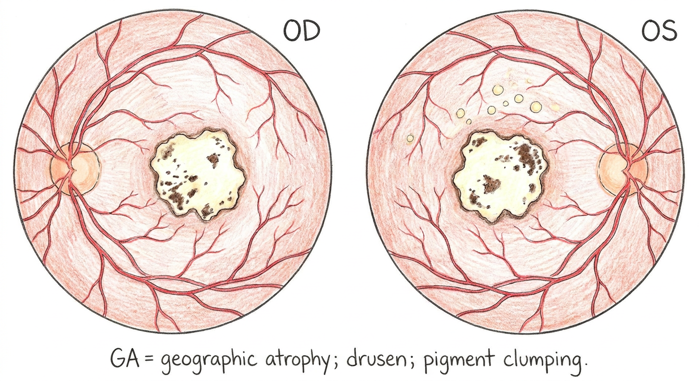
A clean hand-drawn ophthalmology fundus diagram based on bilateral color fundus photographs with geographic atrophy. Show two circular fundus views side by side on a white background. In each eye, draw the optic disc nasal to the macula, retinal vessels, and a central sharply demarcated irregular geographic atrophy lesion at the macula, colored pale yellow-white with visible dark pigment clumping and a thin surrounding hyperpigmented rim. The left fundus additionally has a few small drusen superior to the macula. Label each eye clearly: OD and OS. Add a concise key below: GA = geographic atrophy; drusen; pigment clumping. Do not include arrows, asterisks, or photographic labels.
Fundus diagram: Bilateral geographic atrophy

Make fundus diagram
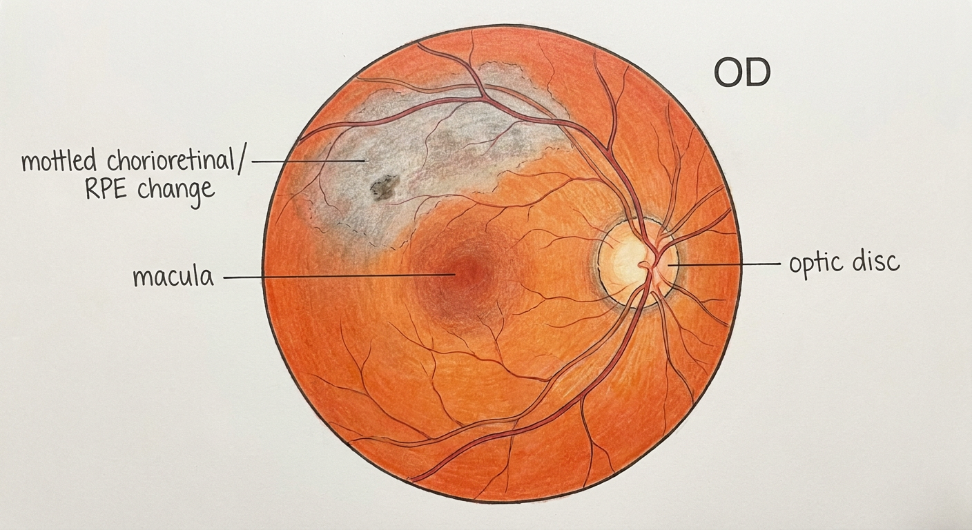
A clean ophthalmology hand-drawn fundus diagram of a right eye based on the provided clinical image. Draw one circular orange-red fundus on a white background. Place the optic disc nasal on the right side, with normal branching retinal vessels. Mark the macula temporal to the disc, slightly left of center. Show a broad subtle grayish mottled area of retinal pigment epithelial change or chorioretinal atrophy in the superotemporal posterior pole, extending from above the macula toward the superior arcade, with a small darker pigment clump within it. The inferior retina is otherwise unremarkable. Add labels: OD, optic disc, macula, and “mottled chorioretinal/RPE change.” Do not include a diagnosis, camera reflections, hands, lens rim, or photographic artifacts.
Fundus diagram: Right eye