
Write a short note on synovial joint. With diagram and clinical approached
labeled diagram of a typical synovial joint articular cartilage joint capsule synovial membrane synovial cavity ligaments
"synovial joint" AND anatomy
synovial joints anatomy structure clinical examination Merck Manual
A clear educational labeled cross-sectional diagram of a typical synovial joint. Include two articulating bone ends, smooth articular hyaline cartilage, a central synovial cavity containing synovial fluid, synovial membrane lining the capsule but not cartilage, outer fibrous capsule, periosteum, and a reinforcing ligament. Use accurate anatomy with concise labels suitable for undergraduate medical notes.
https://www.ncbi.nlm.nih.gov/sites/books/NBK507893?report=pr…
Synovial Joint
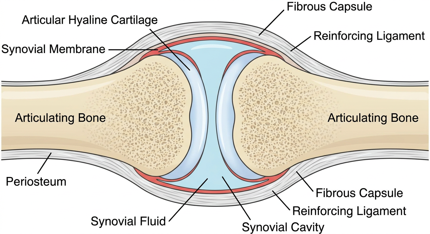
Essential components
-
Articular surfaces
The ends of articulating bones are usually covered by smooth hyaline cartilage. This reduces friction and spreads compressive load. Articular cartilage is avascular and has no perichondrium. -
Joint cavity
A potential space between the articular surfaces that contains a small amount of synovial fluid. -
Articular capsule
It encloses the joint cavity and has two layers:- Outer fibrous capsule: dense connective tissue that holds the bones together and stabilizes the joint.
- Inner synovial membrane: vascular lining that covers the non-articular internal surfaces of the capsule. It secretes synovial fluid.
-
Synovial fluid
A viscous lubricating fluid that reduces friction, nourishes the avascular articular cartilage, and assists shock absorption. -
Ligaments
Capsular, extracapsular, or intracapsular ligaments reinforce the joint and limit excessive movement. -
Accessory structures
Present in some joints: articular discs or menisci, labra, bursae, fat pads, and tendon sheaths. Discs and menisci improve congruity and distribute load.
Classification with examples
| Type | Main movement | Example |
|---|---|---|
| Plane | Gliding | Intercarpal joints |
| Hinge | Flexion and extension | Elbow, interphalangeal joints |
| Pivot | Rotation | Superior radioulnar joint |
| Condyloid/Ellipsoid | Flexion, extension, abduction, adduction | Wrist |
| Saddle | Biaxial movement | First carpometacarpal joint |
| Ball-and-socket | Multiaxial movement | Shoulder, hip |
| Bicondylar | Mainly flexion and extension with slight rotation | Knee |
Clinical approach to a painful synovial joint
- Onset: acute or chronic
- Trauma, fever, recent infection, rash, urethral symptoms
- Pain pattern: inflammatory pain often causes morning stiffness and improves with activity
- Number and distribution of joints involved
- Previous attacks, gout, psoriasis, autoimmune disease, or family history
- Look: swelling, erythema, deformity, scars, muscle wasting.
- Feel: warmth, tenderness, effusion, synovial thickening, crepitus.
- Move: assess active and passive range of motion.
- Function: gait, grip, ability to bear weight or use the limb.
- Examine the corresponding joint on the other side and assess nearby joints.
- Osteoarthritis: mechanical pain, brief morning stiffness, bony enlargement, crepitus.
- Inflammatory arthritis: warmth, tenderness, prolonged morning stiffness, synovitis.
- Crystal arthritis: sudden severe pain and swelling, commonly gout or calcium pyrophosphate disease.
- Septic arthritis: acute severe pain, hot swollen joint, restricted movement, fever may occur. This is an emergency.
- Trauma/internal derangement: ligament, meniscal, cartilage, or fracture-related injury.
- X-ray for fracture, joint-space narrowing, osteophytes, erosions, or alignment.
- Ultrasound for effusion, synovial thickening, active synovitis, and needle guidance. It may identify effusion and synovial thickening more sensitively than clinical examination. Rheumatology, 2-Volume Set, 2022, pp. 2492-2525.
- MRI for cartilage, menisci, ligaments, marrow lesions, and early inflammatory disease.
- Blood tests as indicated: CBC, ESR/CRP, uric acid, rheumatoid factor, anti-CCP.
- Arthrocentesis for a new unexplained effusion or suspected infection/crystal arthritis: send fluid for cell count, Gram stain, culture, and polarized microscopy for crystals. The Merck clinical guidance stresses prompt aspiration to exclude septic arthritis.