So I am final year PG resident in the department of pathology. I am going to have my session exams in about a month and then after one more month I will be having university exams. I want to prepare using my previous year question papers and would want to practice those questions. So I will be uploading questions and I believe being an expert you can provide beautiful answers to those questions which I would replicate in my MD exams. Is that doable ?
Absolutely, that's very doable! Here's what you can expect:
What I'll do for each question you upload:
- Provide a structured, exam-ready answer formatted the way MD/MS pathology examiners expect (introduction, classification, morphology, microscopy, clinical correlations, tables/diagrams where relevant)
- Draw from authoritative pathology textbooks (Robbins, Rosai & Ackerman, Sternberg, etc.) available in my medical library
- Supplement with recent evidence where relevant
- Tailor the depth and length to match what's appropriate for a university exam answer (typically 8-10 marks or 5-mark short answers)
A few things that will help me give you the best answers:
- Upload the question as text or image - either works
- Let me know the marks allotted if visible (helps calibrate answer length)
- Mention any specific focus your university tends to emphasize (e.g., morphology-heavy, lots of IHC/molecular, lab diagnosis, etc.)
- Let me know your university/exam board if you're comfortable - different universities (AIIMS, MCI-affiliated, state universities) sometimes have different answer expectations
What you'll get:
- Clean, point-format answers ready to write in an exam
- Key diagrams/flowcharts described where examiners expect them
- Important buzzwords and morphological descriptors that fetch marks
- Differential diagnoses and clinicopathological correlations where needed
Go ahead and upload your first question whenever you're ready - we'll get started right away!
Role of PDL1 and PD1 testing in lung carcinoma
Reading File
Finding Sources
Finding Sources
Searching PubMed
PD-L1 PD-1 testing lung cancer immunotherapy
Reading File
Reading File
Finding Sources
Reading File
Reading File
Finding Sources
Reading File
Reading File
Reading File
Finding Sources
Reading File
Reading File
Reading File
Loading Image
I now have all the material needed for a comprehensive, exam-ready answer. Here it is:
Role of PD-L1 and PD-1 Testing in Lung Carcinoma
I. Introduction
Lung carcinoma, particularly Non-Small Cell Lung Cancer (NSCLC), has undergone a therapeutic revolution with the introduction of immune checkpoint inhibitors (ICIs). The PD-1/PD-L1 axis is the most clinically actionable immune checkpoint pathway in lung cancer. Testing for PD-L1 expression by immunohistochemistry (IHC) on tumor tissue is now mandatory in the workup of all patients with newly diagnosed advanced NSCLC, as it directly guides first-line treatment decisions.
II. Biology of the PD-1/PD-L1 Axis
PD-1 (Programmed Death-1)
- A co-inhibitory receptor expressed on activated T cells, B cells, monocytes, and NK cells
- A member of the CD28 superfamily
PD-L1 (Programmed Death Ligand-1 / CD274)
- Expressed on tumor cells, macrophages, dendritic cells, and other stromal cells
- Under normal physiology: PD-L1 - PD-1 interaction prevents autoimmunity (peripheral tolerance)
- In cancer: Tumor cells hijack this pathway - overexpressing PD-L1 to ligate PD-1 on tumor-infiltrating T cells, leading to:
- T-cell exhaustion
- Inhibition of cytotoxic effector function
- Immune evasion
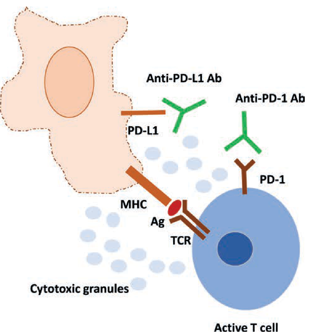
Blockade of PD-1 (by nivolumab/pembrolizumab) or PD-L1 (by atezolizumab/durvalumab) releases T-cell suppression and restores anti-tumor cytotoxic activity
III. Approved ICIs in Lung Cancer
| Drug | Target | Approved Indication |
|---|---|---|
| Pembrolizumab (Keytruda) | Anti-PD-1 | First-line NSCLC (PD-L1 TPS ≥1%) |
| Nivolumab (Opdivo) | Anti-PD-1 | Second-line NSCLC (all comers) |
| Atezolizumab (Tecentriq) | Anti-PD-L1 | First-line NSCLC; SCLC |
| Durvalumab (Imfinzi) | Anti-PD-L1 | Stage III NSCLC (adjuvant post-CRT) |
Note: Pembrolizumab and nivolumab target the receptor (PD-1) on T cells; atezolizumab and durvalumab target the ligand (PD-L1) on tumor cells.
IV. PD-L1 Testing - The Pathologist's Role
A. Why Test?
PD-L1 IHC is the companion diagnostic used to select patients most likely to benefit from ICI therapy. It is classified as a predictive biomarker (not merely prognostic).
B. When to Test?
- All patients with advanced/metastatic NSCLC - testing is mandatory at diagnosis
- Squamous cell carcinoma patients should also be tested (ICI benefit is histology-independent for PD-L1-high tumors)
- Testing before initiating first-line therapy is essential
C. Specimen Requirements
- Formalin-Fixed Paraffin-Embedded (FFPE) tissue
- Core biopsy preferred over FNA to ensure adequate tissue
- May be performed on primary tumor, lymph node, or metastatic site
D. FDA-Approved IHC Assays and Antibody Clones
| IHC Assay | Antibody Clone | Drug Paired With | Platform |
|---|---|---|---|
| PD-L1 IHC 22C3 pharmDx (Dako) | 22C3 | Pembrolizumab | Autostainer Link 48 |
| PD-L1 IHC 28-8 pharmDx (Dako) | 28-8 | Nivolumab | Autostainer Link 48 |
| Ventana PD-L1 (SP142) | SP142 | Atezolizumab | BenchMark |
| Ventana PD-L1 (SP263) | SP263 | Durvalumab | BenchMark |
Key exam point: Each drug uses a different clone on a potentially different platform - this creates challenges for interassay comparability.
V. Scoring Systems
1. Tumor Proportion Score (TPS)
- Used for NSCLC (pembrolizumab/22C3)
- Defined as: the percentage of viable tumor cells showing partial or complete membrane staining at any intensity
- Formula: TPS = (No. of PD-L1 positive tumor cells / Total viable tumor cells) × 100%
TPS Cut-offs and Clinical Implications:
| TPS | Category | Clinical Action |
|---|---|---|
| < 1% | Negative | Chemotherapy + ICI combination preferred |
| 1 - 49% | Low positive | ICI + chemotherapy combination recommended |
| ≥ 50% | High positive | Pembrolizumab monotherapy first-line (KEYNOTE-024) |
2. Combined Positive Score (CPS)
- Used for gastric, cervical, HNSCC, esophageal cancers (not standard for NSCLC)
- Formula: CPS = (PD-L1 staining cells [tumor cells + lymphocytes + macrophages] / Total viable tumor cells) × 100
3. Immune Cell (IC) Score
- Used with SP142 assay (atezolizumab)
- Measures PD-L1 expression on tumor-infiltrating immune cells (not tumor cells)
- IC ≥ 1% qualifies for atezolizumab in some settings
VI. Clinical Significance of TPS in NSCLC - Key Trials
| Trial | Drug | TPS Threshold | Key Outcome |
|---|---|---|---|
| KEYNOTE-024 | Pembrolizumab vs chemo | TPS ≥ 50% | OS: 30 months vs 14.2 months (HR 0.63) |
| KEYNOTE-042 | Pembrolizumab vs chemo | TPS ≥ 1% | Improved OS in all groups, greatest benefit at TPS ≥ 50% |
| IMPOWER 110 | Atezolizumab vs chemo | TC ≥ 50% or IC ≥ 10% | Improved OS |
| CheckMate 227 | Nivolumab + Ipilimumab | TPS ≥ 1% | OS: 17.1 vs 14.9 months vs chemo |
VII. Limitations of PD-L1 Testing
- Interassay variability - Different clones (22C3 vs 28-8 vs SP142) give discordant results; SP142 is known to stain less than the others
- Intratumoral heterogeneity - PD-L1 expression varies within a tumor and between primary and metastatic sites
- Dynamic expression - PD-L1 levels change over time and with prior therapy
- Imperfect predictor - PD-L1-negative patients can respond to ICI (especially with high Tumor Mutational Burden); PD-L1-high patients may not respond
- Driver-mutated tumors - Patients with EGFR mutations or ALK rearrangements have lower tumor mutational burden and derive less benefit from ICI despite high PD-L1; TKIs remain preferred even in PD-L1-high EGFR/ALK-positive NSCLC
VIII. Other Biomarkers Used Alongside PD-L1
| Biomarker | Method | Significance |
|---|---|---|
| Tumor Mutational Burden (TMB) | NGS | High TMB (≥10 mutations/Mb) predicts ICI benefit independently of PD-L1 |
| Microsatellite Instability (MSI-H) | IHC/PCR/NGS | Pan-tumor ICI predictor; less common in NSCLC |
| EGFR/ALK/ROS1 | NGS/FISH/IHC | TKI targets; ICI benefit minimal even if PD-L1 high |
| KRAS G12C | NGS | Sotorasib/adagrasib target; can co-exist with PD-L1 positivity |
IX. Treatment Algorithm (NSCLC - First Line, No Driver Mutation)
Advanced NSCLC - No EGFR/ALK/ROS1
         |
    PD-L1 TPS testing
         |
  ┌──────┴──────┐──────────────┐
TPS ≥ 50%   TPS 1-49%    TPS < 1%
   |              |             |
Pembrolizumab  Pembro +     Chemo +
monotherapy    Platinum     Pembro/Atezo
(KEYNOTE-024)  chemo        combination
X. Testing in Special Scenarios
- Small cell lung cancer (SCLC): PD-L1 testing is less established; atezolizumab + chemotherapy is approved for extensive-stage SCLC regardless of PD-L1 status (IMpower 133)
- Neoadjuvant setting: ICI use in early-stage NSCLC (Stage I-IIIA) is being actively studied; PD-L1 testing is relevant here too
- Liquid biopsy: ctDNA-based PD-L1 analysis is emerging but not yet standard
XI. Summary
PD-L1/PD-1 testing in lung carcinoma represents the interface between pathology and precision oncology. The pathologist's role is to accurately quantify PD-L1 expression using the correct IHC assay (clone 22C3 for pembrolizumab), apply the TPS scoring method correctly, and communicate results in a clinically actionable manner. While PD-L1 TPS ≥50% is the strongest predictor of benefit from ICI monotherapy, it must be interpreted alongside driver mutation testing (EGFR, ALK, ROS1) and emerging biomarkers like TMB to guide optimal therapy selection in NSCLC.
Sources: Harrison's Principles of Internal Medicine 22E (2025); Murray & Nadel's Textbook of Respiratory Medicine; Fishman's Pulmonary Diseases and Disorders; Tietz Textbook of Laboratory Medicine 7E
Molecular testing in lung cancer another question
Finding Sources
Finding Sources
Reading File
Finding Sources
Reading File
Reading File
Reading File
Reading File
Reading File
Finding Sources
Reading File
Loading Image
Excellent - I have comprehensive material from multiple textbooks plus the pie chart. Now I'll compose the full exam-ready answer.
Molecular Testing in Lung Cancer
I. Introduction
Lung cancer, particularly Non-Small Cell Lung Cancer (NSCLC), has been at the forefront of precision oncology. The discovery of actionable "driver mutations" - somatic mutations that drive neoplastic transformation and sustain tumor growth - has transformed management from a one-size-fits-all chemotherapy approach to a molecularly guided, personalized treatment strategy.
Molecular testing is now standard of care for all patients with advanced/metastatic NSCLC, and the pathologist plays a central role in tissue handling, assay selection, and result reporting.
II. Why Molecular Testing? - Rationale
- Driver mutations are present in ~50% of nonsquamous NSCLC - identifying them allows matched targeted therapy with dramatically superior outcomes
- TKIs (Tyrosine Kinase Inhibitors) targeting driver mutations yield response rates of 60-80% vs ~30% with chemotherapy
- Precludes unnecessary immunotherapy - EGFR/ALK-mutated tumors respond poorly to ICIs despite high PD-L1 expression (lower tumor mutational burden, fewer neoantigens)
- Resistance profiling - molecular testing at progression identifies acquired resistance mechanisms and guides second-line selection
III. Relative Frequency of Driver Mutations in NSCLC
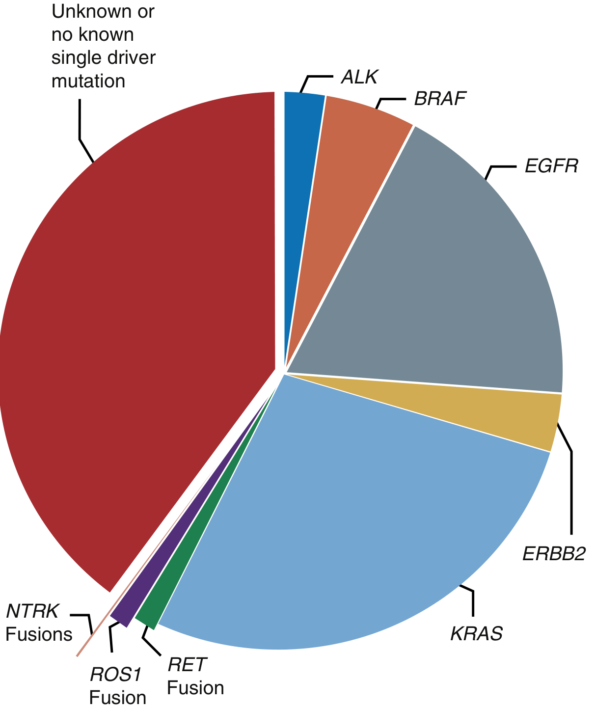
Distribution of targetable driver mutations in NSCLC (AACR GENIE Consortium). ~50% of nonsquamous NSCLC carry a known driver mutation. Frequency varies by geographic origin, tobacco exposure, and patient population.
| Driver | Frequency | Key Features |
|---|---|---|
| KRAS | ~32% adenocarcinoma | Most common; smokers; KRAS G12C targetable with sotorasib |
| EGFR | 10-15% (West), 25-30% (Asia) | Women, never/light smokers; exon 19 del + L858R most common |
| ALK | 3-7% | Young, never-smokers; EML4-ALK fusion |
| ERBB2 (HER2) | ~2-3% | Exon 20 insertions; adenocarcinoma |
| BRAF | ~2% | V600E most common; more in smokers |
| ROS1 | ~2% | Gene rearrangement; adenocarcinoma |
| RET | ~2% | Gene rearrangement; adenocarcinoma |
| MET | ~3-4% | Exon 14 skipping mutation; also MET amplification |
| NTRK 1/2/3 | <1% | Gene fusions; pan-tumor target |
IV. Specimen Requirements
- Tissue type: FFPE (Formalin-Fixed Paraffin-Embedded) core biopsy is preferred over FNA - provides adequate tumor cellularity for both histology and molecular studies
- Minimum tumor content: Generally ≥20-30% tumor nuclei required for reliable NGS
- Sites: Primary tumor, mediastinal lymph node biopsy (EBUS-guided), or metastatic site (bone, liver, adrenal, pleural effusion cell block)
- Repeat biopsy: Required at disease progression to identify acquired resistance mutations
V. Recommended Molecular Targets and Testing Methods
A. EGFR (Epidermal Growth Factor Receptor)
- Gene: Chromosome 7p11.2; receptor tyrosine kinase
- Alterations tested:
- Exon 19 deletions (most common, ~45%)
- Exon 21 L858R point mutation (~40%)
- Exon 20 insertions (resistance; treated differently)
- T790M (acquired resistance mutation; ~60% of 1st/2nd gen TKI failures)
- Less common: G719X (exon 18), S768I (exon 20), L861Q (exon 21)
- Testing method: PCR-based assays, Sanger sequencing, or preferably NGS
- Targeted drugs:
- 1st generation: Erlotinib, Gefitinib (reversible binders)
- 2nd generation: Afatinib, Dacomitinib (irreversible)
- 3rd generation (preferred 1st line): Osimertinib - targets L858R/exon 19 del AND T790M; superior CNS penetration (FLAURA trial: median PFS 18.9 vs 10.2 months)
B. ALK (Anaplastic Lymphoma Kinase)
- Alteration: EML4-ALK gene fusion (chromosomal inversion 2p); results in constitutively active kinase
- Clinical profile: Young, never-smokers, adenocarcinoma; predilection for CNS metastases
- Testing methods:
- FISH (Fluorescence In Situ Hybridization) - gold standard historically; uses break-apart probe
- IHC - D5F3 clone (Ventana) is highly sensitive and specific; used as screening tool
- NGS - preferred for comprehensive fusion detection
- Targeted drugs:
- 1st line: Alectinib (preferred; superior CNS penetration) or Brigatinib
- After resistance: Lorlatinib (3rd generation; broad resistance coverage)
- Others: Ceritinib, Crizotinib (1st generation; less preferred now)
C. ROS1
- Alteration: Gene rearrangement (multiple fusion partners); ~2% NSCLC
- Similar profile to ALK - young, never-smokers, adenocarcinoma
- Testing: FISH (break-apart probe), IHC (D4D6 clone), NGS
- Drugs: Crizotinib, Entrectinib, Ceritinib; Lorlatinib for resistance
D. BRAF
- Alteration: V600E point mutation (exon 15); ~2% NSCLC adenocarcinoma
- More common in smokers (unlike EGFR/ALK)
- Testing: PCR, allele-specific assays, NGS
- Drug: Dabrafenib + Trametinib combination (BRAF inhibitor + MEK inhibitor; required combination due to MAPK pathway feedback)
E. RET
- Alteration: Gene rearrangement (~2%); multiple fusion partners (KIF5B most common)
- Testing: FISH, IHC (not reliable), NGS (preferred)
- Drugs: Selpercatinib, Pralsetinib (highly selective; superior to older multikinase inhibitors)
F. MET
- Alterations:
- Exon 14 skipping mutation (~3-4%): Splicing alteration; removes regulatory juxtamembrane domain; targetable
- MET amplification: Primary (de novo) or secondary (acquired resistance to EGFR TKI)
- Testing: RNA-based NGS preferred for exon 14 skipping; FISH for amplification
- Drugs: Capmatinib, Tepotinib (for exon 14 skipping); Crizotinib (historical)
G. KRAS
- Alteration: Point mutations (~32% adenocarcinoma); G12C, G12V, G12D most common
- KRAS G12C (~13% of NSCLC) - now targetable with Sotorasib, Adagrasib
- Testing: NGS; allele-specific PCR
- Important: Other KRAS variants (G12D, G12V) are not yet reliably targetable
H. NTRK (1/2/3)
- Alteration: Gene fusions; <1% NSCLC but pan-tumor target (agnostic approval)
- Testing: IHC (screening), FISH, RNA-based NGS (confirmatory)
- Drugs: Larotrectinib, Entrectinib - tumor-agnostic approvals
I. HER2 (ERBB2)
- Alteration: Exon 20 insertions (~2-3%)
- Testing: NGS (preferred); IHC and FISH can assess amplification
- Drugs: Trastuzumab deruxtecan (T-DXd) - antibody drug conjugate
VI. Testing Platform Comparison
| Method | What it Detects | Advantages | Limitations |
|---|---|---|---|
| PCR / RT-PCR | Specific mutations/fusions | Rapid, sensitive, cheap | Limited to known variants |
| FISH | Fusions, amplifications | Gold standard for ALK/ROS1 | Labor intensive, 1 gene at a time |
| IHC | Protein overexpression | Rapid, cheap, widely available | Surrogate only; must confirm by molecular |
| Sanger Sequencing | Known mutations | Simple | Low sensitivity (~20% allele freq) |
| Next-Generation Sequencing (NGS) | Mutations, fusions, CNVs, TMB, MSI | Comprehensive; single test; multiple genes | Cost, turnaround time, bioinformatics |
Key exam point: NGS (panel-based) is now the preferred and most cost-effective approach as it detects all targets simultaneously in a single tissue sample - critical when tissue is limited.
VII. Next-Generation Sequencing (NGS) - Details
Definition: Massively parallel sequencing technology that sequences hundreds of genes simultaneously
Types of NGS used in NSCLC:
- DNA-based panel: Detects SNVs (single nucleotide variants), indels, CNVs, MSI, TMB
- RNA-based panel: Required for fusion/rearrangement detection (ALK, ROS1, RET, NTRK) - DNA-based NGS may miss some fusions
- Comprehensive genomic profiling (CGP): e.g., FoundationOne CDx (FDA-approved companion diagnostic; detects alterations in 324 genes + TMB + MSI)
What NGS reports additionally:
- Tumor Mutational Burden (TMB): Measured in mutations/megabase; high TMB (≥10 mut/Mb) predicts ICI response independently of PD-L1
- Microsatellite Instability (MSI-H): Predicts pembrolizumab response (pan-tumor approval)
- Co-mutations: e.g., TP53 co-mutation with EGFR worsens prognosis
VIII. Liquid Biopsy
Definition: Analysis of circulating tumor DNA (ctDNA) from peripheral blood
Principle: Tumor cells shed DNA into circulation; ctDNA carries tumor-specific somatic mutations detectable by sensitive NGS or PCR (ddPCR)
Uses in lung cancer:
| Scenario | Role |
|---|---|
| Tissue unavailable / insufficient | Primary molecular testing |
| High-risk for invasive biopsy | Alternative to tissue |
| Monitoring treatment response | Serial ctDNA decline = response |
| Detecting acquired resistance | e.g., T790M at EGFR TKI progression |
| Minimal residual disease (MRD) | Early detection of relapse |
Limitations:
- Sensitivity is lower than tissue biopsy (~60-70% sensitivity for known mutations)
- Specificity is high
- A negative liquid biopsy does NOT exclude a mutation - tissue biopsy must follow
- Preferred technique: Cobas EGFR Mutation Test v2 (FDA approved for plasma); ddPCR; NGS-based ctDNA panels
IX. Clinical Testing Algorithm (Pathologist Perspective)
Newly diagnosed advanced NSCLC (adenocarcinoma / adenosquamous / NSCLC NOS)
                    |
         Adequate tissue biopsy (FFPE core)
                    |
         SIMULTANEOUS REFLEX TESTING
              /              \
     NGS Panel             PD-L1 IHC (22C3)
  (DNA + RNA based)         TPS scoring
        |
 Reports: EGFR, ALK, ROS1, BRAF,
 RET, MET, NTRK, HER2, KRAS,
 TMB, MSI
        |
  If tissue insufficient → Liquid biopsy (plasma ctDNA)
  If liquid biopsy negative → Repeat tissue biopsy
X. Important CAP/IASLC/AMP Guideline Points
- Reflexive testing should be performed at diagnosis for all advanced NSCLC without waiting for oncology request
- Turnaround time: ≤10 working days recommended
- ALK IHC with D5F3 clone has sufficient sensitivity/specificity to guide alectinib therapy in resource-limited settings
- RNA-based fusion testing is mandatory - DNA-based NGS alone may miss up to 20-30% of ALK/ROS1 fusions
- Squamous cell carcinoma: Molecular testing recommended if never/light smoker, small biopsy (cannot exclude adenocarcinoma component), or clinician request
- At disease progression on TKI, re-biopsy (tissue or liquid) is mandatory to identify resistance mechanisms
XI. Resistance Mechanisms and Retesting
| Drug | Common Acquired Resistance | Next Step |
|---|---|---|
| 1st/2nd gen EGFR TKI | T790M mutation (~60%) | Osimertinib |
| Osimertinib | C797S mutation, MET amplification, SCLC transformation | Lorlatinib; MET inhibitor; re-biopsy |
| ALK TKI (alectinib) | ALK kinase domain mutations, MET bypass | Lorlatinib |
| Any TKI | SCLC transformation (~5%) | Requires tissue biopsy (morphology + IHC: synaptophysin, chromogranin) |
XII. Summary for Exam
Molecular testing in lung cancer is the cornerstone of precision oncology. The pathologist must ensure adequate tissue procurement, select the appropriate testing platform (NGS preferred), and report results with turnaround time ≤10 days. Key actionable alterations include EGFR mutations (TPS: osimertinib), ALK/ROS1 fusions (alectinib/crizotinib), BRAF V600E (dabrafenib+trametinib), RET fusions (selpercatinib), MET exon 14 skipping (capmatinib), KRAS G12C (sotorasib), and NTRK fusions (larotrectinib). Liquid biopsy using ctDNA is a valuable adjunct, particularly when tissue is limited, but cannot replace tissue biopsy due to lower sensitivity. All molecular testing should be performed alongside PD-L1 IHC to complete the biomarker profile needed for first-line treatment decisions.
Sources: Fishman's Pulmonary Diseases and Disorders; Murray & Nadel's Textbook of Respiratory Medicine; Harrison's Principles of Internal Medicine 22E (2025); Tietz Textbook of Laboratory Medicine 7E; Goldman-Cecil Medicine
Can you make it concise it’s tough for me to remember
Absolutely. Here's the concise, high-yield version - structured for quick recall and exam writing.
Molecular Testing in Lung Cancer
1. Why Test?
- ~50% of nonsquamous NSCLC carry actionable driver mutations
- Matched targeted therapy → response rates 60-80% vs ~30% chemo
- Guides against immunotherapy in EGFR/ALK+ tumors (they respond poorly to ICIs)
2. Who to Test?
- All patients with advanced/metastatic adenocarcinoma, adenosquamous carcinoma, NSCLC-NOS
- Squamous: test if never/light smoker or small biopsy
3. Specimen
- FFPE core biopsy preferred (FNA often insufficient for NGS)
- Re-test at disease progression (resistance mechanism profiling)
4. Key Targets - One Line Each
| Target | Frequency | Clue | Drug |
|---|---|---|---|
| KRAS G12C | ~13% | Smoker | Sotorasib, Adagrasib |
| EGFR exon 19 del / L858R | 10-15% (West), 25-30% (Asia) | Women, never-smoker | Osimertinib (3rd gen, preferred) |
| ALK fusion (EML4-ALK) | 3-7% | Young, never-smoker | Alectinib (1st line) |
| ROS1 fusion | ~2% | Young, never-smoker | Crizotinib, Entrectinib |
| BRAF V600E | ~2% | Smoker | Dabrafenib + Trametinib |
| RET fusion | ~2% | Adenocarcinoma | Selpercatinib, Pralsetinib |
| MET exon 14 skip | ~3-4% | Older, smoker | Capmatinib, Tepotinib |
| HER2 exon 20 ins | ~2-3% | Adenocarcinoma | Trastuzumab deruxtecan (T-DXd) |
| NTRK fusion | <1% | Pan-tumor | Larotrectinib, Entrectinib |
5. Testing Methods - Simplified
| Method | Best for | Limitation |
|---|---|---|
| PCR | EGFR (rapid) | Known variants only |
| FISH | ALK, ROS1 fusions | One gene at a time |
| IHC | ALK screening (D5F3), ROS1 (D4D6) | Surrogate only - confirm molecularly |
| NGS (preferred) | All targets at once | Needs adequate tissue |
Bottom line: Use NGS (DNA + RNA panel) to detect everything in one shot. RNA-based component is essential - DNA-based NGS alone misses fusions.
6. NGS Also Reports
- TMB (Tumor Mutational Burden) ≥10 mut/Mb → predicts ICI response
- MSI-H → pembrolizumab eligible (pan-tumor)
7. Liquid Biopsy (ctDNA)
- Use when: Tissue insufficient or biopsy risky
- Best for: EGFR T790M detection at progression
- Limitation: Lower sensitivity (~60-70%) → negative result does NOT rule out mutation → follow with tissue biopsy
8. Key Resistance Mutations to Know
| TKI | Resistance Mechanism |
|---|---|
| 1st/2nd gen EGFR TKI | T790M (~60%) → switch to Osimertinib |
| Osimertinib | C797S, MET amplification, SCLC transformation |
| ALK TKI | ALK kinase domain mutations → Lorlatinib |
9. Mnemonic for Targets
"Every ABLE KID Makes New Hits"
- EGFR, ALK, BRAF, Liquid biopsy (ctDNA), KRAS, Immunotherapy (TMB/MSI), Drivers, MET, NTRK, HER2 (+ RET + ROS1)
10. Exam One-liner Summary
Molecular testing (NGS preferred) is mandatory in all advanced nonsquamous NSCLC to identify actionable driver mutations (EGFR, ALK, ROS1, BRAF, RET, MET, NTRK, HER2, KRAS G12C), guide matched targeted therapy, and determine ICI eligibility alongside PD-L1 testing. Liquid biopsy (ctDNA) is a useful adjunct but cannot replace tissue biopsy.
This version covers everything an examiner expects in about 1-1.5 pages of writing. Let me know if you want any target explained in more depth.
I don’t think it’s enough for 10 marks question
Fair point. Here's the sweet spot - detailed enough for 10 marks, but structured so it's easy to memorize and reproduce quickly.
Molecular Testing in Lung Cancer (10 Marks)
I. Introduction (~1 mark)
Lung carcinoma, especially Non-Small Cell Lung Cancer (NSCLC), has been revolutionized by the concept of "driver mutations" - somatic mutations in oncogenes that drive tumor growth and are amenable to targeted therapy. Molecular testing identifies these mutations, allowing personalized, precision-based treatment with superior outcomes compared to conventional chemotherapy.
- ~50% of nonsquamous NSCLC carry an actionable driver mutation
- Mandatory for all advanced/metastatic NSCLC at diagnosis (CAP/IASLC/AMP guidelines)
II. Specimen Requirements (~1 mark)
| Parameter | Requirement |
|---|---|
| Preferred specimen | FFPE core biopsy (adequate cellularity) |
| Minimum tumor content | ≥20-30% tumor nuclei |
| Sites | Primary tumor / lymph node (EBUS) / metastatic site |
| Alternative | Liquid biopsy (ctDNA) if tissue insufficient |
| At progression | Re-biopsy mandatory (resistance profiling) |
FNA alone is usually insufficient for comprehensive molecular testing.
III. Testing Methods (~1 mark)
| Method | Detects | Key Use |
|---|---|---|
| PCR / RT-PCR | Specific known mutations | Rapid EGFR testing |
| FISH | Gene fusions, amplifications | ALK, ROS1 fusions (gold standard historically) |
| IHC | Protein overexpression | ALK screening (D5F3 clone); surrogate only |
| Sanger Sequencing | Point mutations | Low sensitivity (~20%); largely replaced |
| NGS (preferred) | Mutations, fusions, CNV, TMB, MSI simultaneously | Comprehensive; single test; tissue-sparing |
NGS is the preferred platform - DNA panel detects mutations; RNA panel is essential for fusion/rearrangement detection (ALK, ROS1, RET, NTRK). DNA-based NGS alone misses up to 30% of fusions.
IV. Key Molecular Targets and Targeted Therapies (~4 marks)
A. EGFR (Most important target)
- Frequency: 10-15% Western; 25-30% Asian; women, never/light smokers
- Sensitizing mutations: Exon 19 deletion (~45%), L858R point mutation (~40%), others (G719X, S768I, L861Q)
- Resistance mutation: T790M (exon 20) - acquired in ~60% after 1st/2nd gen TKIs
- Exon 20 insertion: Resistant to standard TKIs; treated differently
- Drugs:
| Generation | Drugs | Mechanism |
|---|---|---|
| 1st | Erlotinib, Gefitinib | Reversible binding |
| 2nd | Afatinib, Dacomitinib | Irreversible binding |
| 3rd (preferred 1st-line) | Osimertinib | Irreversible; targets T790M + sensitizing mutations; superior CNS penetration |
FLAURA trial: Osimertinib → median PFS 18.9 vs 10.2 months (1st gen TKI)
B. ALK Fusion
- Frequency: 3-7%; young, never-smokers, adenocarcinoma; EML4-ALK most common
- Predilection for CNS metastases
- Testing: FISH (break-apart probe), IHC (D5F3 clone - highly sensitive), NGS
- Drugs: Alectinib / Brigatinib (1st line preferred) → Lorlatinib (resistance)
C. ROS1 Fusion
- Frequency: ~2%; similar clinical profile to ALK (young, never-smoker, adenocarcinoma)
- Testing: FISH, IHC (D4D6 clone), NGS
- Drugs: Crizotinib, Entrectinib; Lorlatinib for resistance
D. Other Actionable Targets (summarize in table)
| Target | Frequency | Key Feature | Drug(s) |
|---|---|---|---|
| KRAS G12C | ~13% | Smoker, adenocarcinoma | Sotorasib, Adagrasib |
| BRAF V600E | ~2% | Smoker | Dabrafenib + Trametinib |
| RET fusion | ~2% | Adenocarcinoma | Selpercatinib, Pralsetinib |
| MET exon 14 skip | ~3-4% | Older patient, smoker | Capmatinib, Tepotinib |
| HER2 (ERBB2) | ~2-3% | Exon 20 insertion | Trastuzumab deruxtecan (T-DXd) |
| NTRK 1/2/3 fusion | <1% | Pan-tumor target | Larotrectinib, Entrectinib |
V. Additional Biomarkers from NGS (~1 mark)
Tumor Mutational Burden (TMB)
- Measured as mutations/megabase (mut/Mb)
- High TMB (≥10 mut/Mb) predicts benefit from immune checkpoint inhibitors independent of PD-L1
- Reported by comprehensive NGS panels (e.g., FoundationOne CDx)
Microsatellite Instability (MSI-H)
- Less common in NSCLC but pan-tumor predictor of pembrolizumab response
- Detected by IHC (MMR proteins) or PCR/NGS
VI. Liquid Biopsy (~1 mark)
- Sample: Peripheral blood → plasma → ctDNA (circulating tumor DNA)
- Method: ddPCR or NGS-based assays
Uses:
- When tissue is insufficient or biopsy too risky
- Detecting T790M acquired resistance at EGFR TKI progression
- Monitoring treatment response (serial ctDNA)
- Minimal residual disease (MRD) detection
Limitations:
- Sensitivity ~60-70% (lower than tissue)
- Negative result does NOT exclude a mutation → must confirm with tissue biopsy
- Sensitivity varies with tumor burden and shedding
VII. Resistance Mechanisms (~1 mark)
| TKI | Acquired Resistance | Next Line |
|---|---|---|
| 1st/2nd gen EGFR TKI | T790M mutation (~60%) | Osimertinib |
| Osimertinib | C797S, MET amplification, SCLC transformation | Lorlatinib; MET inhibitor; rebiopsy |
| ALK TKI (alectinib) | ALK domain mutations | Lorlatinib |
| Any TKI | SCLC transformation (~5%) | Rebiopsy essential (morphology + IHC) |
At progression: Tissue re-biopsy OR liquid biopsy must be performed to identify resistance mechanism and guide next-line therapy.
VIII. Summary / Closing Statement
Molecular testing is the cornerstone of NSCLC management. NGS (DNA + RNA based) is the preferred platform as it simultaneously identifies all actionable mutations (EGFR, ALK, ROS1, BRAF, RET, MET, NTRK, HER2, KRAS G12C), reports TMB and MSI, and spares limited tissue. Testing must be performed at diagnosis AND at progression. The pathologist's role is central - from ensuring adequate tissue procurement to selecting the correct assay, interpreting results, and integrating molecular findings with PD-L1 IHC for a complete biomarker report that directly guides therapy.
How to write this in 10 marks: Introduction (3-4 lines) → Specimen (table) → Methods (table) → EGFR in detail → ALK/ROS1 → Other targets (summary table) → TMB/MSI → Liquid biopsy → Resistance → Closing line. That gives you a well-structured, mark-fetching answer covering breadth + depth.
This is a shared conversation. Sign in to Orris to start your own chat.