
95 years female with known case of stroke 12 years back on aspirin with COPD with hypothyrodism with hypertension came with complain of bilateral weekness of limb and headache for 1 day on examination power was 3/5 sensation intact bilateral planter down going how to manage this case
Reading File
Finding Sources
Finding Sources
Finding Sources
Searching PubMed
recurrent stroke elderly management aspirin
Finding Sources
Finding Sources
Finding Sources
Reading File
Reading File
Reading File
Reading File
Finding Sources
Finding Sources
Reading File
Reading File
I now have sufficient information to provide a comprehensive clinical response. Let me compile the full management plan for this complex geriatric case.
Clinical Assessment and Management
Rapid Differential Diagnosis - This is NOT a Simple Recurrent Stroke
This 95-year-old female presents with a highly atypical pattern that demands careful analysis before assuming "another stroke." The combination of:
- Bilateral limb weakness (not unilateral)
- Headache for 1 day
- Known aspirin use (antiplatelet therapy)
- Age 95 with prior stroke + cerebral atrophy
- Plantar responses DOWN-GOING (indicating UMN pattern, but bilateral)
...raises a critical differential that must be prioritized before assuming ischemic stroke.
Step 1: Immediate Stabilization (Emergency)
ABC + Vitals
- Secure airway, administer O2 (target SpO2 92-96% in COPD - avoid over-oxygenation)
- IV access, cardiac monitoring, pulse oximetry
- Check blood glucose immediately (hypoglycemia mimics stroke)
- BP measurement (both arms)
- 12-lead ECG (AF? new arrhythmia?)
- Temperature (infection?)
Urgent Bloods:
- CBC (thrombocytopenia on aspirin?)
- Coagulation screen (PT/INR, APTT)
- BMP/electrolytes (sodium, glucose, calcium, renal function)
- TSH (hypothyroid crisis causing neurological symptoms?)
- ABG if COPD concern
- Troponin
Step 2: Urgent Imaging - CRITICAL PRIORITY
Non-Contrast CT Head - STAT
This is the most important immediate investigation. Key differentials to exclude:
TOP DIFFERENTIAL: Chronic Subdural Hematoma (CSDH)
This patient is at very high risk for CSDH based on:
- Age 95 - significant cerebral atrophy (bridging veins stretched and vulnerable)
- Long-term aspirin use - impairs hemostasis, promotes venous ooze
- Bilateral involvement (CSDH is bilateral in ~20% of patients)
- Headache + bilateral limb weakness is a classic CSDH presentation
- No mention of trauma needed - minor or no trauma can cause CSDH in the elderly
As per Plum and Posner's Diagnosis and Treatment of Stupor and Coma: "Chronic subdural hematomas usually occur in elderly patients or those on anticoagulants... A history of trauma can be elicited in only about one-half of patients, and then the trauma is usually minor." The bilateral pattern occurs in ~20% of cases.
The same text notes: "A useful rule when faced with a comatose or neurologically impaired patient is that 'it could always be a subdural,' and hence imaging is needed even in cases where focal signs are absent."
Other Differentials on CT Head:
| Diagnosis | Key Clues |
|---|---|
| Chronic subdural hematoma (bilateral) | Age, aspirin, cerebral atrophy, bilateral signs |
| Intracerebral hemorrhage | Sudden onset, HTN history |
| New ischemic stroke (bilateral = basilar or bilateral hemisphere) | Rule out by imaging |
| Subdural hygroma | Similar presentation to CSDH |
| Cerebral venous sinus thrombosis | Headache + bilateral signs, dehydration in elderly |
| Metabolic encephalopathy | Bilateral signs, no focal imaging changes |
| Hypertensive emergency (PRES) | HTN + bilateral weakness + headache |
If CT is non-diagnostic or CSDH suspected but CT equivocal:
- MRI brain with FLAIR/DWI - superior for CSDH dating, small infarcts, posterior fossa lesions, venous thrombosis
Step 3: Aspirin - HOLD Pending Imaging
Do NOT give additional antithrombotic loading until hemorrhage is excluded. The patient is already on aspirin - this drug may in fact be contributing to CSDH expansion. Hold aspirin until CT result is reviewed.
Do NOT give IV tPA until:
- Hemorrhage is excluded
- Exact diagnosis is established
- Time window is confirmed
- Age/benefit assessment completed (95 years - high bleeding risk)
Step 4: Diagnosis-Specific Management
If CSDH confirmed:
Neurosurgical referral immediately
Management options per textbook evidence (Plum & Posner):
- Burr hole drainage / craniotomy - for symptomatic CSDH causing neurological deficit (standard of care)
- Hold aspirin perioperatively
- Corticosteroids (dexamethasone) - some evidence supports this as an adjunct or alternative in patients unfit for surgery; recent meta-analyses show no superiority of surgery over steroids in selected low-risk cases
- Consider patient's frailty, comorbidities, and goals of care at age 95
If Acute Ischemic Stroke confirmed (no hemorrhage):
IV tPA (alteplase) consideration:
- Age 95 is not an absolute contraindication, but risk-benefit must be weighed carefully
- Window: within 4.5 hours of symptom onset
- NIHSS score assessment required
- Bilateral hemiplegia suggests basilar territory or bilateral hemisphere involvement - may need CT angiography to look for basilar artery occlusion
- Pre-existing aspirin use is noted; does not preclude thrombolysis
- The Washington Manual notes: "Thrombolytic therapy should not be delayed while waiting for PT/PTT/platelet unless bleeding abnormality suspected or patient has been taking warfarin, heparin, dabigatran, rivaroxaban, or apixaban" - since she is only on aspirin, this is less of a concern
BP management in acute ischemic stroke:
- Per Family Medicine Textbook: "During the first day after an ischemic stroke, most patients with elevated blood pressure should not be treated unless SBP is consistently >220 mmHg or DBP consistently >120 mmHg"
- Exception: if tPA is being administered, target BP <185/110 before and <180/105 after
Endovascular thrombectomy:
- Consider if large vessel occlusion on CTA (basilar, MCA, ICA)
- Window up to 24 hours in selected patients
- Core infarct volume ≤20 mL if age ≥80 years
If Intracerebral Hemorrhage:
- Stop aspirin immediately
- Neurosurgical consult
- BP target: SBP <140 mmHg
- Reversal of antiplatelet effect: consider platelet transfusion if surgical intervention planned
If Cerebral Venous Sinus Thrombosis:
- Anticoagulation (LMWH or IV heparin) even if hemorrhagic infarct present
- Hydration
Step 5: Comorbidity-Specific Management
COPD
- O2 target SpO2 88-92% (controlled oxygen therapy; avoid respiratory drive suppression)
- Continue bronchodilators
- Avoid over-sedation
Hypothyroidism
- Check TSH - myxedema can mimic and worsen neurological outcomes
- Ensure thyroxine is continued
- Myxedema coma is rare but can present with altered mentation and bilateral weakness
Hypertension
- Context-dependent BP management (see above)
- Avoid aggressive lowering in acute ischemic stroke
Age 95 - Frailty Considerations
- Early discussion with family/surrogate regarding goals of care and resuscitation status
- Frailty assessment (Clinical Frailty Scale)
- Multi-disciplinary team: neurology, neurosurgery, geriatrics, physiotherapy
Step 6: Secondary Prevention Review
Once acute phase is stabilized:
Per Goldman-Cecil Medicine: antiplatelet therapy is a first-line agent for secondary stroke prevention. The Family Medicine Textbook notes: "Both extended-release dipyridamole plus aspirin (Aggrenox) and clopidogrel are reasonable first-line antiplatelet agents for secondary stroke prevention, shown to be more effective than aspirin alone."
- If ischemic stroke: consider switching from aspirin alone to aspirin + dipyridamole or clopidogrel monotherapy
- If AF discovered on ECG: anticoagulation (DOAC preferred over warfarin in elderly, subject to bleeding risk assessment; PMID 39921198 meta-analysis supports DOACs over aspirin for embolic stroke)
- If CSDH: antiplatelet therapy should be restarted cautiously post-drainage, typically after 4-6 weeks
Summary Checklist
| Priority | Action |
|---|---|
| 1 | Stabilize ABC, check glucose, ECG, vitals |
| 2 | STAT non-contrast CT head |
| 3 | Hold aspirin until hemorrhage excluded |
| 4 | Labs: CBC, coag, electrolytes, TSH, ABG |
| 5 | Controlled O2 for COPD (88-92%) |
| 6 | Call neurosurgery if CSDH/ICH found |
| 7 | Thrombolysis/thrombectomy if ischemic and eligible |
| 8 | Goals of care discussion given age 95 |
| 9 | Neurology + geriatrics consult |
| 10 | Physiotherapy, dysphagia screen, DVT prophylaxis |
Key Teaching Point: In a 95-year-old on aspirin with bilateral headache and weakness, chronic bilateral subdural hematoma must be excluded first before assuming recurrent stroke. The clinical picture does not fit a classic unilateral hemispheric stroke, and this patient has multiple risk factors for CSDH. The CT head result is the pivot point for all further management decisions.
- Plum and Posner's Diagnosis and Treatment of Stupor and Coma, pp. 248-254
- Textbook of Family Medicine 9e, pp. 1220-1226
- The Washington Manual of Medical Therapeutics, pp. 1019-1022
This patient gcs was 15/15 ct was done no evidence of haemorrhage seen power on bilateral leg 3/5 sensation intact planter was bilateral down going her sodium is 122
Finding Sources
Finding Sources
Finding Sources
Reading File
Reading File
Reading File
Finding Sources
Reading File
Reading File
Loading Image
This is now very clear. Here is the complete, structured management plan:
Revised Diagnosis: Severe Symptomatic Hyponatremia (Na 122 mEq/L)
The clinical picture is now clarified. The bilateral leg weakness, headache, GCS 15 with no hemorrhage on CT - this is a hyponatremic encephalopathy/neuromyopathy picture, NOT a recurrent stroke. Sodium of 122 mEq/L is severe (normal 135-145) and is fully capable of producing bilateral limb weakness, headache, and altered neuromuscular function. The plantar responses being down-going are consistent with upper motor neuron dysfunction from cerebral swelling from hypo-osmolality.
Step 1: Identify the TYPE of Hyponatremia (Mandatory Before Treatment)
Use this diagnostic flowchart:
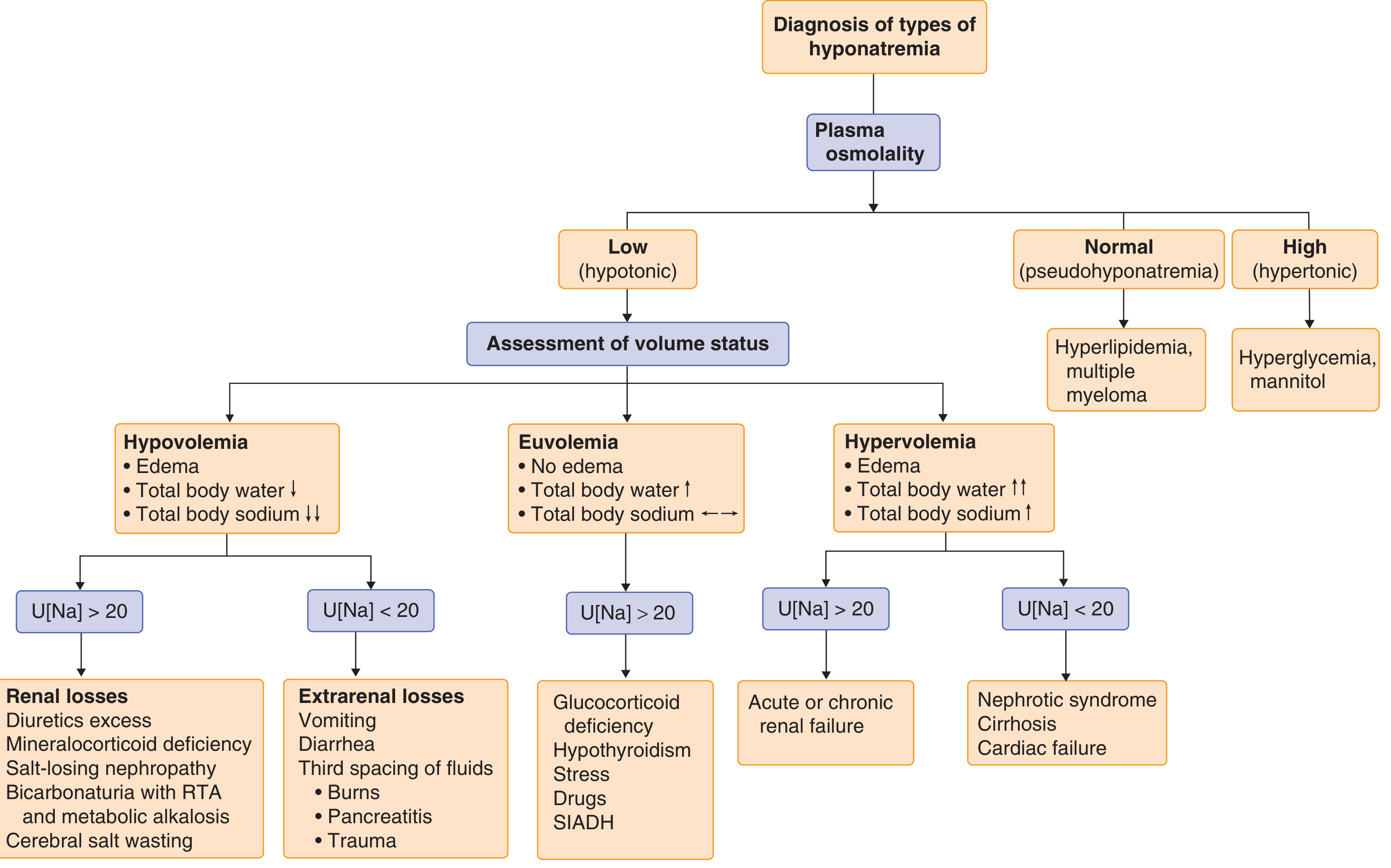
In this patient, the most likely causes given her comorbidities are:
| Cause | Why Likely |
|---|---|
| SIADH | Most common cause of euvolemic hyponatremia; can be triggered by COPD (pulmonary disease stimulates ADH), stroke history, age, drugs |
| Hypothyroidism | This patient HAS known hypothyroidism - a direct cause of hyponatremia that perfectly mimics SIADH. Check: is she compliant with thyroxine? What is her TSH? |
| Thiazide diuretics | If she is on hydrochlorothiazide for hypertension (very common) - this is a major cause |
| Cerebral salt wasting | Less likely with Na 122 (usually more profound volume depletion) |
Immediately check:
- Serum osmolality (should be low - hypotonic)
- Urine osmolality
- Urine sodium (U[Na])
- TSH (urgently - hypothyroidism in this patient is a treatable cause)
- Review all her medications (diuretics? SSRIs? PPIs? carbamazepine?)
- Assess volume status clinically (JVP, skin turgor, mucous membranes, BP, oedema)
- Cortisol (to exclude adrenal insufficiency)
Step 2: Determine Acuity - CRITICAL for Treatment Rate
Duration unknown = treat as CHRONIC (Goldman-Cecil: "When duration is unknown, assume chronic and treat with a longer correction time")
At age 95 with no prior sodium documented, this hyponatremia is likely chronic (>48 hours). Chronic hyponatremia carries very high risk of osmotic demyelination syndrome (ODS) if corrected too fast.
Step 3: Treatment - The Correction Rate Rule
The Golden Rule: Do NOT exceed 6-8 mEq/L in 24 hours in high-risk patients
This patient is at maximum risk for ODS because she is:
- Elderly (age 95)
- Female
- Likely chronic hyponatremia
- Malnourished/hypothyroid baseline
ODS (central pontine myelinolysis) is irreversible and can cause locked-in syndrome, quadriparesis, and death. The risk from over-correction exceeds the risk from the hyponatremia itself given her GCS is 15.
Treatment Strategy:
Is she symptomatic enough to need hypertonic saline?
With GCS 15, no seizures, no coma - she has moderate symptoms (weakness, headache). Per Tintinalli's Emergency Medicine:
- "Raising serum sodium by 5 mEq/L is typically all that is required to see an improvement in severe neurologic symptoms"
- Initial target: raise Na by 4-6 mEq/L over first few hours to stabilize
- Then slow down significantly
Practical Treatment Plan:
| Symptom Severity | Treatment |
|---|---|
| Severe (seizure, coma, respiratory arrest) | 3% NaCl 100 mL IV bolus, repeat x2 PRN |
| Moderate (this patient - weakness, headache, GCS 15) | 3% NaCl at 0.5-1 mL/kg/h with close monitoring |
| Mild (nausea, confusion only) | Fluid restriction ± treat underlying cause |
For this patient (moderate symptoms, likely chronic):
- 3% Hypertonic saline at 0.5 mL/kg/h (approximately 25-35 mL/h for 50-60 kg woman)
- Check serum Na every 2-4 hours
- Target: +4 to +6 mEq/L in first 6 hours to improve symptoms
- Strict ceiling: No more than 6-8 mEq/L in 24 hours total (given chronic, elderly, female = high ODS risk)
- Once symptoms improve - switch to treating underlying cause
Do NOT use:
- Normal saline (0.9%) as primary treatment if SIADH - can paradoxically worsen hyponatremia if urine osmolality > 308 mOsm/kg
- Glucose-containing fluids (hypotonic - will worsen sodium further)
Step 4: Treat the Underlying Cause
Hypothyroidism (this patient has it - check TSH NOW)
- If TSH is very elevated - this is the cause
- Give levothyroxine - IV if severely hypothyroid/myxedematous
- Sodium will correct as thyroid function normalises
- Per Goldman-Cecil: "Hypothyroidism can be associated with hypotonic hyponatremia that mimics SIADH"
If SIADH confirmed (euvolemic, urine Na >40, urine osmolality high):
- Fluid restriction to 500-800 mL/day (strict)
- Identify and treat precipitant (COPD exacerbation? infection? drug?)
- Consider urea or tolvaptan (vasopressin receptor antagonist) in refractory cases
- Per Frameworks for Internal Medicine: "Fluid restriction is key to treating chronic hyponatremia related to SIADH"
If thiazide-induced (hypovolemic):
- Stop thiazide immediately
- Normal saline (0.9% NaCl) for volume repletion
- Sodium will self-correct as volume is restored
If hypovolemic from any cause:
- Normal saline 0.9% cautiously
Step 5: Coexisting Issues to Manage Simultaneously
| Issue | Action |
|---|---|
| Hypertension | Caution - do not aggressively lower BP; may be reactive to hyponatremia |
| COPD | O2 target 88-92%; avoid hypoxia which worsens ADH release |
| Hypothyroidism | Urgent TSH; restart/adjust thyroxine |
| Aspirin (stroke secondary prevention) | Continue unless other contraindication; aspirin is NOT causing hyponatremia |
| DVT prophylaxis | Compression stockings; avoid LMWH until sodium stabilised and mobility assessed |
| Nutrition | Monitor - poor intake common cause of hyponatremia in elderly; dietitian review |
Step 6: Monitoring Protocol
- Serum Na every 2-4 hours while on hypertonic saline
- Urine output hourly
- Strict fluid balance
- Daily weight
- Repeat neurological exam every 4-6 hours
- Renal function, electrolytes (K+) daily
- ECG monitoring (hyponatremia can cause QT prolongation and arrhythmias)
Key Teaching Points for This Case
-
Bilateral weakness + headache + Na 122 = Hyponatremia is the diagnosis, not stroke. CT confirmed no hemorrhage, and the bilateral symmetrical pattern never fit a single vessel stroke territory.
-
Hypothyroidism + hyponatremia is a classic combination in elderly women - this is the first thing to exclude given her known history. Myxoedema can cause SIADH-like hyponatremia.
-
Never correct faster than 6-8 mEq/L per 24 hours in elderly chronic cases - osmotic demyelination is catastrophic and irreversible.
-
GCS 15 = no emergency bolus needed - controlled slow correction with 3% saline or treat the cause (hypothyroidism/fluid restriction) is the right approach.
-
The stroke history from 12 years ago and the chronic antiplatelet therapy are NOT the cause of today's presentation - they remain part of ongoing secondary prevention.
- Goldman-Cecil Medicine, p. 1202 (Diagnostic approach to hyponatremia)
- Tintinalli's Emergency Medicine, pp. 578-580 (Hyponatremia treatment)
- Frameworks for Internal Medicine, pp. 515-516 (Osmotic demyelination)
- Fischer's Mastery of Surgery, p. 214 (Hyponatremia types and correction principles)
This is a shared conversation. Sign in to Orris to start your own chat.