
Water soluble vitamins in detail seminar
water soluble vitamins B complex vitamin C deficiency signs
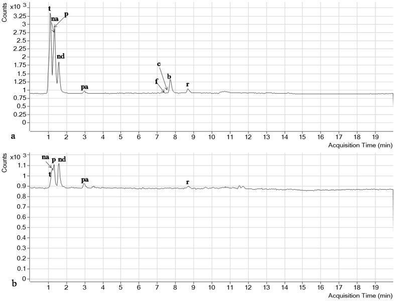
This Comparison Chart displays two RRLC-MS/MS Total Ion Current (TIC) chromatograms illustrating the separation and quantification of water-soluble vitamins. Panel (a) shows the chromatogram for vitamin standards, exhibiting high-intensity, well-defined peaks for thiamine (t), nicotinic acid (na), pyridoxine (p), nicotinamide (nd), pantothenic acid (pa), folic acid (f), cyanocobalamin (c), biotin (b), and riboflavin (r). Panel (b) depicts the chromatogram for a complex 'phytococktail' sample, where the same analytes are present but with significantly lower peak intensities and reduced resolution due to matrix effects. The x-axis represents 'Acquisition Time (min)' ranging from 0 to 19 minutes, and the y-axis represents 'Counts.' This diagnostic and analytical visual demonstrates the efficacy of mass spectrometry in identifying specific nutritional components—specifically B-vitamins—within a biological or botanical extract, serving as a reference for nutritional biochemistry and laboratory diagnostics.
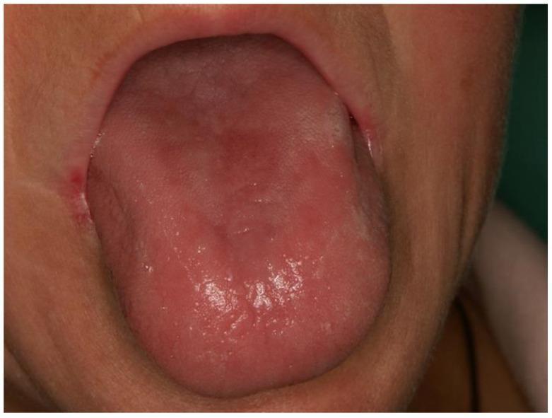
This clinical photograph shows intraoral manifestations of vitamin deficiency, specifically exfoliative glossitis and angular cheilitis associated with hypovitaminosis of B-group vitamins (B2 and B6). The tongue exhibits a diffuse, erythematous (reddish) appearance with a predominantly smooth, depapillated surface, characteristic of atrophic glossitis. Patchy areas of irregular texture and mucosal thinning are visible across the dorsal surface. At the left labial commissure (corner of the mouth), there is evident redness, inflammation, and superficial skin breakdown consistent with angular cheilitis. These findings are clinical markers for nutritional deficiencies, often presenting with burning sensations in the oral mucosa. The image serves as an educational example of the systemic relationship between water-soluble vitamin status and soft tissue oral health, highlighting diagnostic signs that may be encountered in pediatric or malnourished populations.
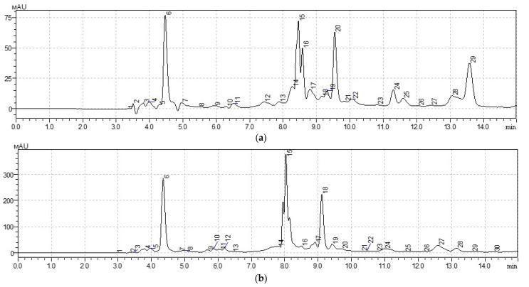
This diagnostic image consists of two high-performance liquid chromatography (HPLC) chromatograms, labeled (a) and (b), illustrating the qualitative and quantitative analysis of water-soluble vitamins in medicinal plant extracts. The x-axis represents retention time in minutes (0.0 to 14.0 min), and the y-axis indicates detector response in milli-absorbance units (mAU). Chromatogram (a) displays the chemical profile of Taraxacum officinale with peak heights ranging up to 75 mAU. Chromatogram (b) displays Arctium lappa with significantly higher absorbance peaks reaching approximately 350 mAU, suggesting higher concentrations of specific compounds. Both charts feature numbered peaks corresponding to specific bioactive compounds: peak 6 indicates Vitamin C (ascorbic acid), peaks 15 and 16 correspond to Vitamin B1 (thiamine), peak 20 corresponds to Vitamin B3 (niacin), and peak 24 corresponds to Vitamin B6 (pyridoxine). The visual data serves as an educational tool for pharmacognosy and nutritional chemistry, demonstrating the separation and identification of essential vitamins through HPLC peak morphology and retention time.
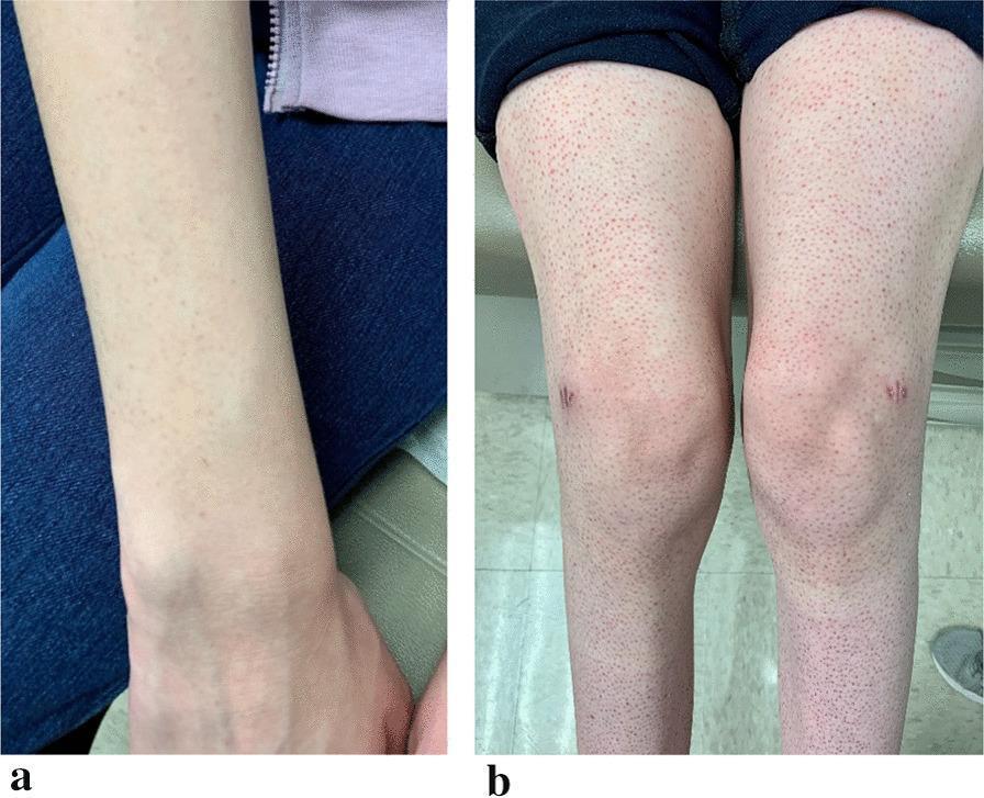
Clinical photograph consisting of two panels (a and b) demonstrating dermatologic and musculoskeletal signs of vitamin C deficiency (scurvy). Panel a shows the anterior forearm with a faint, erythematous perifollicular rash characterized by small, reddish macules centered around hair follicles. Panel b displays the bilateral lower extremities, exhibiting a more prominent and diffuse perifollicular petechial rash extending from the thighs to the lower legs. Significant bilateral knee swelling (effusions) is visible, along with small, horizontal, linear scars on the anterior knees consistent with healed biopsy incisions. The skin findings illustrate classic hemorrhagic manifestations of scurvy, where capillary fragility leads to perifollicular hemorrhage. This visual evidence is used in medical education to identify nutritional deficiencies in patients with restrictive eating habits, highlighting the triad of perifollicular petechiae, corkscrew hairs (as described in clinical context), and joint swelling due to hemarthrosis or effusions.
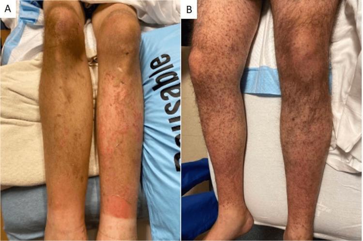
Clinical photograph of bilateral lower extremities from two different patients demonstrating dermatologic manifestations of Vitamin C deficiency (scurvy). Panel A shows extensive non-blanching perifollicular purpura and petechiae across the pretibial areas and calves. There is a large, confluent erythematous rash on the lower right calf, suggestive of ecchymosis or secondary inflammation. Panel B displays diffuse, folliculocentric hyperkeratotic papules and petechiae extending from the shins to the thighs. Characteristic 'corkscrew' hairs (coiled, malformed hair shafts) are visible within the hyperkeratotic follicles. These findings are classic pathognomonic signs of scurvy, resulting from impaired collagen synthesis and capillary fragility. The images serve as an educational resource for identifying nutritional deficiency-related skin disorders in vulnerable populations.
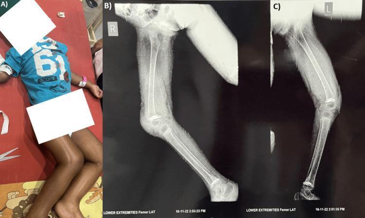
This composite educational material consists of a clinical photograph and lateral radiographs illustrating pediatric scurvy (Vitamin C deficiency). Panel A is a clinical photograph of a pediatric patient exhibiting signs of severe acute malnutrition (SAM), including muscle wasting and thinning of the extremities. Panels B and C are lateral X-ray images of the right and left lower limbs, respectively. The radiographs demonstrate pathognomonic radiological signs of scurvy at the knee joint. Key findings include the 'white line of Frankel,' which is a dense, sclerotic band at the metaphysis representing a zone of provisional calcification. Visible at the metaphyseal margins are 'corner signs' (Pelkan spurs), which are small, triangular bony outgrowths. The epiphyses of the distal femur and proximal tibia show a 'Wimberger ring sign,' characterized by a dense sclerotic periphery surrounding a radiolucent center. Additionally, generalized osteopenia and cortical thinning are observed in the long bones. These images serve as a classic diagnostic reference for the musculoskeletal manifestations of vitamin C deficiency in a pediatric population.
pellagra niacin deficiency dermatitis skin lesions
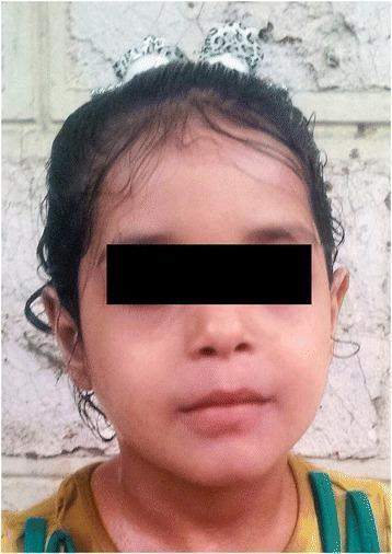
This clinical photograph shows the frontal view of a pediatric patient following treatment for niacin deficiency (pellagra). The image demonstrates a complete resolution of previously severe dermatological symptoms. The facial skin appears smooth and healthy, with an even tone and a total absence of the scaly, desquamating rash and hyperpigmentation characteristic of pellagra. The forehead and cheeks show no signs of inflammation or photosensitivity-induced lesions. The scalp and hairline appear normal with no evidence of alopecia or dermatitis. This visual serves as an educational comparison to show therapeutic success in managing Hartnup disease or secondary nutritional deficiencies through oral niacin supplementation. The patient's eyes are masked for privacy, while the remaining facial features highlight the restoration of normal skin integrity and texture in a clinical dermatology context.
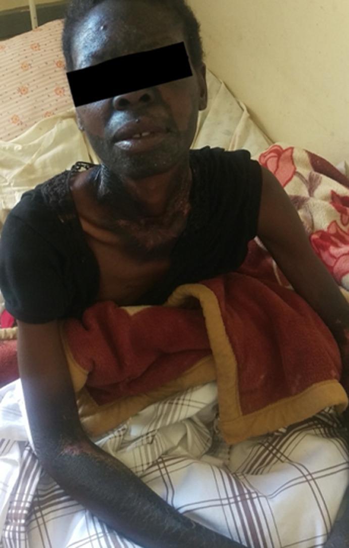
This clinical photograph displays classic dermatological manifestations of pellagra (niacin deficiency) in a patient with darker skin tones. The image depicts extensive, symmetric hyperpigmented and hyperkeratotic plaques distributed in sun-exposed areas. A prominent finding is 'Casal's necklace,' a well-demarcated band of hyperpigmented, thickened skin encircling the lower neck. Similar hyperpigmented lesions with visible scaling and fissuring are present on the face (forehead, nose, and malar regions) and the extensor surfaces of the forearms. The skin in these areas appears dry, rough, and desquamating, characteristic of photodermatitis secondary to nutritional deficiency. This visual record serves as a classic educational example of the '3 Ds' (dermatitis, diarrhea, dementia) presentation, specifically illustrating the characteristic cutaneous distribution patterns used to diagnose pellagra in a clinical setting.
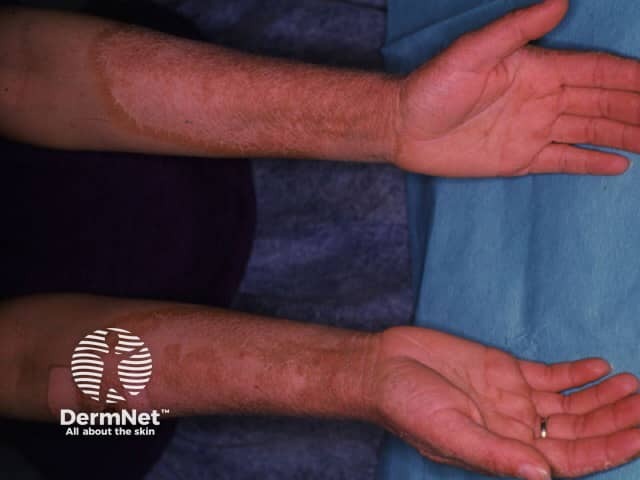
Clinical photography of both forearms demonstrating pellagra dermatitis due to niacin (nicotinamide) deficiency. Imaging modality: standardized dermatologic photography using noninvasive external illumination; views capture the ventral and dorsal surfaces of the bilateral forearms for symmetry assessment. The skin exhibits bilateral, sun-exposed distribution with erythematous, dry, scaly plaques and subtle hyperkeratosis. Lesions are arranged primarily on the extensor aspects of the forearms and dorsum of the hands, with well‑defined margins and occasional superficial desquamation. The texture is rough, with atrophic or macularly pigmented areas where scaling is more prominent; color ranges from erythema to brownish hyperpigmentation. No acute vesiculation is evident. The overall pattern is characteristic of photosensitive dermatitis in nutritional deficiency, supported by accompanying symptoms in typical clinical contexts. Clinically, pellagra dermatitis reflects niacin deficiency and can accompany diarrhea and cognitive changes if untreated. Differential diagnosis includes eczema/atopic dermatitis, contact dermatitis, photodermatoses, and arsenic-related dermatitis; clinical correlation with dietary history, gastrointestinal symptoms, and neurocognitive status is essential. This image serves educational purposes for dermatology and medical education, illustrating classic symmetry, solar-exposed distribution, and coarse keratosis that guide diagnosis and treatment planning with niacin supplementation and dietary modification. Correlation with systemic signs reinforces severity and guides rapid nutritional therapy.
Wernicke encephalopathy beriberi thiamine deficiency MRI brain
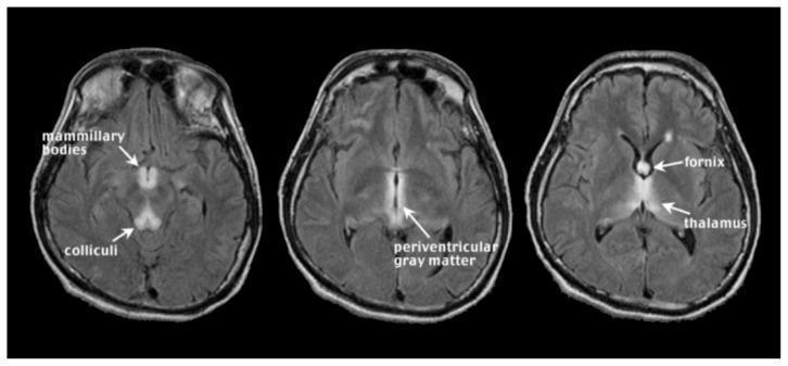
This diagnostic image consists of three axial MRI slices of the brain using Fluid Attenuated Inversion Recovery (FLAIR) sequences, illustrating the classic neuroimaging findings of acute Wernicke encephalopathy. The images demonstrate bilateral, symmetrical hyperintense (bright) signals in specific anatomical regions characteristic of thiamine deficiency. Key structures labeled include the mammillary bodies in the posterior hypothalamus and the colliculi in the midbrain. Higher slices show prominent hyperintensities within the periventricular gray matter surrounding the third ventricle, as well as involvement of the fornix and the medial aspects of the thalamus. These findings represent edema or glial changes associated with the acute clinical manifestation of the disease. The primary educational focus is to teach the radiological signature of Wernicke encephalopathy in the context of chronic alcoholism and nutritional deficiency, emphasizing the typical distribution of lesions in midline brain structures.
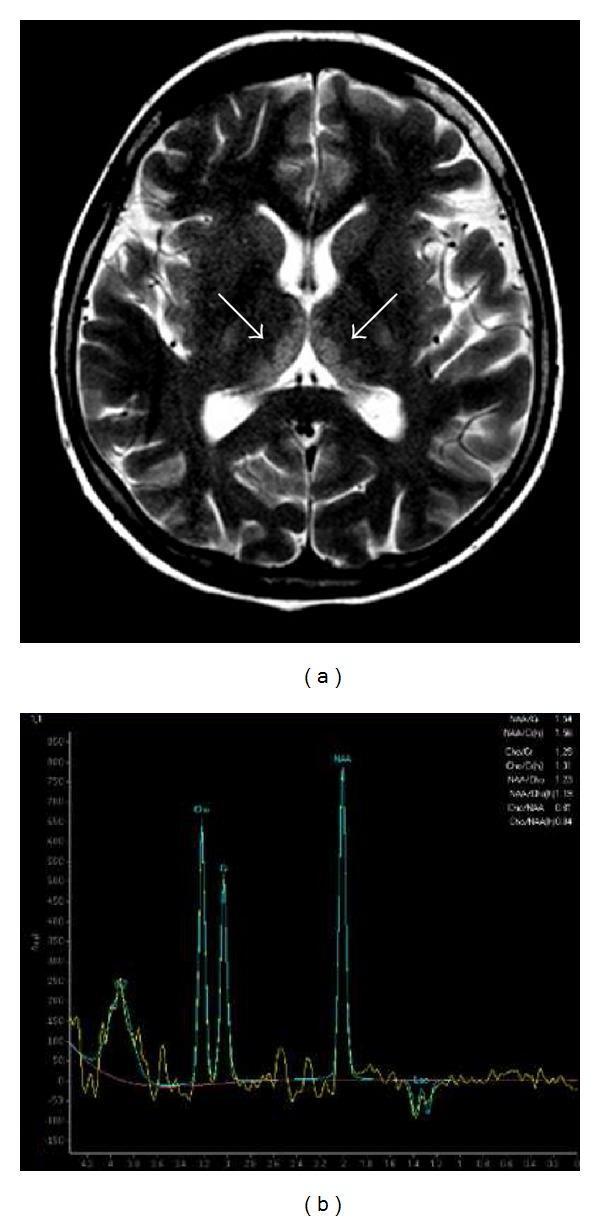
This composite diagnostic image features a T2-weighted axial MRI and an MR spectroscopy (MRS) plot, illustrating the classic neuroimaging findings of Wernicke’s encephalopathy in a patient with chronic alcohol abuse. Image (a) is an axial T2-weighted brain MRI demonstrating bilateral, symmetric hyperintense signal abnormalities within the medial thalami (indicated by arrows), representing vasogenic or cytotoxic edema typical of thiamine deficiency. Image (b) shows a single-voxel MR spectroscopy (MRS) profile acquired from the affected thalamic region. The spectrum exhibits biochemical markers of neuronal injury, specifically a reduced N-acetylaspartate (NAA) peak relative to Creatine (Cr) and Choline (Cho), indicating neuronal loss or dysfunction. Additionally, a notable lactate (Lac) peak, seen as an inverted doublet at 1.3 ppm, suggests a shift to anaerobic metabolism and localized lactic acidosis. This combination of structural MRI and metabolite quantification serves as a critical diagnostic tool for identifying metabolic encephalopathies and assessing the extent of reversible versus irreversible brain damage.
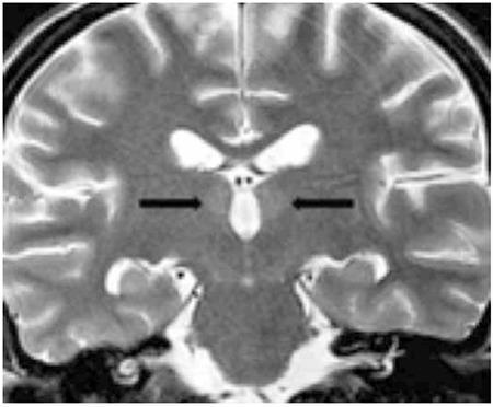
This diagnostic image is a coronal T2-weighted magnetic resonance (MRI) scan of the brain, demonstrating characteristic radiological findings associated with Wernicke encephalopathy. The image reveals symmetrical, weak, and limited increased signal intensity (hyperintensity) localized within the medial thalami, indicated by bilateral black arrows. Additional signal changes are noted along the surfaces facing the third ventricle. These findings are highly suggestive of thiamine deficiency-related metabolic derangement. The scan captures key anatomical structures including the lateral ventricles, the third ventricle, and the temporal lobes. This imaging is clinically significant for medical education in neurology and radiology, illustrating the classic distribution of lesions in acute Wernicke encephalopathy, which can occur in patients with nutritional deficiencies secondary to conditions like leukemia or long-term parenteral nutrition without vitamin supplementation.
megaloblastic anemia folate B12 deficiency peripheral blood smear hypersegmented neutrophil
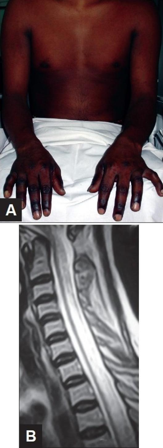
This composite figure demonstrates clinical and diagnostic findings associated with Vitamin B12 deficiency (Subacute Combined Degeneration of the spinal cord). Image A is a clinical photograph of an 18-year-old male showing significant cutaneous hyperpigmentation of the distal upper extremities. The darkening is most pronounced on the dorsal surfaces of the hands and fingers, contrasting with the lighter skin tone of the proximal arms and chest. This is a common dermatologic manifestation of megaloblastic anemia. Image B is a sagittal T2-weighted MRI of the cervical and upper thoracic spine. It reveals a long-segment, linear intramedullary hyperintense signal within the posterior aspect of the spinal cord. This finding represents edema and demyelination characteristic of Subacute Combined Degeneration, which typically affects the posterior and lateral columns while sparing the anterior column. The combination of these visual findings—distal hyperpigmentation and longitudinal posterior cord hyperintensity—is highly suggestive of severe Vitamin B12 deficiency in the context of progressive paraplegia.
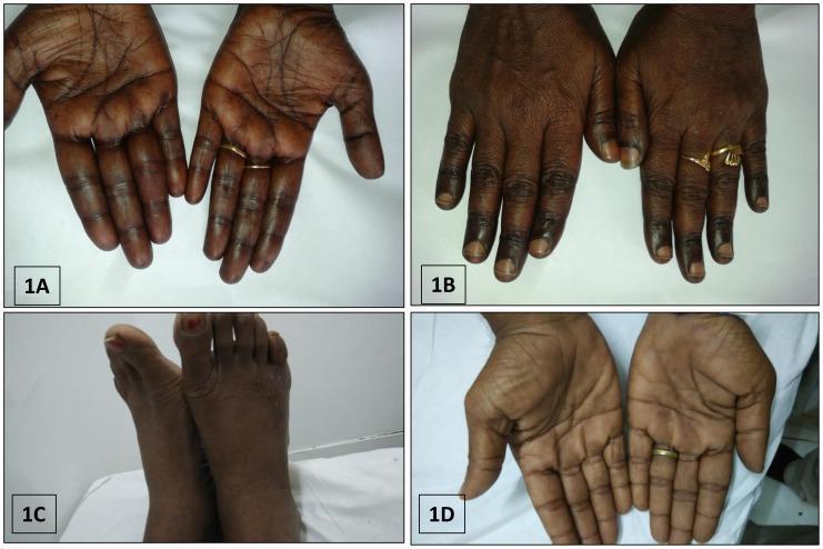
This composite of clinical photographs illustrates various patterns of cutaneous hyperpigmentation associated with megaloblastic anemia due to Vitamin B12 deficiency. Image 1A and 1D display the palmar surfaces of the hands, showing diffuse, brownish-black hyperpigmentation with prominent accentuation along the palmar creases. Image 1B focuses on the dorsal aspect of the hands, highlighting localized brownish-black pigmentation specifically over the knuckle pads and phalangeal joints. Image 1C depicts the dorsal aspect of the feet, exhibiting a similar diffuse, dusky, brownish-black discoloration across the skin surface. These dermatological findings are classic external markers of severe B12 deficiency and often present alongside systemic symptoms like pancytopenia and macrocytosis. The collection serves as an educational tool for recognizing the cutaneous manifestations of nutritional deficiencies and metabolic disorders in hematology and dermatology.
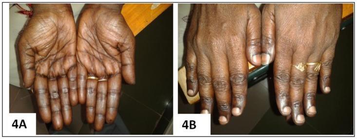
Clinical photograph comparison (Figure 4A and 4B) demonstrating the cutaneous manifestations of Vitamin B12 deficiency and its resolution following treatment. Image 4A (Pre-treatment): Palmar view showing diffuse, brownish-black hyperpigmentation with prominent accentuation along the palmar creases and longitudinal hyperpigmentation of the fingers. The knuckles exhibit characteristic darkening, a classic sign of megaloblastic anemia-associated pigmentary changes. Image 4B (Post-treatment): Dorsal view of the same patient's hands 12 weeks after initiating parenteral cyanocobalamin (Vitamin B12) therapy. There is a marked reversal of the hyperpigmentation, with the skin returning to a more uniform, lighter tone. The previously dusky knuckles and digits show significant clearing, and the nail beds appear healthy. This comparison serves as an educational tool for identifying dermatological markers of nutritional deficiencies and monitoring therapeutic response to Vitamin B12 supplementation.
scurvy vitamin C deficiency bleeding gums corkscrew hairs
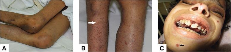
A composite of three clinical photographs demonstrating the systemic manifestations of Vitamin C deficiency (Scurvy). Panel A shows the lower extremities with prominent knee flexion contractures and diffuse ecchymoses around the joints. Panel B provides a close-up of the skin on the legs, highlighting perifollicular purpura and follicular hyperkeratosis; a white arrow points to characteristic 'corkscrew' hairs. Panel C displays the facial and oral features, including hemorrhagic gingivitis with swollen, friable, and bleeding gums. A black arrow in Panel C indicates a neurotic excoriation on the chin. The clinical findings represent the classic triad of scurvy: follicular hyperkeratosis with perifollicular hemorrhage, gingival bleeding, and musculoskeletal involvement. This content is intended for medical education regarding nutritional deficiencies and dermatologic manifestations of systemic disease.
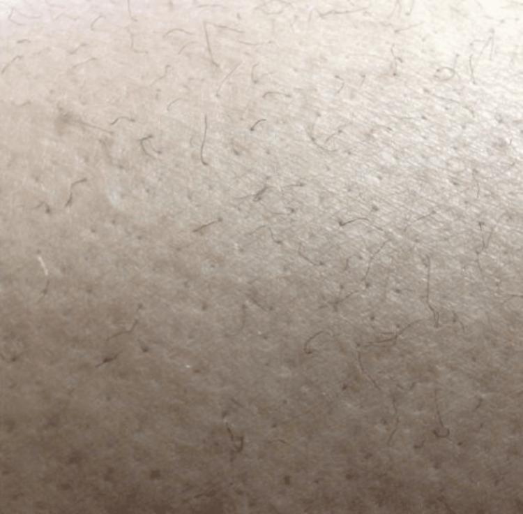
Clinical photograph of a skin surface, likely from the lower extremity, showing characteristic dermatological findings of scurvy (Vitamin C deficiency). The image displays 'corkscrew hairs,' which are hair shafts that appear abnormally coiled, twisted, and fragile in a spiral or helical pattern. Additionally, there is evidence of follicular hyperkeratosis and perifollicular changes, where the skin around the hair follicles appears slightly raised and textured. These visual markers are classic pathognomonic signs used in the diagnosis of ascorbic acid deficiency, often occurring alongside other systemic symptoms like ecchymosis and gingival changes. The educational focus is on identifying specific hair morphology associated with nutritional deficiencies and metabolic disorders.
folate trap methylation homocysteine methionine synthesis B12 cobalamin biochemistry
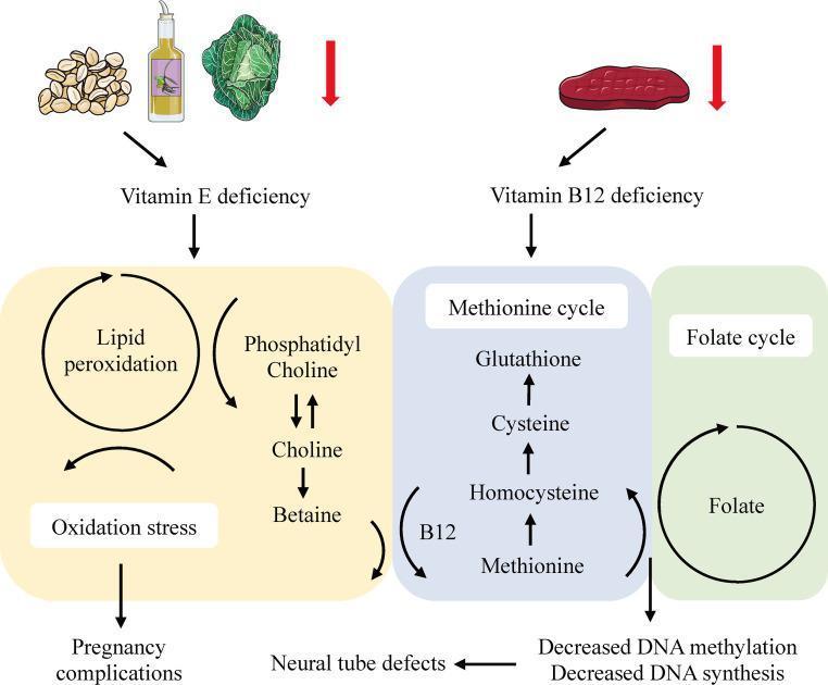
A pathophysiology diagram illustrating the systemic effects of Vitamin E and Vitamin B12 deficiencies on lipid peroxidation, one-carbon metabolism, and associated pregnancy complications. The diagram is divided into two primary functional pathways: 1. Vitamin E Pathway (yellow-shaded area): Depicts dietary sources (nuts, oil, leafy greens) leading to a deficiency state. This state triggers lipid peroxidation and oxidative stress, involving the Phosphatidyl Choline-Choline-Betaine pathway. The ultimate clinical consequence shown is pregnancy complications. 2. Vitamin B12 and Folate Pathway (blue and green shaded areas): Shows a deficiency originating from meat-based sources. It illustrates the 'Methionine cycle' where B12 acts as a cofactor in the conversion of Homocysteine to Methionine. The pathway also includes Cysteine and Glutathione production. The 'Folate cycle' is shown adjacent, contributing to DNA health. Impairment in these cycles due to B12 deficiency leads to decreased DNA methylation and decreased DNA synthesis, which are linked to the development of neural tube defects. The diagram serves as an educational summary of micronutrient biochemistry and its relevance to maternal-fetal health and clinical obstetrics.
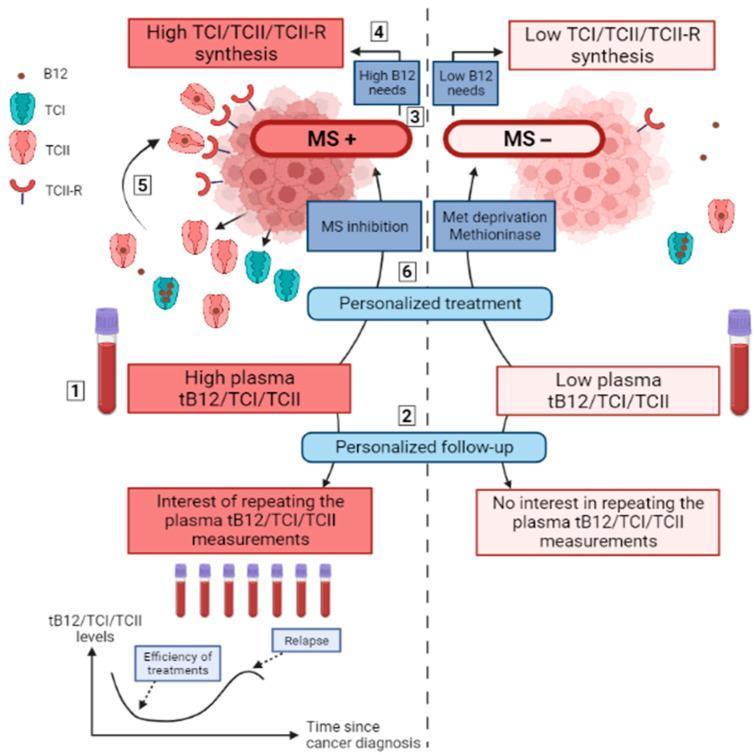
A medical flowchart and conceptual diagram detailing the relationship between cobalamin (B12) metabolic pathways and solid cancer management. The diagram is split into two comparative phenotypes: MS+ (high Methionine Synthase activity) and MS- (low activity). The MS+ phenotype is characterized by high B12 needs and high synthesis of transport proteins Transcobalamin I (TCI), II (TCII), and the TCII-receptor (TCII-R), leading to elevated plasma tB12/TCI/TCII levels. Clinical management for MS+ suggests MS inhibition and longitudinal personalized follow-up using serial plasma measurements to monitor treatment efficiency and relapse. Conversely, the MS- phenotype is associated with low B12 needs, low synthesis of transport proteins, and lower plasma levels, with personalized treatment focusing on methionine deprivation or methioninase. The diagram highlights the educational concept that plasma cobalamin markers serve as diagnostic and monitoring tools for metabolic singularities in cancer cells, specifically identifying which tumors are dependent on exogenous methionine versus endogenous synthesis.
Water-Soluble Vitamins - Detailed Seminar
Overview
- Vitamin B1 (Thiamine)
- Vitamin B2 (Riboflavin)
- Vitamin B3 (Niacin)
- Vitamin B5 (Pantothenic acid)
- Vitamin B6 (Pyridoxine)
- Vitamin B7 (Biotin)
- Vitamin B9 (Folate / Folic acid)
- Vitamin B12 (Cobalamin)
- Vitamin C (Ascorbic acid)
1. Vitamin B1 - Thiamine
Structure & Active Form
Biochemical Roles
| Enzyme Complex | Pathway | Reaction |
|---|---|---|
| Pyruvate dehydrogenase | Glycolysis → TCA cycle | Pyruvate → Acetyl-CoA |
| α-Ketoglutarate dehydrogenase | TCA cycle | α-KG → Succinyl-CoA |
| Branched-chain keto acid dehydrogenase | BCAA catabolism | Leucine, Isoleucine, Valine metabolism |
Dietary Sources
- Enriched/whole grain cereals and breads
- Pork, legumes, seeds, nuts
- Destroyed by cooking (heat-labile)
Deficiency States
- Symmetric peripheral neuropathy
- Motor weakness, loss of reflexes
- "Glove and stocking" sensory loss
- High-output cardiac failure
- Dilated cardiomyopathy
- Peripheral edema, tachycardia
- Acute fulminating form = Shoshin beriberi (life-threatening, predominant metabolic/cardiac features without peripheral neuropathy)
- Associated with alcohol abuse and narcotic abuse
- Wernicke's encephalopathy (acute): Classic triad:
- Confusion/altered consciousness
- Ophthalmoplegia (nystagmus, lateral rectus palsy)
- Ataxia (cerebellar)
- Korsakoff's psychosis (chronic): Anterograde + retrograde amnesia, confabulation
- MRI: Bilateral symmetric FLAIR hyperintensities in mammillary bodies, periaqueductal gray, medial thalami
Assessment of Status
Treatment
2. Vitamin B2 - Riboflavin
Structure & Active Forms
- FMN (Flavin Mononucleotide) - formed by ATP-dependent phosphorylation
- FAD (Flavin Adenine Dinucleotide) - formed by further reaction with ATP
Biochemical Roles
- Mitochondrial respiratory chain (Complex I uses FMN; Complex II uses FAD)
- Fatty acid β-oxidation (acyl-CoA dehydrogenase uses FAD)
- Amino acid oxidation
- TCA cycle
- Glutathione reductase (antioxidant defense)
Dietary Sources
- Dairy products (primary source)
- Meat, poultry, fish
- Enriched/whole grain cereals
Deficiency - Ariboflavinosis
- Cheilosis (cracking/fissuring at corners of mouth = angular cheilitis)
- Angular stomatitis
- Glossitis (magenta/purplish-red tongue)
- Seborrheic dermatitis (nasolabial folds, perinasal, perianal areas)
- Sore throat, hyperemia, edema of oral mucosa
- Normochromic normocytic anemia
Assessment
3. Vitamin B3 - Niacin
A Note on Classification
- Nicotinic acid (niacin)
- Nicotinamide (niacinamide)
Active Forms
- NAD (Nicotinamide Adenine Dinucleotide)
- NADP (Nicotinamide Adenine Dinucleotide Phosphate)
Additional Role of NAD
- ADP-ribosylation of proteins (e.g., by bacterial toxins like cholera toxin, pertussis toxin)
- Poly-ADP-ribosylation of nucleoproteins involved in DNA repair
- Cyclic ADP-ribose formation - acts as second messenger increasing intracellular calcium in response to neurotransmitters and hormones
Dietary Sources
- Meat, poultry, fish
- Enriched/whole grain cereals
- Note: Niacin in cereals is largely biologically unavailable (bound as niacytin)
Deficiency - Pellagra
- Photosensitive, symmetric, affecting sun-exposed areas
- Casal's necklace - hyperpigmented band around neck
- Progresses from erythema → bullae → hyperpigmentation → hyperkeratosis
- Hartnup disease - genetic defect in membrane transport of tryptophan → intestinal malabsorption + renal loss
- Carcinoid syndrome - up to 60% of tryptophan diverted to serotonin (5-HT) synthesis
- Isoniazid use - inhibits B6 (needed for tryptophan → niacin conversion)
- Corn-based diets (low in tryptophan + niacin bound as niacytin)
Pharmacological Use of Niacin
4. Vitamin B5 - Pantothenic Acid
Structure & Active Forms
- Coenzyme A (CoA) - the major acyl-group carrier in metabolism
- Acyl Carrier Protein (ACP) - involved in fatty acid synthesis
Biochemical Roles
- Fatty acid synthesis (as acetyl-CoA and malonyl-CoA)
- Fatty acid β-oxidation (as acyl-CoA)
- TCA cycle (acetyl-CoA enters as starting substrate)
- Synthesis of cholesterol, steroid hormones, ketone bodies
- Amino acid metabolism
Dietary Sources & RDA
- Widely distributed in animal tissues
- Whole grain cereals, legumes, eggs, liver
- "Pantothenic" = from Greek "pantos" meaning everywhere - reflects its wide distribution
Deficiency
5. Vitamin B6 - Pyridoxine
Forms
- Pyridoxine, pyridoxal, pyridoxamine, and their respective 5'-phosphates
- Active coenzyme form: Pyridoxal 5'-phosphate (PLP)
Body Distribution
Biochemical Roles
| Role | Detail |
|---|---|
| Transamination | Aminotransferases (AST, ALT) - amino acid interconversion |
| Decarboxylation | Amino acid decarboxylases - synthesis of neurotransmitters (serotonin from 5-HTP, dopamine from DOPA, GABA from glutamate) |
| Glycogen phosphorylase | Phosphate group catalytically important in glycogenolysis |
| Tryptophan → niacin | Required for this conversion pathway |
| Steroid hormone modulation | PLP removes hormone-receptor complexes from DNA binding, terminating steroid action |
| Heme synthesis | Aminolevulinic acid (ALA) synthase requires PLP |
Dietary Sources
- Meat, poultry, fish; eggs
- Fortified cereals, unmilled rice, oats
- Starchy vegetables, noncitrus fruits, nuts
Deficiency
- Seborrheic dermatitis
- Microcytic, hypochromic anemia (impaired heme synthesis)
- Epileptiform convulsions (especially in neonates - due to reduced GABA synthesis)
- Depression and confusion (reduced serotonin and dopamine)
- Peripheral neuropathy
Toxicity
Assessment
6. Vitamin B7 - Biotin
Structure
Biochemical Roles
| Enzyme | Reaction | Pathway |
|---|---|---|
| Acetyl-CoA carboxylase | Acetyl-CoA → Malonyl-CoA | Fatty acid synthesis (rate-limiting step) |
| Pyruvate carboxylase | Pyruvate → OAA | Gluconeogenesis |
| Propionyl-CoA carboxylase | Propionyl-CoA → Methylmalonyl-CoA | Odd-chain FA catabolism |
| Methylcrotonyl-CoA carboxylase | BCAA catabolism (leucine) | Leucine oxidation |
Dietary Sources & RDA
- Liver, egg yolk, widespread in foods
- Also synthesized by intestinal flora (in excess of requirements)
Deficiency
- Total parenteral nutrition (TPN) without biotin supplementation
- Excess raw egg white consumption - contains avidin, a heat-stable protein that binds biotin with very high affinity (K_d ~10⁻¹⁵ M) → prevents absorption. Cooking denatures avidin.
- Alopecia (hair loss)
- Dry, scaly dermatitis (similar to seborrheic dermatitis)
- Conjunctivitis
- CNS abnormalities (hypotonia, seizures, developmental delay)
- Glossitis
7. Vitamin B9 - Folate (Folic Acid)
Forms
DFE = μg food folate + 1.7 × μg synthetic folic acid (from supplements/enrichment)
Biochemical Role - One-Carbon Transfer
| THF derivative | C1 group | Function |
|---|---|---|
| 5,10-Methylene-THF | -CH₂- | dTMP synthesis (thymidylate synthase) |
| 5-Methyl-THF | -CH₃ | Methionine synthesis (requires B12) |
| 10-Formyl-THF | -CHO | Purine ring synthesis |
| 5-Formimino-THF | -CH=NH | Histidine catabolism |
The Folate Trap & Methyl Trap (Crucial Concept)
5-Methyl-THF + Homocysteine → THF + Methionine (B12-dependent)
Dietary Sources
- Citrus fruits, leafy green vegetables ("folate" from "folium" = leaf)
- Fortified cereals and breads
- Legumes
- Heat-labile - destroyed by cooking
Deficiency
- Megaloblastic anemia - large, immature red cells (macrocytes), hypersegmented neutrophils (≥5 lobes), hypercellular bone marrow with megaloblastic change
- Neural tube defects (NTDs) - spina bifida, anencephaly (periconceptional deficiency - critical in first 28 days of pregnancy, often before pregnancy recognized)
- Impaired cell division and growth
- Poor diet (commonest)
- Malabsorption (celiac disease, tropical sprue)
- Increased demand: pregnancy, hemolytic anemia
- Drugs: methotrexate (DHFR inhibitor), phenytoin, trimethoprim, sulfonamides
- Alcohol (impairs absorption and increases excretion)
Folate and Cancer
Public Health Note
8. Vitamin B12 - Cobalamin
Structure
- Cyanocobalamin (pharmaceutical form)
- Methylcobalamin (active metabolic form)
- Adenosylcobalamin (active metabolic form)
- Hydroxocobalamin
Unique Feature
Absorption - Two Binding Proteins
- Gastric acid + pepsin releases B12 from food proteins
- Haptocorrin (Cobalophilin/R-proteins) from saliva binds B12 in stomach
- In duodenum, pancreatic proteases degrade haptocorrin → B12 released
- Intrinsic factor (IF) - secreted by gastric parietal cells - binds free B12 with high affinity; accepts only active vitamers (not other corrinoids)
- IF-B12 complex absorbed in distal ileum via specific cubilin receptors
- Significant enterohepatic circulation - excreted in bile → reabsorbed → explains why deficiency takes years to develop even on a vegan diet (hepatic stores last 3-5 years)
B12-Dependent Enzymes (Only Two!)
| Enzyme | Coenzyme form | Reaction |
|---|---|---|
| Methionine synthase | Methylcobalamin | 5-Methyl-THF + Homocysteine → THF + Methionine |
| Methylmalonyl-CoA mutase | Adenosylcobalamin | L-Methylmalonyl-CoA → Succinyl-CoA |
Deficiency
| Mechanism | Examples |
|---|---|
| Dietary lack | Vegans (most common worldwide) |
| Impaired absorption - IF absent | Pernicious anemia (autoimmune - anti-parietal cell Ab + anti-IF Ab); total gastrectomy |
| Impaired release from food | Atrophic gastritis (elderly - failure of gastric acid) |
| Ileal disease | Crohn's disease, ileal resection, tropical sprue |
| Pancreatic insufficiency | Failure to degrade haptocorrin |
| Drug-induced | Metformin (reduces absorption), PPIs, H2 blockers |
| Bacterial overgrowth | B12 consumed by bacteria before absorption |
| Fish tapeworm (Diphyllobothrium latum) | Competes for B12 |
- Megaloblastic anemia - identical to folate deficiency on blood film (macrocytosis, hypersegmented neutrophils, pancytopenia)
- Subacute combined degeneration (SCD) of the spinal cord - pathognomonic to B12 deficiency:
- Demyelination of dorsal (posterior) columns → loss of vibration sense, proprioception, Romberg positive
- Demyelination of lateral (corticospinal) columns → upper motor neuron signs, spasticity
- Peripheral neuropathy may also occur
- Due to failure of methylation of arginine in myelin basic protein (methionine deficiency in CNS, not folate deficiency)
- Cutaneous hyperpigmentation - especially of hands, knuckles, palmar creases (seen more in darker-skinned individuals)
- Glossitis - smooth, red tongue
- Neuropsychiatric features - "megaloblastic madness"
- Serum/plasma B12 levels
- Serum methylmalonic acid (elevated - sensitive and specific)
- Plasma homocysteine (elevated - less specific, also elevated in folate, B6 deficiency)
- Schilling test (historical - to distinguish causes)
Pernicious Anemia
9. Vitamin C - Ascorbic Acid
Structure & Chemistry
Biochemical Roles
- Dopamine β-hydroxylase - converts dopamine → norepinephrine (catecholamine synthesis in adrenal medulla/CNS). Ascorbate reduces Cu²⁺ → Cu⁺ after each reaction cycle.
- Peptidylglycine hydroxylase - amidation of peptide hormones (vasopressin, oxytocin, CRH, etc.)
- Prolyl hydroxylase - hydroxylation of proline residues in procollagen → hydroxyproline → stabilizes collagen triple helix via hydrogen bonds
- Lysyl hydroxylase - hydroxylation of lysine in collagen → hydroxylysine → provides sites for crosslinking and glycosylation
- Also required for osteocalcin, C1q complement component
- Aspartate β-hydroxylase - modification of protein C (vitamin K-dependent anticoagulant)
- Trimethyllysine hydroxylase + γ-butyrobetaine hydroxylase - synthesis of carnitine (fatty acid transport into mitochondria)
- Enhances iron absorption from gut (reduces Fe³⁺ to Fe²⁺, the absorbable form)
- Antioxidant - quenches superoxide, hydroxyl radicals; regenerates vitamin E
- Immune function
Dietary Sources
- Citrus fruits, strawberries, kiwi
- Broccoli, spinach, peppers
- Potatoes
- Very heat-labile - destroyed by cooking, storage, exposure to air
Deficiency - Scurvy
- Blood vessel walls → capillary fragility → bleeding
- Bone matrix → perifollicular hemorrhages, bone pain
- Wound healing failure
- Skin changes: Perifollicular hemorrhage (petechiae around hair follicles)
- Corkscrew hairs (pathognomonic - coiled, malformed hair shafts)
- Bleeding gums (gingivitis, tooth loosening and loss)
- Subperiosteal hemorrhage (severe bone pain, hemarthrosis)
- Poor wound healing (old wounds may reopen)
- Anemia (from hemorrhage + impaired iron absorption)
- Psychological changes - depression, irritability (impaired catecholamine synthesis)
- Children: Infantile scurvy (Barlow's disease) - pseudoparalysis, "frog-leg" position, tender bones; X-ray: Frankel's white line, Wimberger ring sign, Pelkan spurs
High Intake
Summary Comparison Table
| Vitamin | Active Coenzyme | Key Metabolic Role | Deficiency Disease | Key Assessment |
|---|---|---|---|---|
| B1 Thiamine | Thiamine diphosphate (ThDP) | Oxidative decarboxylations; pentose phosphate | Beriberi (wet/dry), Wernicke-Korsakoff | Erythrocyte transketolase activation |
| B2 Riboflavin | FMN, FAD | Electron transport, redox reactions | Ariboflavinosis (cheilosis, glossitis) | Erythrocyte glutathione reductase activation |
| B3 Niacin | NAD, NADP | Redox reactions, DNA repair | Pellagra (3-4 Ds) | Urine N-methylnicotinamide |
| B5 Pantothenic acid | CoA, ACP | Acyl group carrier | Burning feet syndrome (rare) | Urinary pantothenic acid |
| B6 Pyridoxine | Pyridoxal phosphate (PLP) | Transamination, decarboxylation, glycogenolysis | Seborrheic dermatitis, microcytic anemia, convulsions | Erythrocyte transaminase activation |
| B7 Biotin | Biocytin | Carboxylation reactions | Alopecia, dermatitis (rare; raw eggs) | Serum biotinidase |
| B9 Folate | Tetrahydrofolate (THF) | One-carbon transfer; DNA synthesis | Megaloblastic anemia, NTDs | Serum/RBC folate; homocysteine |
| B12 Cobalamin | Methylcobalamin, Adenosylcobalamin | Methionine synthesis, propionate metabolism | Megaloblastic anemia + SCD of spinal cord | Serum B12, MMA, homocysteine |
| Vitamin C | Ascorbate (cofactor, not true coenzyme) | Hydroxylations (collagen, catecholamines) | Scurvy | Plasma ascorbate |
Key Clinical Distinctions to Remember
-
Folate vs B12 deficiency: Both cause megaloblastic anemia, but only B12 deficiency causes neurological damage (SCD). Folic acid supplements treat the anemia in either case but will mask ongoing B12 neurological degeneration - this is a critical clinical pitfall.
-
Wernicke's triad: Confusion + ophthalmoplegia + ataxia - always give IV thiamine before IV glucose/dextrose.
-
Pellagra with normal diet: Think Hartnup disease (tryptophan malabsorption) or carcinoid syndrome (tryptophan diversion) or isoniazid therapy.
-
Biotin deficiency: Think raw egg whites (avidin) or prolonged TPN.
-
B12 deficiency without anemia: Neurological damage can precede anemia - never rely on normal MCV to rule out B12 deficiency. Measure serum B12 and MMA directly.
-
Niacin toxicity: Flushing and hepatotoxicity at pharmacological doses; aspirin pretreatment reduces flushing.
-
B6 toxicity: Sensory neuropathy at 2-7 g/day - one of few water-soluble vitamins with significant toxicity.
- Harper's Illustrated Biochemistry 32nd Ed, Chapter 44 (pp. 553-559)
- Basic Medical Biochemistry: A Clinical Approach 6th Ed
- Fitzpatrick's Dermatology Vol 1&2
- Sleisenger and Fordtran's Gastrointestinal and Liver Disease
Convert into 60slides
mkdir -p /tmp/workspace/water-soluble-vitamins && ls /tmp/workspace/water-soluble-vitamins/