
Sclerosis of the bone
bone sclerosis radiograph X-ray
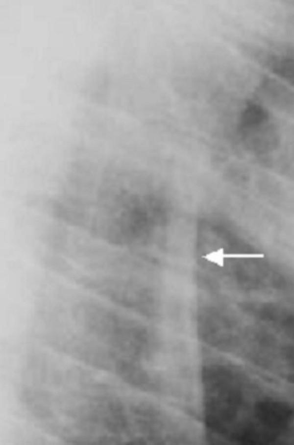
This lateral X-ray radiograph of the thoracic spine demonstrates advanced bone pathology characteristic of a systemic sclerosing bone disorder. The primary diagnostic finding is marked sclerosis of the vertebral rim circumference and the anterior longitudinal ligaments. A white arrow highlights a region where progressive ossification of these ligaments has resulted in bony ankylosis, appearing as radiopaque bridging and fusion between adjacent vertebral bodies. The vertebral bodies themselves exhibit increased bone density but lack signs of platyspondyly (vertebral flattening). These visual findings indicate a significant reduction in spinal mobility and flexibility due to the loss of distinct intervertebral spaces. The image serves as a clinical example of how progressive sclerosis in skeletal dysplasias or inflammatory spondyloarthropathies leads to structural fusion and altered biomechanical integrity of the spinal column.
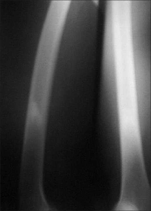
This diagnostic image is a plain radiograph (X-ray) of the right thigh, specifically focusing on the lower shaft of the femur. The radiograph demonstrates a focal area of irregular-shaped, predominantly endosteal sclerosis. The sclerosis is characterized by increased radiopacity (whiteness) within the medullary space of the bone. Notably, there is an absence of an identifiable radiolucent nidus, and there is no visible overlying periosteal reaction or cortical thickening. The surrounding soft tissues appear unremarkable with no signs of inflammation or masses. This visual presentation is typical of certain benign bone lesions, such as an osteoid osteoma where the nidus is obscured by dense reactive sclerosis, though differential diagnoses could include chronic osteomyelitis or a stress fracture. The educational focus is on identifying patterns of bone sclerosis and the importance of further imaging (like SPECT or CT) when a primary lesion is radiographically occult.
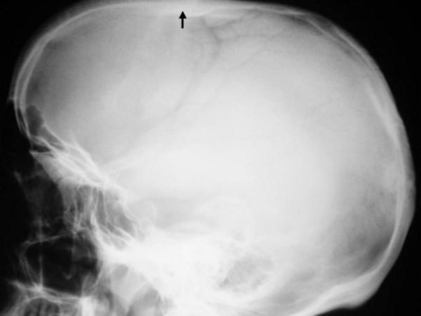
This lateral skull radiograph (X-ray) demonstrates significant skeletal abnormalities. The primary finding is a generalized, marked thickening and sclerosis of the skull vault (calvarium). An arrow highlights a specific area of prominent cortical thickening along the superior aspect of the cranium. The overall bone density is significantly increased, resulting in a more opaque appearance of the cranial bones compared to normal standards. The posterior aspect of the skull appears flattened, consistent with brachycephaly. Additionally, convolutional markings are somewhat obscured by the diffuse sclerosis. These radiographic features are characteristic of generalized sclerosing bone dysplasias, such as osteopetrosis or endosteal hyperostosis syndromes. The image provides clinical evidence for diagnosing metabolic or genetic bone disorders affecting cranial development and mineralization.
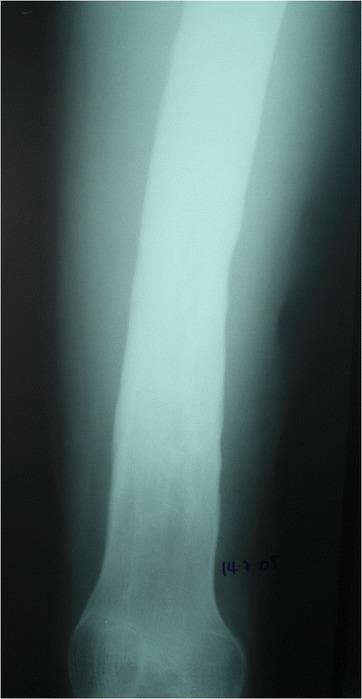
This diagnostic X-ray displays a long bone segment, likely the femur, demonstrating hallmark signs of Type C childhood chronic haematogenous osteomyelitis (CCHOM) according to the Beit CURE classification. The primary radiological feature is diffuse, dense sclerosis involving a significant portion of the diaphysis and metaphysis. The bone appears uniformly radiopaque with a loss of the normal corticomedullary differentiation, indicating extensive reactive bone formation. Notably, there is an absence of visible sequestra (detached necrotic bone fragments) or discrete involucrum formations on this plain radiograph. The cortical margins appear thickened and irregular. This pattern represents a late-stage manifestation of chronic infection where the bone has responded with generalized osteosclerosis. While not explicitly visible as a radiolucent area in this specific view, the clinical context of Type C classification suggests that an intraosseous abscess may frequently coexist with these sclerotic changes. This image serves as a teaching example for identifying advanced sclerotic responses in pediatric chronic bone infections.
Sclerosis of Bone (Osteosclerosis)
Definition & Pathophysiology
- Excess bone - osteosclerosis, osteopetrosis, hyperostosis
- Reduced bone - osteoporosis and osteolytic syndromes
Classification: Causes of Increased Bone Density
1. Inherited / Genetic Disorders
| Condition | Key Feature |
|---|---|
| Osteopetrosis (Albers-Schonberg / "marble bone disease") | Failure of osteoclastic activity; entire skeleton sclerotic |
| Pycnodysostosis | Short stature, open fontanelles |
| Diaphyseal dysplasia (Camurati-Engelmann disease) | Myopathy, delayed puberty |
| Endosteal hyperostosis / Sclerosteosis | Large jaw, facial palsy, large forehead, syndactyly |
| Osteopoikilosis | Spotted sclerotic lesions at epiphyses |
| Melorheostosis | "Dripping candlewax" pattern |
2. Metabolic / Toxic
| Condition | Key Feature |
|---|---|
| Renal osteodystrophy | "Rugger-jersey spine"; diagnostic biochemistry |
| Paget disease | Bones enlarged with disordered structure |
| Osteoblastic metastases | From prostate and breast carcinoma |
| X-linked hypophosphatemia (XLH) | Enthesopathy, low serum phosphate |
| Fluorosis | Rare except in endemic areas |
| Hepatitis C-associated osteosclerosis | Generalized increased bone mass, achy limbs |
3. Hematologic
| Condition | Key Feature |
|---|---|
| Myelofibrosis | Hepatosplenomegaly, leukoerythroblastic anemia |
| Lymphoma | Biopsy useful |
| Mastocytosis | Mast cells in bone, urticaria pigmentosa |
- Rheumatology, 2-Volume Set (Elsevier, 2022), Table 216.6
Major Conditions in Detail
Osteopetrosis ("Marble Bone Disease")
- Radiograph: Generalized symmetric increase in bone mass; thickening of cortical AND trabecular bone; diaphyses and metaphyses broadened; alternating sclerotic and lucent bands possible
- Dense, brittle bones prone to fracture despite appearing hard
- Bone marrow encroachment causes anemia, thrombocytopenia, cranial nerve palsies (compressed foramina)
- Treatment: bone marrow transplantation (in severe childhood form)
Camurati-Engelmann Disease (Progressive Diaphyseal Dysplasia)
- Autosomal dominant; mutations in TGF-beta1 gene
- New bone gradually envelops periosteal AND endosteal surfaces of long bone diaphyses
- Clinical: limping/waddling gait in childhood, enlarged head, prominent forehead, proptosis, thin limbs
- Severe cases: osteosclerosis also affects axial skeleton
Sclerosteosis
- Autosomal dominant; mutation causing loss of sclerostin regulation
- Osteosclerosis + syndactyly, facial palsy from cranial nerve compression
- Radiograph: poorly modeled, enlarged, dense bones (see hands X-ray below)
Pycnodysostosis
- Autosomal recessive; loss-of-function mutations in CTSK (cathepsin K gene)
- Bone collagen degradation and skeletal turnover diminished
- Clinical: large cranium, fronto-occipital prominence, proptosis, bluish sclerae, beaked nose, short stature, repeated fractures causing knock-knee deformity, acro-osteolysis of terminal phalanges
- Diagnosis: uniform osteosclerosis, thick cortices, narrow medullary canals, sclerotic skull base, wormian bones
- Goldman-Cecil Medicine, 2nd International Edition
Osteopoikilosis ("Spotted Bones")
- Highly penetrant autosomal dominant; deactivating mutation of LEMD3
- Usually a radiographic curiosity - asymptomatic
- Important: can be confused with metastatic disease on imaging
- Radiograph: numerous small, round/oval foci of bone sclerosis in cancellous bone at metaepiphyseal regions of tubular, tarsal, carpal, and pelvic bones
- Some patients have connective tissue nevi (Buschke-Ollendorff syndrome)
Melorheostosis
- Somatic mutations in MAP2K1 oncogene in mesenchymal bone progenitor cells
- Asymmetric thickening on surface of one or more bones in segmental distribution
- Radiograph: classic "dripping candlewax" appearance
- Can produce mechanical problems; associated with local soft tissue, vascular, and skin involvement
- Rheumatology, 2-Volume Set (Elsevier, 2022)
Fibrogenesis Imperfecta Ossium
- Rare, usually sporadic; coarsening of trabeculae
- Clinical: intractable skeletal pain beginning in middle age, spontaneous fractures, bone tenderness
- Radiograph: skull spared; corticomedullary junctions indistinct; fishnet pattern of mixed lytic and sclerotic areas; elevated serum alkaline phosphatase
Reactive / Secondary Bone Sclerosis
Subchondral Sclerosis in Osteoarthritis
- Occurs beneath articular cartilage in weight-bearing joints
- Graded radiographically: subchondral tibial bone sclerosis appears early; progresses with joint space narrowing and osteophyte formation
- In Grade 4 (severe): subchondral bone sclerosis with cysts, bone deformity, flattening of femoral condyle contours
- Miller's Review of Orthopaedics, 9th Edition
Osteoblastic Metastases
- Primarily from prostate and breast carcinoma
- Reactive bone formation around tumor deposits
- Contrast with multiple myeloma: purely lytic lesions without any surrounding reactive sclerosis
Osteoid Osteoma/Osteoblastoma
- Central radiolucent nidus surrounded by variable reactive bone sclerosis
- In spine: the reactive sclerosis can obscure the nidus, making detection difficult
- Grainger & Allison's Diagnostic Radiology
Chronic Osteomyelitis
- Dense diffuse diaphyseal sclerosis representing the bone's chronic response to infection
- Loss of normal corticomedullary differentiation; cortical margins thickened and irregular
Renal Osteodystrophy
- Chronic renal failure disrupts vitamin D activation and phosphate regulation
- Leads to hyperparathyroidism-driven bone changes including sclerosis
- Classic sign: "rugger-jersey spine" (alternating sclerotic end-plates and lucent vertebral bodies)
Radiographic Patterns to Recognize
| Pattern | Typical Diagnosis |
|---|---|
| Uniform generalized dense bones | Osteopetrosis |
| Diaphyseal cortical thickening | Camurati-Engelmann disease |
| "Dripping candlewax" | Melorheostosis |
| Spotted epiphyseal lesions | Osteopoikilosis |
| Subchondral sclerosis + osteophytes | Osteoarthritis |
| Vertebral end-plate sclerosis ("rugger jersey") | Renal osteodystrophy |
| Vertebral rim sclerosis + ankylosis | Spondyloarthropathy |
| Dense bone + pathological fractures | Osteopetrosis, Paget |
| Mixed lytic/sclerotic lesions | Paget disease, metastases |
| Purely lytic, no surrounding sclerosis | Multiple myeloma |
Radiographic Examples
Key Clinical Pearls
-
Dense bone does NOT mean strong bone - Osteopetrosis is the clearest example: bones appear marble-like on X-ray but are brittle and fracture easily because abnormal woven bone (with no remodeling) lacks structural organization.
-
Bone marrow failure in osteopetrosis - The medullary canal becomes obliterated, displacing hematopoiesis and causing anemia, recurrent infections, and bleeding.
-
Sclerosis vs. lysis - The pattern of a lesion (sclerotic vs. lytic vs. mixed) is one of the most important radiographic clues in bone pathology. Multiple myeloma is characteristically purely lytic with no reactive sclerosis, while prostate metastases are characteristically osteoblastic/sclerotic.
-
Osteopoikilosis misdiagnosis - These benign spotted lesions are often mistaken for skeletal metastases. The symmetrical epiphyseal distribution and asymptomatic nature are distinguishing features.