
Anatomy of hand
Reading File
Now let me search the library for hand anatomy content in parallel.
Finding Sources
Finding Sources
Searching the Web
anatomy of the hand bones muscles tendons nerves overview
Excellent - two strong textbook sources found. Let me read both in parallel.
Reading File
Reading File
Excellent content from both books. Now let me read more from both sources and fetch the key anatomical images in parallel.
Reading File
Reading File
Loading Image
Loading Image
Great content. Let me fetch a few more key images (muscles/tendons, nerves, vessels) and read the neurovascular section.
Loading Image
Loading Image
Reading File
I now have comprehensive content from both textbooks plus images. Let me compile the full answer.
Anatomy of the Hand
Surface Anatomy and Orientation
The hand is described using palmar (volar) for the anterior surface and dorsal for the posterior surface. Rather than medial/lateral (which change with rotation), the terms radial (thumb side) and ulnar (little finger side) are used. Abduction and adduction of the fingers refer to movement away from and toward the middle finger, respectively.
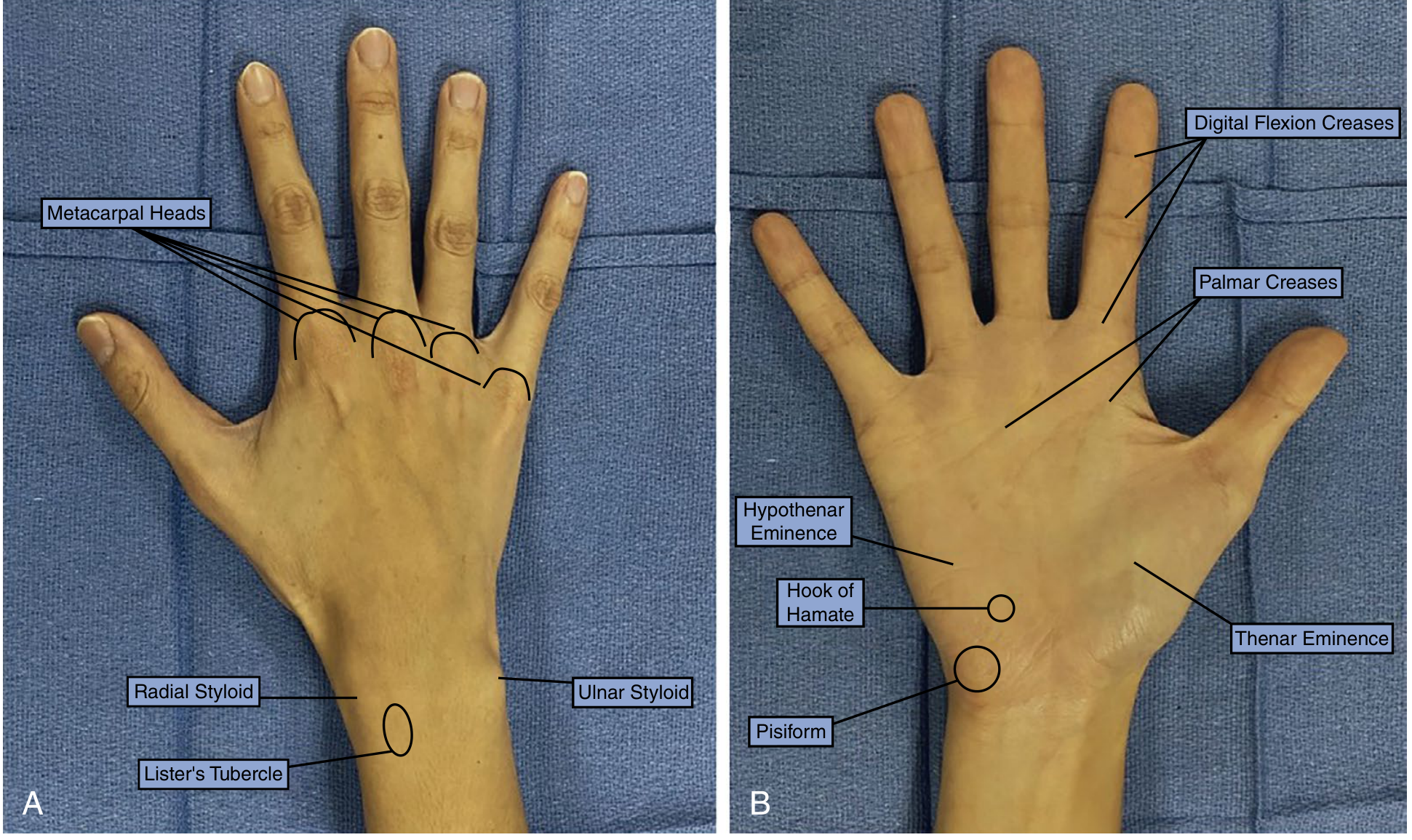
1. Bones
The hand contains 19 bones arranged in 5 rays (numbered 1-5, thumb to little finger). Each ray runs from the metacarpal base to the fingertip.
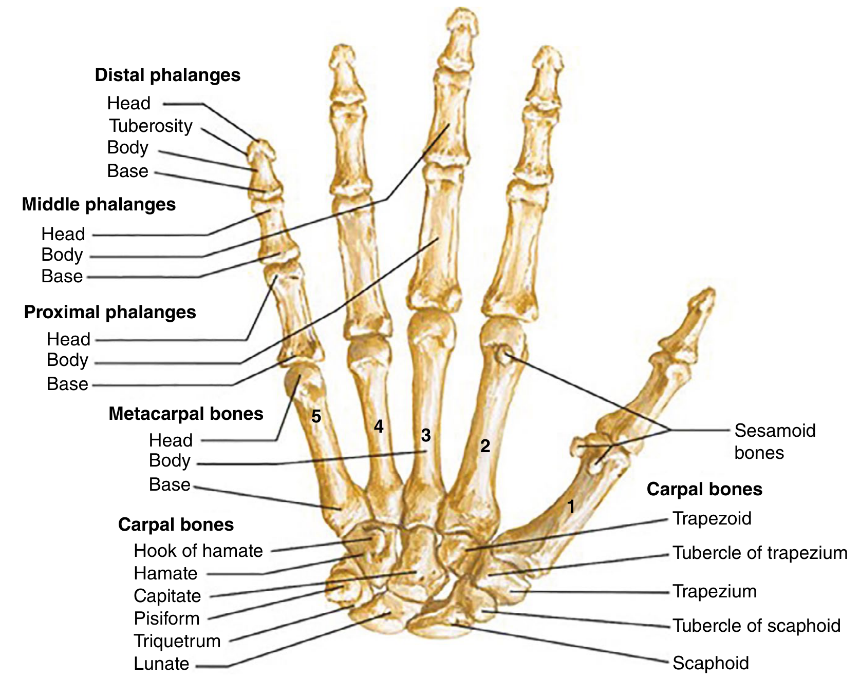
Carpal Bones (8 bones, 2 rows)
| Proximal Row | Distal Row |
|---|---|
| Scaphoid | Trapezium |
| Lunate | Trapezoid |
| Triquetrum | Capitate |
| Pisiform | Hamate (with hook) |
Mnemonic: Some Lovers Try Positions That They Can't Handle
Metacarpals (5 bones)
- Form the visible palm
- Each has a base (proximal), body/shaft, and head (distal - forming the "knuckles")
Phalanges (14 bones)
- Thumb: 2 phalanges (proximal + distal only - no middle phalanx)
- Fingers: 3 phalanges each (proximal, middle, distal)
Joints
- Metacarpophalangeal (MP) joint: 90° of flexion, slight hyperextension; fingers also actively abduct/adduct here
- Proximal interphalangeal (PIP) joint: hinge joint, ~110° flexion
- Distal interphalangeal (DIP) joint: hinge joint, ~80° flexion
- Thumb carpometacarpal (CMC) joint: saddle joint allowing opposition
2. Muscles
Muscles are divided into extrinsic (originating in the forearm, inserting in the hand) and intrinsic (originating and inserting within the hand itself).
Extrinsic Muscles
Flexor (volar) compartment - 3 layers:
| Layer | Muscles |
|---|---|
| Superficial | Pronator teres, Flexor carpi radialis (FCR), Palmaris longus*, Flexor carpi ulnaris (FCU) |
| Intermediate | Flexor digitorum superficialis (FDS) - flexes PIP joints |
| Deep | Flexor digitorum profundus (FDP) - flexes DIP joints; Flexor pollicis longus (FPL) - flexes thumb IP; Pronator quadratus |
*Palmaris longus is absent in ~10-15% of individuals.
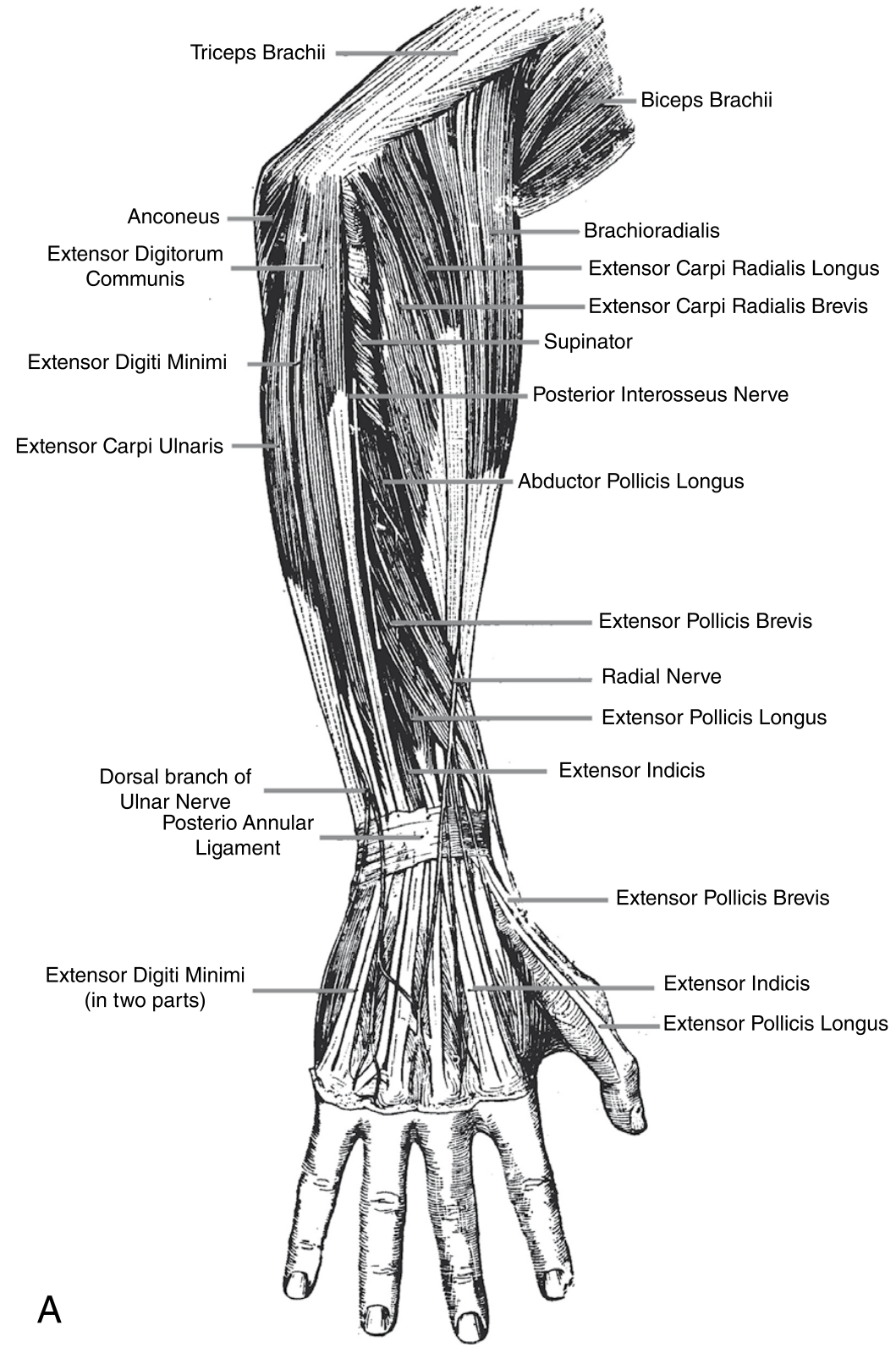
Extensor (dorsal) compartment:
| Subgroup | Muscles |
|---|---|
| Mobile wad (radial) | Brachioradialis, ECRL, ECRB |
| Superficial | EDC (extends fingers), EDM/EDQ (extends little finger), ECU (ulnar deviation + wrist extension) |
| Deep | APL, EPB, EPL (thumb function), EIP (index finger extension) |
Intrinsic Muscles
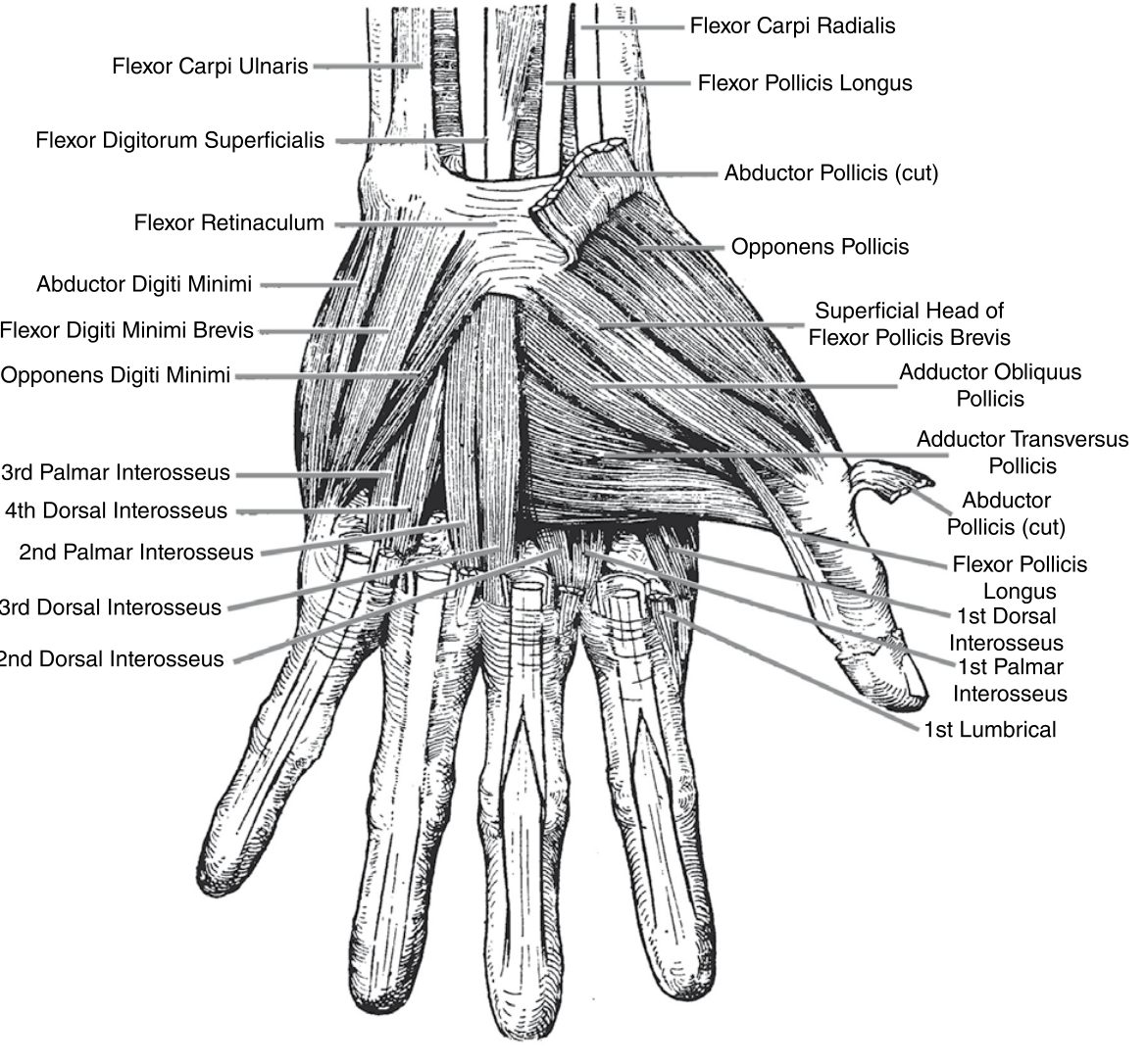
Thenar muscles (thumb - all median nerve except deep FPB head):
- Abductor pollicis brevis (APB) - abducts thumb
- Flexor pollicis brevis (FPB) - flexes thumb MP
- Opponens pollicis (OP) - opposition (most important)
- Adductor pollicis - adducts thumb (ulnar nerve)
Hypothenar muscles (little finger - all ulnar nerve):
- Abductor digiti quinti (ADQ) - abducts little finger
- Opponens digiti quinti (ODQ) - brings little finger across palm
- Flexor digiti quinti (FDQ) - flexes little finger metacarpal
Lumbricals (4):
- Unique: originate from FDP tendons in the palm
- Pass along the radial aspect of each finger
- Action: flex MP joints + extend IP joints
- Lumbricals 1 & 2 (index, middle) - median nerve
- Lumbricals 3 & 4 (ring, little) - ulnar nerve
Interosseous muscles:
- 3 palmar interossei - ADduct fingers (PAD mnemonic)
- 4 dorsal interossei - ABduct fingers (DAB mnemonic)
- All: flex MP joints, extend IP joints
- All innervated by ulnar nerve
3. Tendons and Pulleys
Flexor Tendon Pulleys
Each finger has 5 annular (A1-A5) and 3 cruciate (C1-C3) pulleys. The A2 and A4 pulleys are the most critical - they prevent bowstringing of the tendon. The thumb has its own pulley system (A1, oblique, A2).
Extensor Retinaculum (6 compartments, dorsal wrist)
| Compartment | Tendons |
|---|---|
| 1st | APL, EPB (De Quervain's territory) |
| 2nd | ECRL, ECRB |
| 3rd | EPL |
| 4th | EIP, EDC |
| 5th | EDM/EDQ |
| 6th | ECU |
Carpal Tunnel (Flexor Retinaculum)
Attaches from the scaphoid tubercle and trapezium (radially) to the hook of hamate and pisiform (ulnarly). Contents:
- FDS tendons (4)
- FDP tendons (4)
- FPL tendon (1)
- Median nerve
Extensor Hood (Dorsal Digital Apparatus)
At the MP joint, the long extensor tendon broadens into the extensor hood. The proximal portion at MCPJ is the sagittal band. The intrinsic muscles (interossei and lumbricals) enter as lateral bands, inserting distally to the axis of the PIP joint - this is why intrinsics flex the MP but extend the IP joints. The extensor inserts as the central slip (to base of middle phalanx) and terminal slip (to base of distal phalanx).
4. Blood Supply
Arterial Arches
Two main arteries supply the hand:
Radial artery:
- Passes superficially at wrist (radial to FCR tendon) - the pulse palpation site
- At wrist, splits: superficial branch enters palm; larger branch passes dorsally through anatomic snuffbox (between EPL and APB/EPB), then through first web space to form the deep palmar arch
Ulnar artery:
- Travels through Guyon's canal (between hamate and pisiform, superficial to flexor retinaculum)
- Larger superficial branch forms the superficial palmar arch
Key fact: In 97% of patients, at least one arch is intact, so the entire hand can survive on either the radial or ulnar artery alone.
Digital arteries:
- Each digit receives radial and ulnar proper digital arteries
- Run palmar to the midaxis of the digit, but dorsal to the digital nerves
5. Nerve Supply
Three nerves innervate the hand:
Median Nerve
Passes through the carpal tunnel and gives off the recurrent motor branch immediately distal to it.
- Motor (LOAF muscles): Lumbricals 1 & 2, Opponens pollicis, Abductor pollicis brevis, Flexor pollicis brevis (superficial head)
- Sensory: Palmar surface of thumb, index, middle finger, and radial half of ring finger; dorsal tips of same fingers
Ulnar Nerve
Enters the hand through Guyon's canal (superficial to flexor retinaculum).
- Motor: All interossei (3 palmar + 4 dorsal), lumbricals 3 & 4, hypothenar muscles, adductor pollicis, deep head of FPB
- Sensory: Little finger + ulnar half of ring finger (palmar and dorsal); dorsal ulnar hand via dorsal branch (arises proximal to wrist)
- Responsible for the power grasp function of the hand
Radial Nerve
- Motor: All extrinsic wrist and finger extensors (in forearm, via posterior interosseous nerve)
- Sensory only in hand: Superficial branch supplies dorsal radial hand, dorsum of thumb, index, long finger, and radial half of ring finger proximal to the DIP joints
- No intrinsic motor function in the hand itself
Summary Innervation Table
| Structure | Nerve |
|---|---|
| Thenar muscles (APB, OP, FPB superficial) | Median |
| Adductor pollicis, FPB deep head | Ulnar |
| Lumbricals 1 & 2 | Median |
| Lumbricals 3 & 4 | Ulnar |
| All interossei | Ulnar |
| All hypothenar muscles | Ulnar |
| Wrist/finger extensors (extrinsic) | Radial (PIN) |
6. Skin and Fascia
- The palmar skin is thick, hairless, and tethered to underlying fascia by vertical fibrous septa - forming 8 compartments for flexor tendons and neurovascular bundles
- The palmar fascia (palmar aponeurosis) is the condensation of the deep fascia of the palm, continuous with the palmaris longus tendon proximally - central thickening is important in Dupuytren's contracture
- The dorsal skin is thin and loose, allowing for the large excursion required with finger flexion
Sources: Schwartz's Principles of Surgery, 11th Ed., Chapter 44; Sabiston Textbook of Surgery, Chapter 119
This is a shared conversation. Sign in to Orris to start your own chat.