
Diabetic foot ulcer
"diabetic foot ulcer"[MeSH Terms] AND management
diabetic foot ulcer Wagner classification clinical
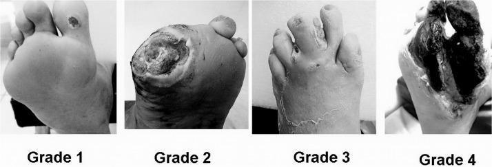
A comparative clinical photograph set illustrating the Wagner Classification System for diabetic foot ulcers across four stages (Grade 1 to Grade 4). Grade 1 (Superficial Ulcer) shows a small, localized skin lesion on the plantar surface near the base of the toes with no deep tissue involvement. Grade 2 (Deep Ulcer) depicts a larger, deeper wound penetrating to the tendon or joint capsule without abscess or osteomyelitis. Grade 3 (Osteitis/Infection) demonstrates a deep ulcer associated with significant inflammation, purulent discharge, and clinical signs of underlying bone infection or deep tissue abscess. Grade 4 (Forefoot Gangrene) shows extensive necrosis and blackening of the tissue localized to the forefoot or toes, indicating severe vascular compromise and tissue death. This series serves as an educational tool for medical professionals to differentiate between stages of diabetic foot pathology, guiding clinical management and prognosis assessment.
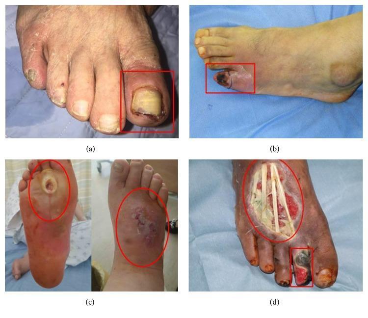
A series of four clinical photographs (a-d) demonstrating the progression of diabetic foot ulcers categorized by the Wagner classification system. (a) Wagner Grade 1: A superficial ulcer on the left hallux (big toe) featuring erythema, periungual inflammation, and a thickened, discolored yellowish toenail, indicative of an ischemic etiology. (b) Wagner Grade 2: A deep ulcer on the fifth digit (pinky toe) showing more extensive tissue involvement and localized cyanosis. (c) Wagner Grade 3: A neuropathic perforating ulcer extending from the plantar surface to the dorsum of the foot, associated with deep infection, abscess, and clinical signs of osteomyelitis. (d) Wagner Grade 4: Advanced diabetic foot pathology exhibiting local gangrene of the second toe and a large, deep dorsal foot ulcer. The necrotic tissue is blackened, and the wound bed is exposed, revealing underlying anatomical structures such as tendons. This set illustrates the clinical manifestations of ischemic, neuropathic, and mixed-type diabetic foot disease for educational use in wound care and endocrinology.
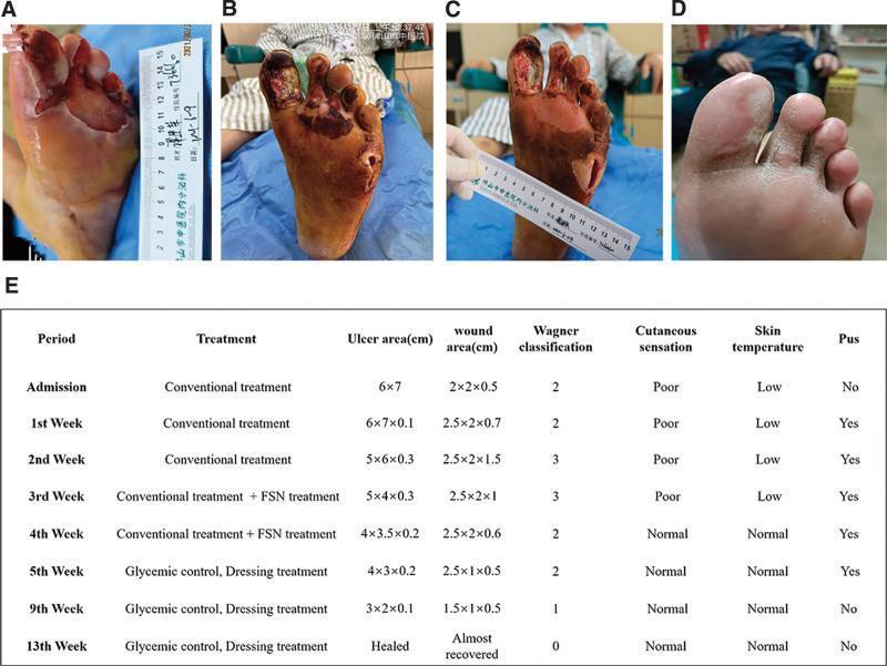
This composite figure presents a clinical progression timeline of a complex diabetic foot ulcer (DFU) on the left foot, categorized under the Wagner classification system. (A) Initial presentation shows a large, 6x7 cm ulcer on the plantar forefoot with surrounding maceration and a lateral abscess. (B) Following initial debridement, the wound exhibits deep tissue involvement (Wagner Grade 3), with necrotic tissue extending to the tendons and musculature of the first through third toes, accompanied by a deep 2x2x0.7 cm abscess cavity. (C) Mid-treatment status demonstrates significant clinical improvement; the wound bed appears ruddy with healthy granulation tissue, reduced exudate, and diminishing necrotic debris. (D) Final follow-up at three months shows complete secondary intention healing with full epithelialization and stable scar tissue formation. (E) An accompanying summary table provides longitudinal data on treatment phases (including Fu's subcutaneous needling), ulcer/wound dimensions, Wagner grade transition (from Grade 3 to 0), and clinical markers such as cutaneous sensation, skin temperature, and presence of pus.
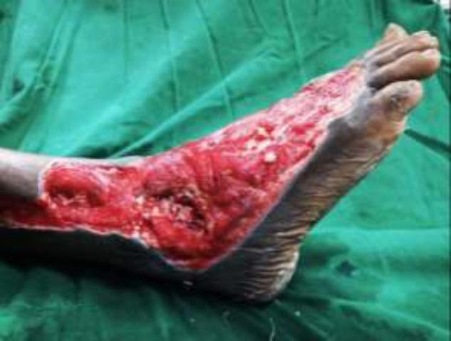
Clinical photograph of a right foot demonstrating a severe, extensive Wagner Grade III or IV diabetic foot ulcer. The wound spans the lateral and dorsal aspects of the foot, extending from the distal lower leg across the ankle joint to the midfoot region. The ulcer bed is characterized by beefy red granulation tissue and deep cavitation, with visible exposure of underlying musculoskeletal structures including the ankle joint. There are focal areas of yellow/white slough and fibrin, indicative of potential infection or chronic inflammatory processes. The wound margins are irregular but well-demarcated against the surrounding hyperpigmented, intact skin. This image serves as a clinical example of complex diabetic wound progression requiring advanced surgical intervention, such as debridement and microvascular flap reconstruction. Key educational concepts include diabetic foot classification, wound bed assessment, and the management of deep tissue infections with joint involvement.
"diabetic foot ulcer" AND treatment
Diabetic Foot Ulcer (DFU)
Epidemiology
- Approximately 1 in 4 patients with diabetes will develop a foot complication during their lifetime
- 15-20% of DFUs result in lower extremity amputation (LEA)
- ~85% of all amputations are preceded by a DFU
- Economic burden in the US: ~$245 billion (medical costs + lost productivity)
Pathophysiology - The Triad
| Factor | Mechanism |
|---|---|
| Neuropathy | Sensory loss → unrecognized trauma; Motor loss → foot deformities (claw toe, hammertoe) altering pressure points; Autonomic loss → dry, cracked skin |
| Ischemia | PAD → poor perfusion → impaired healing and ulcer progression |
| Infection | Impaired immune response + ischemia → polymicrobial colonization, biofilm formation |
Types of DFU
| Type | Features | Common Location |
|---|---|---|
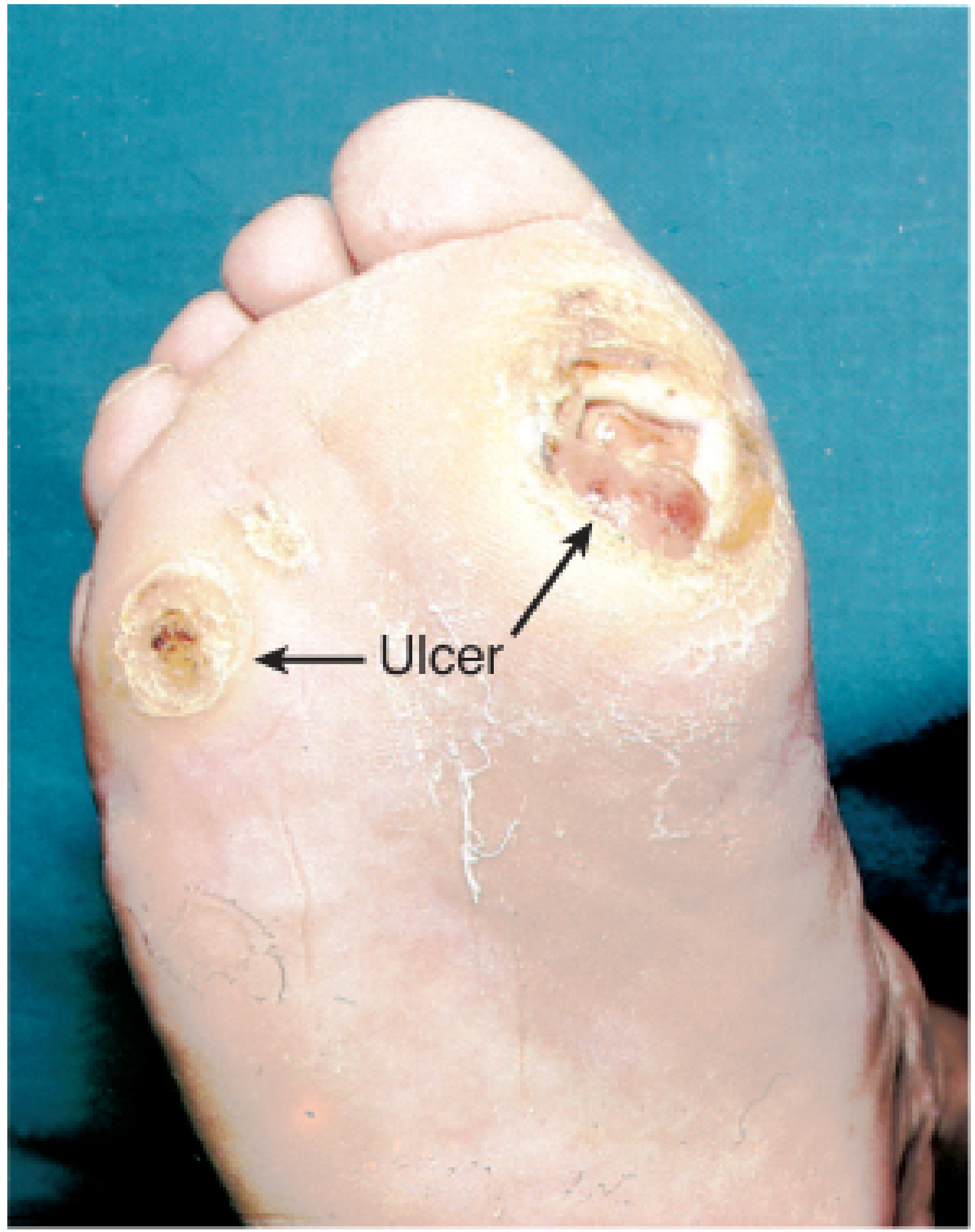
| Neuropathic | Painless, well-defined edges, warm foot, good pulses | Plantar surface - metatarsal heads, pressure points |
| Ischemic | Painful, punched-out, cold/pulseless foot | Tips of toes, margins |
| Neuroischemic | Mixed features - most common in practice | Variable |
Classification Systems
Wagner Classification (widely used)
| Grade | Description |
|---|---|
| 0 | No ulcer; pre-ulcerative lesion or healed ulcer |
| 1 | Superficial ulcer - skin/subcutaneous only |
| 2 | Deep ulcer to tendon, capsule, or bone - no abscess/osteomyelitis |
| 3 | Deep ulcer with abscess, osteomyelitis, or septic arthritis |
| 4 | Localized gangrene (forefoot or heel) |
| 5 | Extensive gangrene of the whole foot |
WIfI Classification (Society for Vascular Surgery - newer, preferred)
- Risk of major amputation at 1 year
- Theoretical benefit of revascularization
IDSA Infection Classification
| Severity | Clinical Criteria |
|---|---|
| Mild | Erythema ≤2 cm, skin/subcutaneous only, ≥2 signs of inflammation (erythema, warmth, tenderness, swelling, purulence) |
| Moderate | Erythema >2 cm OR deep tissue involved (abscess, osteomyelitis, septic arthritis, fasciitis), no SIRS |
| Severe | Local infection + ≥2 SIRS criteria (T >38°C or <36°C, HR >90, RR >20, WBC >12,000 or <4,000) |
Evaluation
History
- How and when the wound started (often unknown due to sensory neuropathy)
- Current wound care and footwear
- Diabetes control (HbA1c), medications, comorbidities
Physical Exam
- Document wound size, depth, edge characteristics, exudate
- Probe to bone (probe-to-bone test positive → ~90% PPV for osteomyelitis)
- Semmes-Weinstein 10-g monofilament + pinprick/vibration/temperature for neuropathy assessment
- Check for structural deformities (calluses, hammertoe, claw toe, Charcot foot)
Vascular Assessment
- Palpate dorsalis pedis and posterior tibial pulses
- ABI (may be falsely elevated due to arterial calcification in diabetics)
- Toe-Brachial Index (TBI) - more reliable: TBI >0.4 or toe pressure >40 mmHg = adequate perfusion
- If toe pressure 60-80 mmHg and wound not healing in 4-6 weeks → angiography
Imaging & Labs
- 3-view plain X-ray of the foot (structural deformities, chronic osteomyelitis, gas)
- MRI: most sensitive/specific for osteomyelitis (preferred over bone scan)
- Bone scan or tagged WBC scan if MRI not available
- Gold standard: bone biopsy (with cultures for antibiotic sensitivities)
- ESR, CRP (elevated but nonspecific for osteomyelitis)
- HbA1c, CBC, BMP
Note: Surface swab cultures have little value in managing DFU infections and should be avoided. Deep cultures through unbroken skin or bone biopsy are preferred. (Textbook of Family Medicine 9e)
Microbiology & Antibiotic Treatment
| Severity | Usual Pathogens | Initial Treatment |
|---|---|---|
| Mild | S. aureus, Streptococcus spp. | Oral cephalexin or amoxicillin-clavulanate (MSSA); Clindamycin if MRSA suspected |
| Moderate | S. aureus, Streptococcus, Enterobacteriaceae, Anaerobes | Parenteral ampicillin-sulbactam, cefoxitin, ertapenem; Add vancomycin if MRSA suspected |
| Severe | As above + Pseudomonas, mixed flora | Broad-spectrum IV (piperacillin-tazobactam ± vancomycin); Guided by bone/deep tissue culture |
- MRSA accounts for ≥15-30% of foot ulcer infections
- Acute infections in non-antibiotic-exposed patients tend to be monomicrobial
- Chronic/recurrent infections are typically polymicrobial with gram-positives, gram-negatives, and anaerobes
Management
1. Glycemic Control
- HbA1c goal <7%
- Tight control halts neuropathy progression; prolonged good control can reverse neuropathy
2. Pressure Off-Loading
- Total Contact Cast (TCC): gold standard - extends above ankle, reduces plantar pressure up to 60%, non-removable
- Fixed ankle walking boot / removable walker boot: allows frequent dressing changes
- Custom orthotics after healing to prevent recurrence
- Surgical options: metatarsal head resection, Achilles tendon lengthening, liquid silicone injections
3. Local Wound Care
| Wound Type | Dressing Choice |
|---|---|
| Healthy granulating | Collagen dressing |
| Fibrinous/sloughy | Santyl (collagenase) or MediHoney |
| Deep healthy wound | VAC (negative pressure wound therapy) |
| Infected | Iodosorb, Bactroban, silver-impregnated alginate |
| Macerated | Absorptive foams, alginate |
4. Debridement
- Sharp debridement of devitalized/necrotic tissue in clinic
- Enzymatic debridement for eschar
- Do not aggressively debride without first ensuring adequate perfusion (unless severe infection demands immediate source control)
5. Revascularization
- Indicated when TBI <0.4, toe pressure <40 mmHg, or wound not healing in 4-6 weeks despite maximal wound care
- Endovascular vs. open bypass: controversial; tibial disease common in diabetics with reduced endovascular patency below knee
- In frankly infected wounds: debride first for source control, then angiography
- In subacute/chronic infection: angiography before debridement to prevent ischemia-mediated necrosis
6. Treatment of Osteomyelitis
- Confirmed osteomyelitis generally requires surgical excision plus prolonged antibiotics (culture-guided)
- Bone biopsy/culture is the gold standard for directing therapy
7. Medical Optimization
- Antihypertensives (ACE inhibitor preferred)
- Statin therapy
- Antiplatelet therapy (aspirin) in PAD patients
- Tobacco cessation at every visit
- Edema control: multilayer compression therapy
8. Surgical/Amputation
- Pedal sepsis is a surgical emergency: fevers, tachycardia, ascending cellulitis, lymphangitis, purulent wound, possible gas on imaging
- Rapid surgical source control to halt sepsis progression
Prevention
- Annual foot examination minimum; more frequent if prior ulcer or structural deformity
- Daily foot hygiene and moisturization
- Professional nail trimming
- No barefoot walking; well-cushioned, well-maintained footwear
- Callus management
- Early wound reporting education
Advanced/Adjunct Therapies (Recent Evidence)
- Platelet-rich plasma (PRP): 2024 systematic review and meta-analysis supports PRP in accelerating DFU healing (PMID 38169990)
- Hydrogel dressings: 2024 systematic review shows hydrogel dressings improve healing rates compared to standard care (PMID 38465784)
- Physical therapy modalities (LLLT, electrical stimulation, ultrasound): emerging supportive evidence (PMID 37095726)
- Hyperbaric oxygen therapy (HBOT): adjunct in selected patients with ischemic DFUs