
Right hemicolectomy explain the procedure in detail
right hemicolectomy surgical procedure diagram anatomy
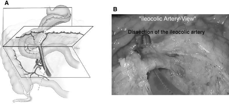
This composite image illustrates the surgical anatomy and intraoperative dissection of the ileocolic artery (ICA) during a right hemicolectomy or similar colonic procedure. Image A is an anatomical diagram featuring a schematic 'open book' model of the abdominal vasculature and mesentery. It depicts the superior mesenteric vein (SMV) as a central axis, with the ileocolic artery branching toward the cecum and ascending colon. A dotted line indicates the planned surgical dissection plane along the SMV axis. Image B is a grayscale intraoperative photograph corresponding to the diagram, labeled as 'ileocolic Artery-View'. It shows the surgical field during the dissection of the ileocolic artery. Laparoscopic or open surgical instruments are visible, including a dissection tool positioned beneath the vessel to isolate it from the surrounding fatty mesenteric tissue. The image demonstrates the clinical application of embryologically defined fascial planes to achieve a complete mesocolic excision (CME), emphasizing the vascular relationship between the ICA and the SMV.
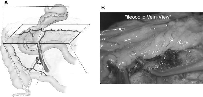
Educational figure illustrating surgical anatomy for right-sided hemicolectomy. Figure A is an anatomical diagram representing the 'open book' model of fascial and vascular planes. It shows the stomach, duodenum, and ascending colon in relation to the mesenteric root. Key vascular structures highlighted include the superior mesenteric artery (SMA) and superior mesenteric vein (SMV) trunk, with a specific focus on the V-shaped confluence of the ileocolic vessels. A curved arrow indicates the incision site of the ileocolic mesentery and the division of the ileocolic vein (ICV). Figure B is an intraoperative clinical photograph (monochrome) providing the 'ileocolic vein-view' post-dissection. The image demonstrates the surgical exposure of the ICV separated from the surrounding fatty mesentery, with surgical retractors and instruments visible in the operative field. This content is designed to teach medical professionals the precise anatomical landmarks and vascular dissection steps required for complete mesocolic excision (CME).
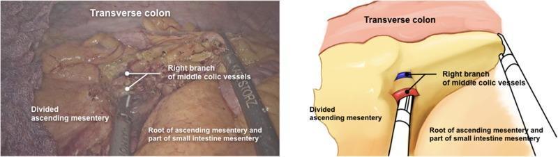
This composite educational resource features an intraoperative laparoscopic photograph (left) paired with a corresponding labeled anatomical diagram (right) depicting a right hemicolectomy procedure. The content illustrates the surgical dissection of the mesocolon and vascular control. Key anatomical structures labeled include the transverse colon at the superior margin and the root of the ascending/small intestine mesentery at the inferior aspect. Centrally, the image highlights the right branch of the middle colic vessels being approached for division. The 'divided ascending mesentery' is shown, demonstrating the mobilization of the colon from the retroperitoneum. A laparoscopic surgical instrument, labeled as a Karl Storz tool, is positioned to manipulate the right branch of the middle colic vessels. This visual is designed to teach laparoscopic surgical anatomy, specifically the identification and ligation of colic vasculature and the mobilization of the right colon using a caudal-to-cranial approach.
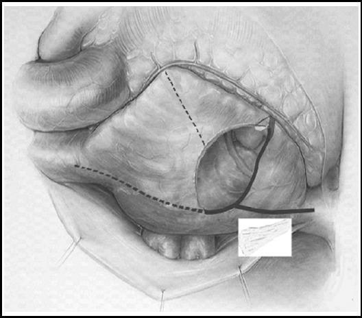
Anatomical illustration and surgical diagram depicting the right atrial cryoablation procedure, likely for the treatment of atrial fibrillation (Cox-Maze IV variant). The diagram shows a grayscale medical illustration of the right atrium and surrounding cardiac anatomy, including the superior vena cava, inferior vena cava, and tricuspid annulus. Superimposed on the anatomy are specific surgical markers: dashed lines represent surgical incisions, specifically the lateral longitudinal incision extending from the interatrial groove toward the tricuspid annulus. Solid black lines delineate the cryoablation lesion set. These ablation lines are strategically placed at the junction of the incision and tricuspid annulus, as well as along the posterior walls of the venae cavae. The educational focus is the spatial mapping of transmural cryo-lesions designed to interrupt macro-reentrant circuits in the right atrium while maintaining surgical access and anatomical integrity of the conduction system.
Right Hemicolectomy - Detailed Procedure
Overview and Indications
- Carcinoma of the cecum, ascending colon, or hepatic flexure
- Crohn's disease of the terminal ileum / ileocecal region
- Cecal volvulus
- Complicated appendiceal pathology (appendiceal neuroendocrine tumors >20 mm, goblet-cell carcinoids, positive margins after appendectomy)
- Severe right-sided colitis refractory to medical therapy
Preoperative Preparation
- Mechanical bowel preparation: controversial - most colorectal surgeons still perform it. Key advantages: intraoperative colonoscopy feasibility, absence of a stool load if a diverting stoma becomes necessary.
- Antibiotic prophylaxis: given within 1 hour of incision, targeting both aerobic (E. coli, Klebsiella) and anaerobic (Bacteroides fragilis) organisms. Options include: a 2nd/3rd-generation cephalosporin + metronidazole, ertapenem, or piperacillin-tazobactam. Duration limited to <24 hours.
- VTE prophylaxis: sequential compression devices and low-molecular-weight heparin.
- Stoma marking: stoma nurse marks the right iliac fossa if a stoma is a possibility.
- Preoperative imaging (CT abdomen/pelvis with contrast) and colonoscopy for tumor staging.
Open Right Hemicolectomy
Positioning and Access
Step 1 - Lateral Mobilization (White Line of Toldt)
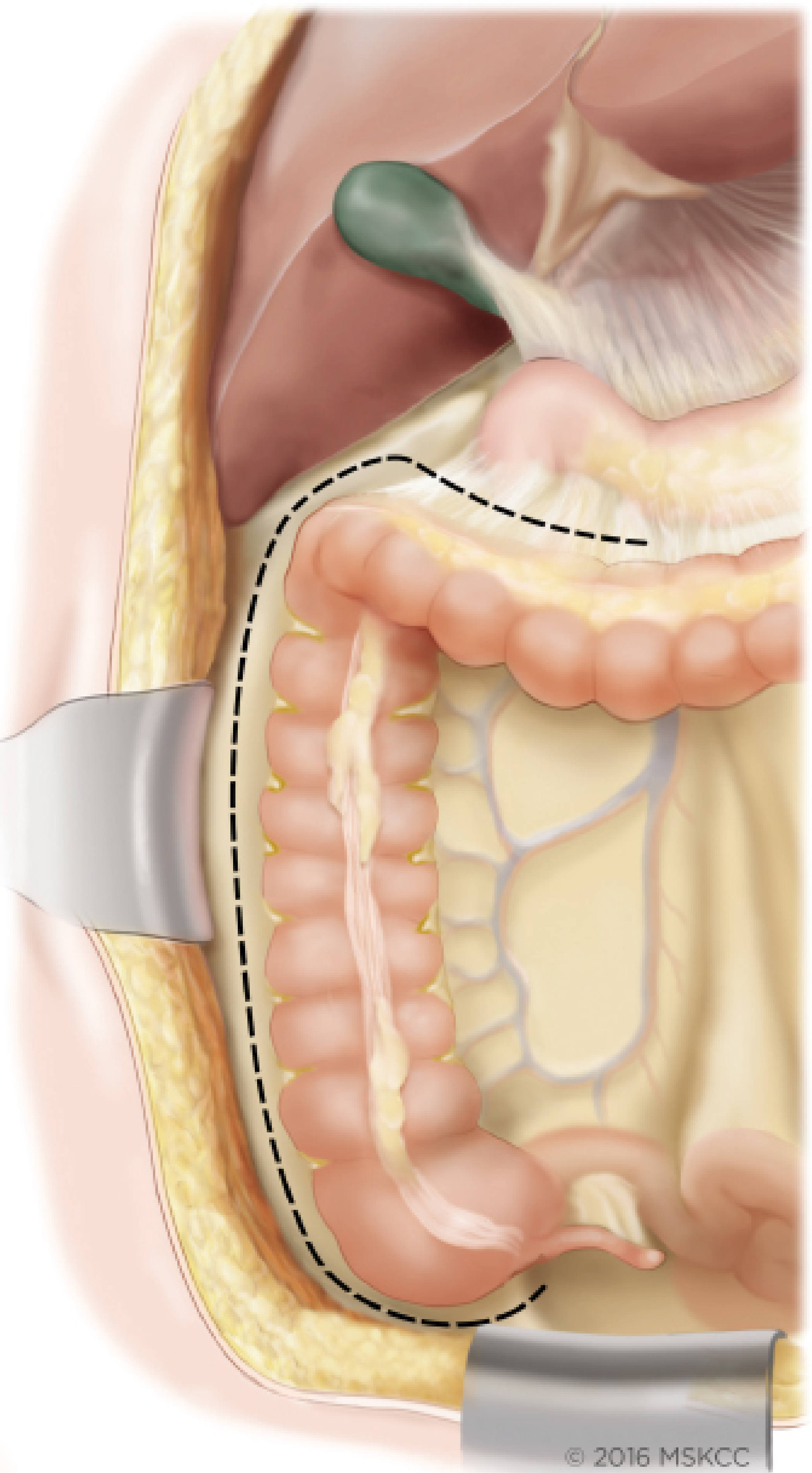
Step 2 - Retroperitoneal Mobilization
- Right gonadal vessels (right ovarian/testicular vein and artery)
- Right ureter - identified crossing the iliac vessels
- Gerota's fascia (covering the right kidney)
- Second and third portions of the duodenum and the head of the pancreas
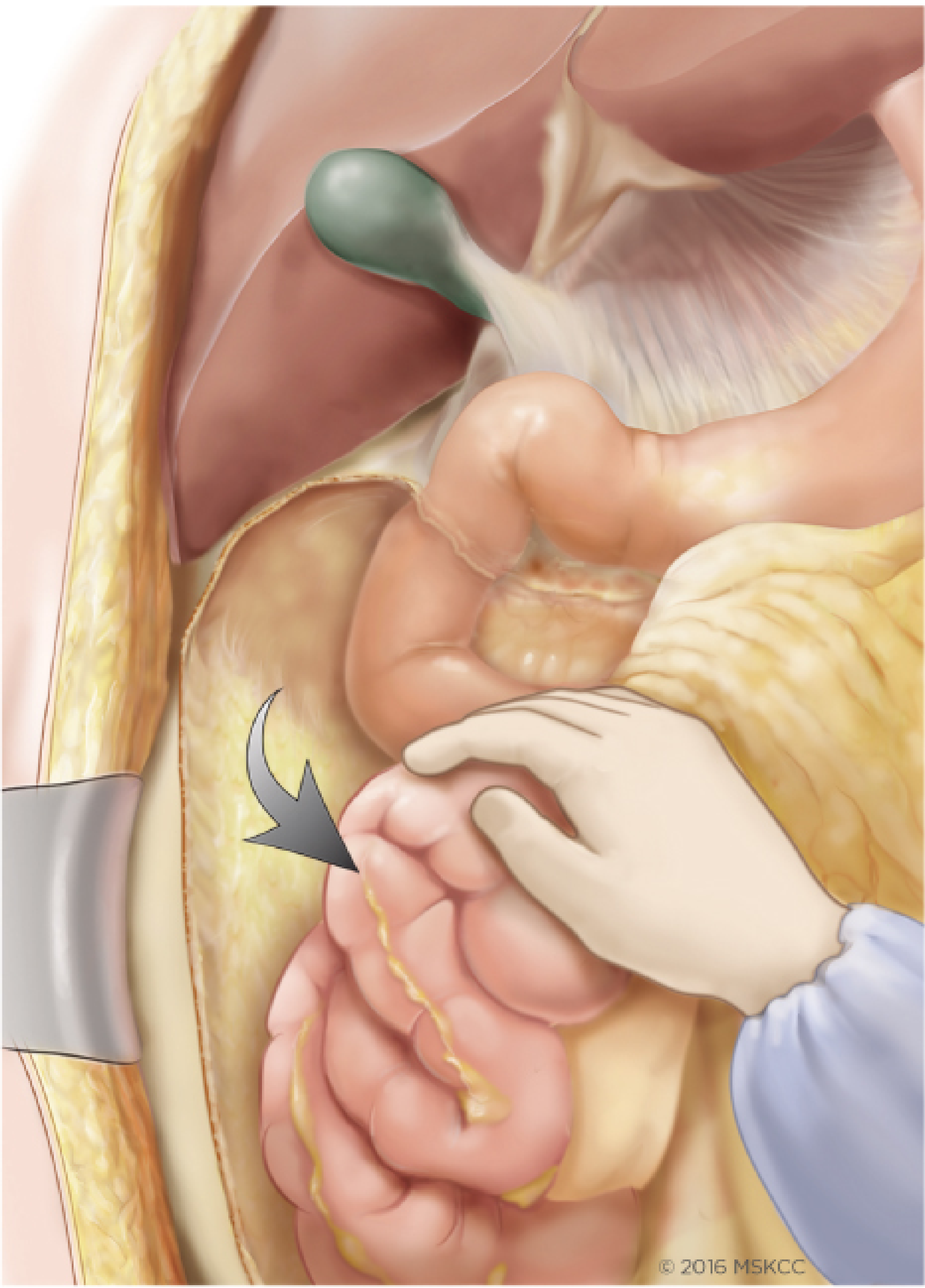
Step 3 - Omentum
Step 4 - Vascular Control (Ileocolic Pedicle)
Step 5 - Division of the Bowel
- Terminal ileum: divided 10-15 cm proximal to the ileocecal valve with a linear stapler (GIA/Endo-GIA).
- Transverse colon: divided at the appropriate level (mid-transverse for standard resection, more distal for extended right hemicolectomy) with a linear stapler.
Step 6 - Ileocolic Anastomosis
- An enterotomy (in the ileum) and a colostomy (in the transverse colon) are made on the antimesenteric borders.
- The GIA-80 stapler is inserted into both lumens and deployed, creating a wide side-to-side anastomosis.
- The common opening is then closed by deploying a TA-90 stapler across the remaining enterotomy/colostomy.
Step 7 - Closure
Laparoscopic Right Hemicolectomy
Positioning and Port Placement
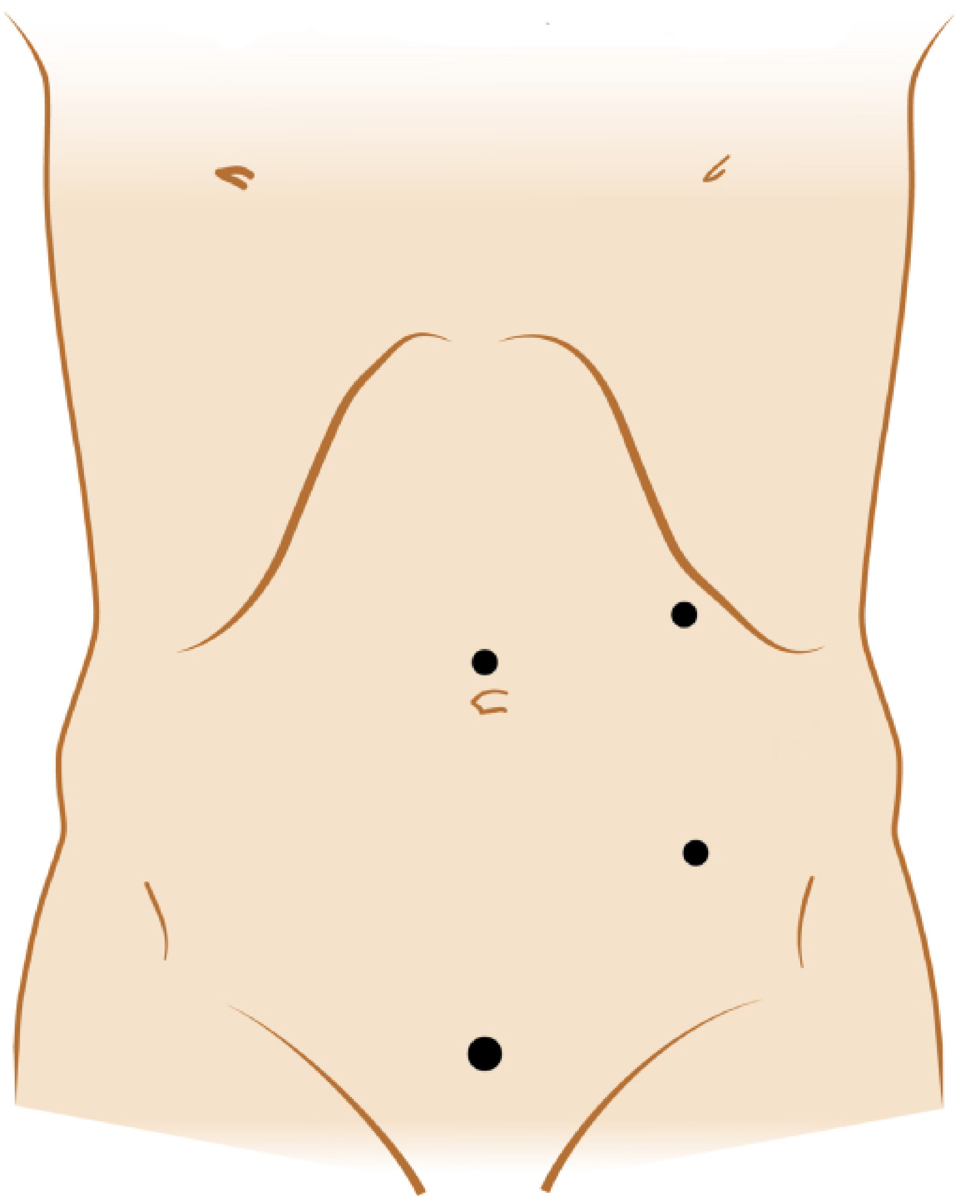
- Supraumbilical port - camera port (also the planned extraction site)
- Suprapubic port - working port
- Left upper quadrant port - working port (lateral to rectus)
- Left lower quadrant port - working port (lateral to rectus)
Medial-to-Lateral Approach (Standard Laparoscopic Technique)
- The terminal ileum and cecum are retracted to the right lower quadrant, placing the ileocolic pedicle under tension.
- The peritoneum is incised below the ileocolic pedicle at the level of the duodenum.
- Submesenteric dissection proceeds from medial to lateral - the right colon mesentery is lifted off the retroperitoneum, exposing the duodenum, Gerota's fascia, gonadal vessels, and ureter. The visceral fascial layer is kept intact (CME principle).
- The ileocolic pedicle is cleared, ligated, and divided at its SMA/SMV origin - using a vessel-sealing device, endoscopic vascular stapler, or clips.
- The transverse colon is placed on tension; the transverse mesocolon is scored; the right branch of the middle colic pedicle is ligated and divided. The mesentery is fully divided to the transverse colon.
- The omentum is dissected off the distal transverse colon with entry into the lesser sac.
- The hepatic flexure attachments are divided medial to lateral - the previous medial-to-lateral dissection has already freed the retroperitoneal plane, keeping the ureter, kidney, and gonadal vessels posteriorly.
- The white line of Toldt is divided laterally to complete mobilization.
- The terminal ileum mesentery is divided to the bowel wall.
Extraction and Extracorporeal Anastomosis
Robotic Right Hemicolectomy
Extent of Resection (Oncological Principles - CME)
| Principle | Detail |
|---|---|
| Plane of dissection | Sharp dissection in the embryological mesocolic plane - intact visceral fascia |
| High vascular ligation | At the SMA/SMV origin for maximum lymph node yield |
| Lymph node harvest | Minimum 12 nodes required for adequate staging |
| Resection margins | Minimum 5 cm proximal and distal to the tumor |
Postoperative Care (Enhanced Recovery After Surgery - ERAS)
- Diet: Early oral fluids same day of surgery; soft diet by day 1-2
- Ambulation: Encouraged from day 0 post-operatively
- Nasogastric tube: Not routinely used
- Urinary catheter: Removed within 24 hours
- Analgesia: Multimodal - non-opioid analgesics (NSAIDs, paracetamol), wound infiltration, epidural or transversus abdominis plane (TAP) block
- VTE prophylaxis: Low-molecular-weight heparin continued for 28 days after cancer surgery
- Hospital stay: Typically 3-5 days laparoscopic, 5-7 days open
Key Anatomical Hazards
| Structure at Risk | Consequence if Injured |
|---|---|
| Right ureter | Ureteric injury, fistula, stricture |
| Right gonadal vessels | Bleeding, gonadal ischaemia |
| Duodenum (2nd/3rd part) | Duodenal fistula - catastrophic |
| Head of pancreas small veins | Troublesome bleeding requiring suture ligature |
| SMV/SMA | Major haemorrhage |
| Right hepatic flexure - bile duct proximity | Bile duct injury (uncommon) |
Anastomotic Leak - Key Points
- Incidence: approximately 2-4% for right-sided anastomoses (lower than left-sided)
- Predisposing factors: tension, poor blood supply, distal obstruction, malnutrition, steroids
- The ileocolic anastomosis has a better blood supply and lower intraluminal pressure than left-sided anastomoses, explaining the lower leak rate
- In elderly patients, loss of the ileocecal valve after right hemicolectomy may increase stool frequency, and in some cases cause fecal incontinence - important in preoperative counseling