
About blood pathways
Reading File
The topic "blood pathways" is broad - it could mean the circulatory pathway (systemic vs. pulmonary circulation), blood flow through the heart chambers, fetal circulation, or even cellular/molecular pathways in blood. Let me clarify before diving in.
Asking for Preferences
Which aspect of blood pathways are you interested in?
I'll provide a general overview covering cardiac, pulmonary, systemic, and fetal circulation. Let me pull from the medical library now.
Finding Sources
Finding Sources
Finding Sources
Finding Sources
Finding Sources
Reading File
Finding Sources
Reading File
Finding Sources
Loading Image
Reading File
Loading Image
Loading Image
Reading File
I now have excellent material from multiple authoritative textbooks. Here is a thorough overview of blood pathways.
Blood Pathways - A Complete Overview
The circulatory system has three main circuits: the pulmonary circulation, the systemic circulation, and the special portal circulation. A completely separate set of pathways exists in fetal life that closes at birth.
1. The Two Main Circuits
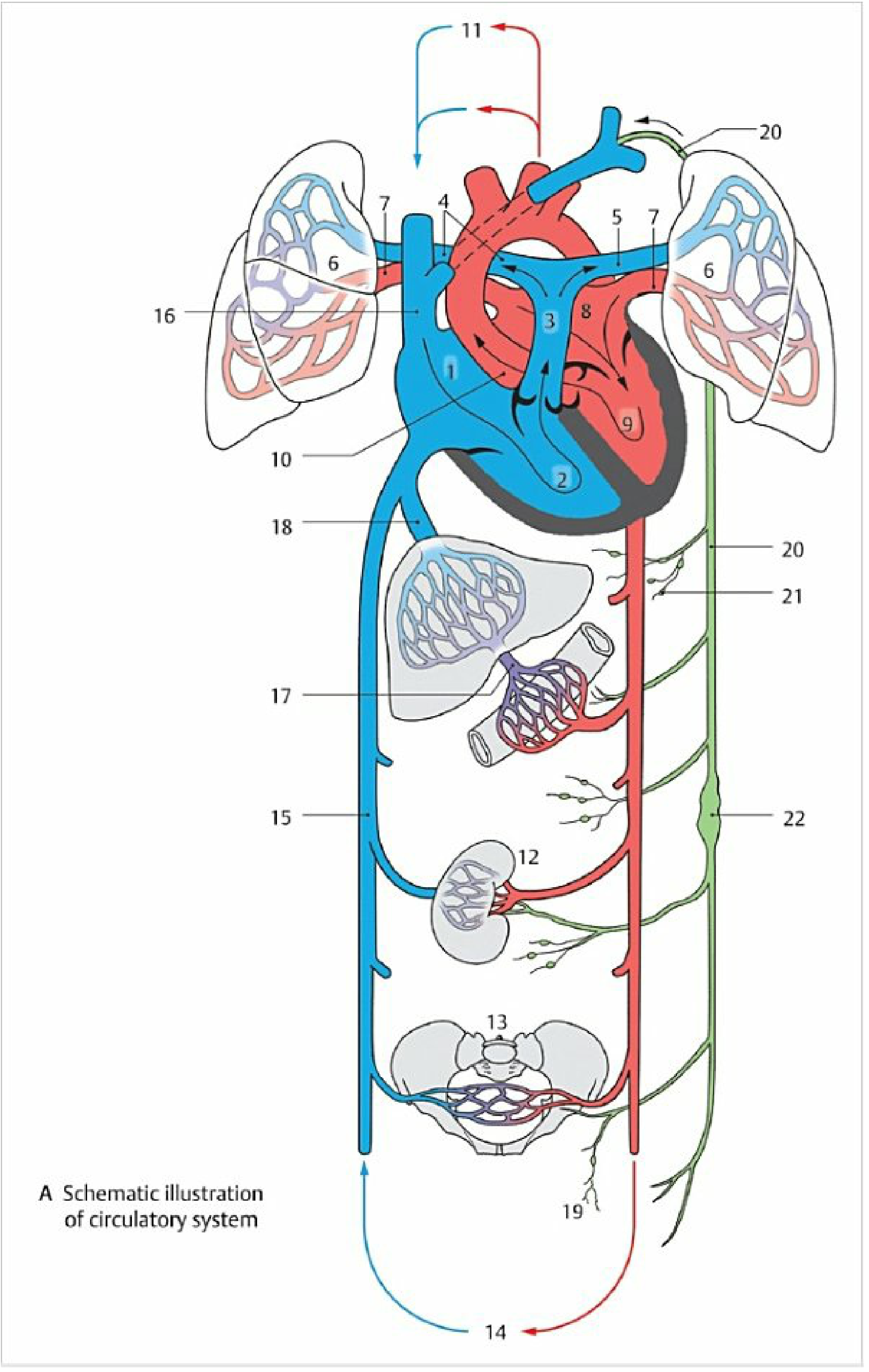
In a resting adult, roughly 70% of blood is in the systemic circulation, 18% in the pulmonary circulation, and 12% in the heart itself. - Junqueira's Basic Histology
Pulmonary Circulation (Right Heart - Lungs - Left Heart)
This is the short, low-pressure circuit for gas exchange.
Pathway: Deoxygenated blood from the body → Right Atrium → (tricuspid valve) → Right Ventricle → (pulmonary valve) → Pulmonary Trunk → splits into Right & Left Pulmonary Arteries → Lung Capillaries (O₂ in, CO₂ out) → Pulmonary Veins (x4) → Left Atrium
Key points:
- The pulmonary circulation is the only circuit where arteries carry deoxygenated blood and veins carry oxygenated blood.
- The driving pressure is only ~15 mmHg (vs. ~95 mmHg in the systemic circuit), making it a low-resistance, high-flow system. - Medical Physiology
Systemic Circulation (Left Heart - Body - Right Heart)
This is the long, high-pressure circuit supplying all organs.
Pathway: Oxygenated blood → Left Atrium → (mitral valve) → Left Ventricle → (aortic valve) → Aorta → Arteries → Arterioles → Capillaries (gas and nutrient exchange) → Venules → Veins → Inferior Vena Cava (lower body) + Superior Vena Cava (head, arms) → Right Atrium
- Color Atlas of Human Anatomy, Vol. 2
Portal Circulation (Special Systemic Circuit)
Venous blood from the stomach, intestine, pancreas, and spleen does NOT drain directly to the inferior vena cava. Instead:
Gut capillaries → Portal Vein → Liver sinusoids (metabolic processing) → Hepatic Veins → Inferior Vena Cava → Right Atrium
This allows the liver to process absorbed nutrients and detoxify substances before they reach the systemic circulation.
2. The Heart as a Pump - Step by Step
| Step | Location | Valve Opens | Valve Closes |
|---|---|---|---|
| 1 | Systemic venous blood fills Right Atrium via SVC + IVC | - | - |
| 2 | Tricuspid valve opens → blood enters Right Ventricle | Tricuspid | - |
| 3 | RV contracts → Pulmonary valve opens → blood enters pulmonary trunk | Pulmonary | Tricuspid |
| 4 | Blood oxygenated in lungs → returns via Pulmonary Veins → Left Atrium | - | - |
| 5 | Mitral valve opens → blood enters Left Ventricle | Mitral | - |
| 6 | LV contracts → Aortic valve opens → blood enters aorta | Aortic | Mitral |
3. Fetal Circulation
Fetal blood pathways are fundamentally different because the lungs are non-functional and the placenta acts as the gas-exchange organ. Three special shunts exist that close at birth.
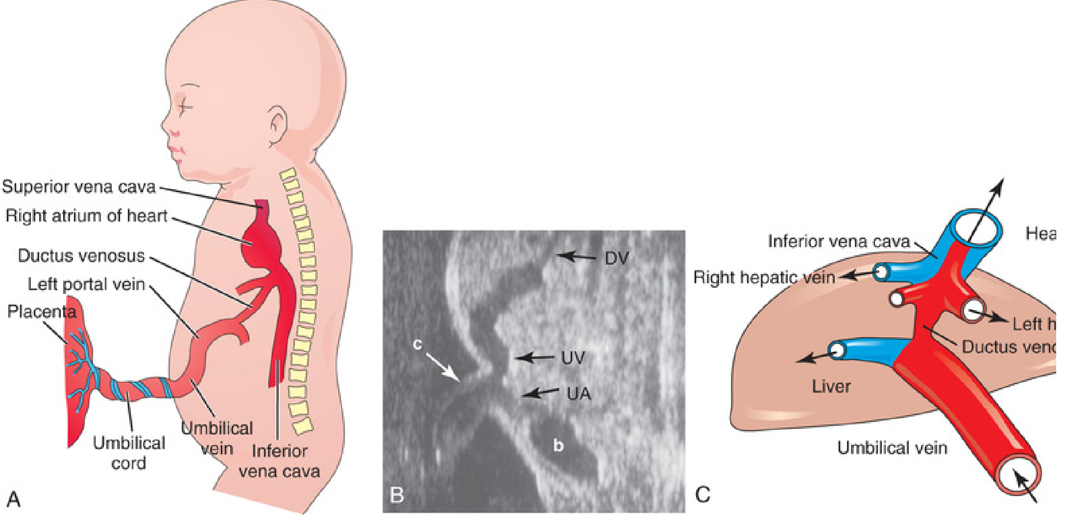
The Three Fetal Shunts
| Shunt | Connects | Purpose | Adult Remnant |
|---|---|---|---|
| Ductus Venosus | Umbilical vein → IVC | Bypasses liver (~50% of umbilical blood) | Ligamentum venosum |
| Foramen Ovale | Right Atrium → Left Atrium | Bypasses pulmonary circuit | Fossa ovalis |
| Ductus Arteriosus | Pulmonary trunk → Descending Aorta | Bypasses non-functional lungs | Ligamentum arteriosum |
Fetal Blood Flow Step by Step
- Oxygenated blood from the placenta travels via the umbilical vein.
- ~50% bypasses the liver via the ductus venosus → IVC → Right Atrium.
- The crista dividens (lower edge of septum secundum) directs most IVC blood through the foramen ovale → Left Atrium → Left Ventricle → Ascending Aorta → head and upper limbs (best-oxygenated blood goes to the brain).
- The remaining blood in the Right Atrium mixes with deoxygenated blood from the SVC → Right Ventricle → Pulmonary Trunk.
- Only ~10% of that blood goes to the (fluid-filled) lungs; the remaining ~90% takes the ductus arteriosus → Descending Aorta → body + umbilical arteries back to the placenta.
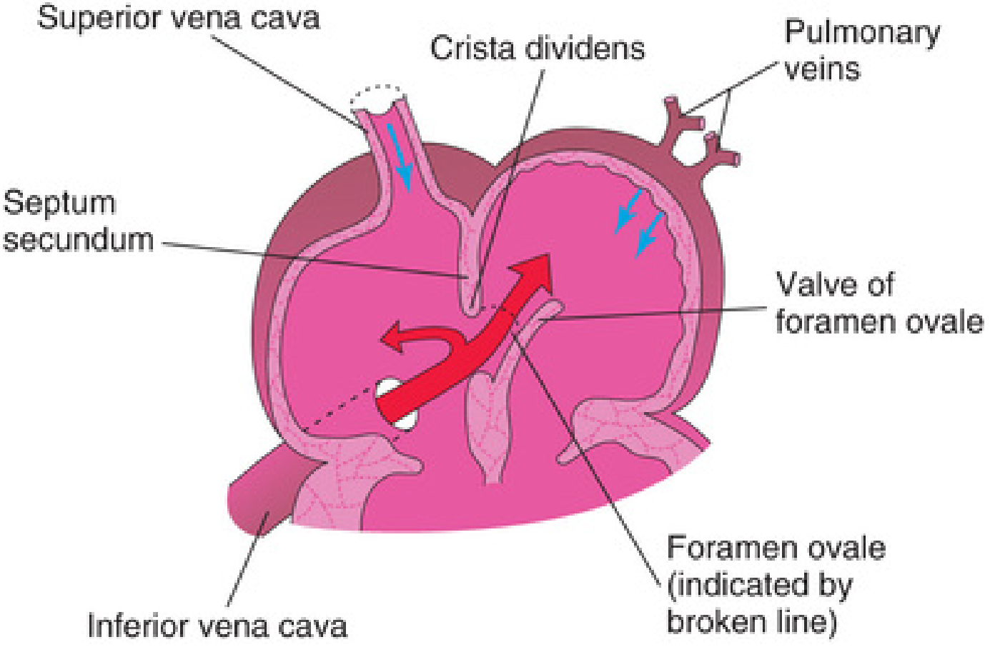
- The Developing Human: Clinically Oriented Embryology
4. Circulatory Changes at Birth
When the baby takes its first breath, lung resistance drops dramatically and four simultaneous changes occur:
- Foramen ovale closes - increased left atrial pressure (from pulmonary blood flow) pushes the valve flap shut → becomes the fossa ovalis.
- Ductus arteriosus closes - rising O₂ tension triggers smooth muscle contraction → becomes the ligamentum arteriosum.
- Ductus venosus closes - loss of umbilical flow triggers sphincter contraction → becomes the ligamentum venosum.
- Umbilical vessels obliterate - cord clamping causes thrombosis → umbilical vein becomes the round ligament of the liver; umbilical arteries become the medial umbilical ligaments.
- Color Atlas of Human Anatomy, Vol. 2
Summary Comparison
| Feature | Systemic | Pulmonary | Fetal (before birth) |
|---|---|---|---|
| Driving pressure | ~95 mmHg | ~15 mmHg | Low (placenta-driven) |
| Arteries carry | Oxygenated blood | Deoxygenated blood | Mixed (shunts active) |
| Gas exchange site | Tissue capillaries | Alveolar capillaries | Placenta |
| Resistance | High | Low | High pulmonary resistance |
This is a shared conversation. Sign in to Orris to start your own chat.