I now have comprehensive, detailed information from multiple authoritative textbooks. Let me compile the full answer.
Oral Cancer: Stages and Treatment in Detail
Overview
Oral cavity cancer is the most common type of head and neck cancer (HNC), with over 377,000 cases reported worldwide in 2020. More than 95% are squamous cell carcinomas (OCSCC). The oral cavity has seven distinct subsites: lips, buccal mucosa, alveolar ridge, retromolar trigone, floor of mouth, oral tongue, and hard palate. Five-year survival rates have improved from 59% (1990) to 70% (2011).
Risk factors: tobacco, betel quid, alcohol (combined use raises risk 45-fold), poor oral hygiene, and repetitive trauma.
Pathology and Grading
The World Health Organization (WHO) histological grading system classifies OCSCC by differentiation:
| Grade | Differentiation | Behavior |
|---|
| G1 | Well differentiated | Less aggressive |
| G2 | Moderately differentiated | Intermediate |
| G3 | Poorly differentiated | Highly aggressive, metastasizes readily |
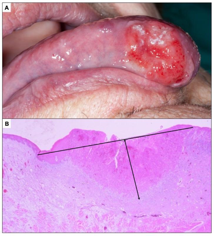
Key histological parameters: tumor grade, pattern of invasion (cohesive vs. non-cohesive), depth of invasion (DOI), perineural invasion (PNI), lymphovascular invasion (LVI), and bone involvement.
Staging (AJCC 8th Edition TNM)
The current AJCC 8th edition introduced two major updates:
- Depth of invasion (DOI) now incorporated into T-staging
- Extranodal extension (ENE) now upstages nodal disease
T (Primary Tumor) Classification
| T Stage | Criteria |
|---|
| TX | Primary tumor cannot be assessed |
| T0 | No evidence of primary tumor |
| Tis | Carcinoma in situ |
| T1 | Tumor ≤2 cm AND DOI ≤5 mm |
| T2 | Tumor ≤2 cm with DOI >5 mm and ≤10 mm, OR tumor >2 cm but ≤4 cm with DOI ≤10 mm |
| T3 | Tumor >4 cm OR any tumor with DOI >10 mm |
| T4a (oral) | Invades adjacent structures: cortical bone, deep tongue musculature, maxillary sinus, or skin of face (moderately advanced) |
| T4b (oral) | Invades masticator space, pterygoid plates, skull base, or encases the internal carotid artery (very advanced, unresectable) |
| T4 (lip) | Invades cortical bone, inferior alveolar nerve, floor of mouth, or skin of face |
DOI is a pathologically determined feature from the surgical specimen; it can be estimated radiologically but is confirmed histologically. It has direct prognostic impact:
- DOI <2 mm: 13% regional metastasis rate, 95% 5-year survival
- DOI 2-9 mm: 46% regional metastasis, 85% 5-year survival
- DOI >9 mm: 65% regional metastasis, 65% 5-year survival
N (Regional Lymph Node) Classification
| N Stage | Criteria |
|---|
| NX | Cannot be assessed |
| N0 | No regional metastasis |
| N1 | Single ipsilateral node <3 cm, ENE-negative |
| N2a | Single ipsilateral node <3 cm + ENE-positive, OR single ipsilateral node >3 cm but <6 cm, ENE-negative |
| N2b | Multiple ipsilateral nodes, all <6 cm, ENE-negative |
| N2c | Bilateral or contralateral nodes, all <6 cm, ENE-negative |
| N3a | Node >6 cm, ENE-negative |
| N3b | Node >3 cm + ENE-positive, OR multiple nodes with any ENE-positive |
ENE (extranodal extension) = tumor extending through the lymph node capsule into surrounding tissue; a major negative prognostic factor introduced in the 8th edition.
M (Distant Metastasis)
| Stage | Meaning |
|---|
| M0 | No distant metastasis |
| M1 | Distant metastasis present (most common sites: lung, liver, bone) |
Overall Stage Grouping
| Stage | T | N | M |
|---|
| Stage 0 | Tis | N0 | M0 |
| Stage I | T1 | N0 | M0 |
| Stage II | T2 | N0 | M0 |
| Stage III | T3 | N0 | M0 |
| Stage IVA | T4a | N0-N2 | M0 |
| Stage IVB | T4b | any N | M0 |
| Stage IVC | Any T | Any N | M1 |
Patterns of Lymph Node Metastasis by Subsite
| Primary Site | Lymph Node Levels at Risk |
|---|
| Upper lip | Levels IB, II; perifacial nodes |
| Lower lip | Levels IA, IB; level II |
| Upper alveolar ridge | Levels IB, II |
| Lower alveolar ridge / floor of mouth | Levels IA, IB, II |
| Retromolar trigone | Levels IIA, IIB, III |
| Hard palate / buccal mucosa | Levels IB, II |
| Oral tongue | Levels I, II, III |
Floor-of-mouth carcinoma has ~50% incidence of cervical metastasis; anterior lesions frequently metastasize bilaterally.
Workup and Diagnosis
- Clinical examination: thorough systematic palpation and inspection of all subsites; leukoplakia and erythroplakia (5-7x higher malignant potential) require biopsy
- Imaging: CT and MRI are mandatory; CT is more sensitive/specific than panorex for mandibular invasion
- EUA (examination under anesthesia): for lesions not fully assessable in clinic
- Sentinel lymph node biopsy (SLNB): used in some centers for staging early (cN0) oral cancers; a positive SLNB prompts completion neck dissection
Warning signs: non-healing ulcer >2 weeks, persistent red/white lesion, unexplained tooth mobility, sensory nerve deficit, trismus, dysphagia, otalgia.
Treatment
Stage I and II (Early Disease - T1-T2, N0)
Primary modality: Surgery
- Wide local excision with adequate margins is the standard of care for early oral cavity cancers
- Oral tongue: partial glossectomy; reconstruction with local flaps, primary closure, or regional flap depending on defect size
- Floor of mouth: transoral resection; marginal mandibulectomy considered when tumor is near periosteum
- Surgical margins: clear margins (typically ≥5 mm) are paramount; close or positive margins mandate re-excision or adjuvant radiation
- Neck: Elective neck dissection (END) is recommended when occult metastasis risk exceeds 20%. Selective neck dissection (SND) of levels I-III is standard for most oral cavity primaries
Adjuvant radiotherapy may be added when:
- Perineural invasion present
- Lymphovascular invasion
- Close surgical margins
- Multiple lymph nodes involved
Stage III (T3N0 or T1-T3 N1)
Surgery + Adjuvant Therapy
- More extensive resection required; mandibular involvement may require marginal or segmental mandibulectomy
- Neck dissection is routinely performed; SND levels I-III for most, extending to levels IV-V for advanced disease
- Adjuvant radiotherapy (60-66 Gy) with or without concurrent cisplatin-based chemotherapy, depending on pathological features
- Reconstruction with regional pedicled flaps (pectoralis major) or microvascular free flaps
Stage IVA (T4a, resectable)
Surgery + Adjuvant Chemoradiotherapy
- Composite resection may include segmental mandibulectomy, infrastructure or total maxillectomy
- Mandibular resection options:
- Marginal mandibulectomy: periosteum involved but not cortical/medullary bone; removes superior alveolar rim
- Segmental mandibulectomy: full-thickness bone removal; mandatory when medullary invasion present
- Reconstruction: microvascular free tissue transfer (e.g., fibula free flap for mandibular reconstruction, anterolateral thigh flap, radial forearm free flap) is the standard for large defects
- Adjuvant concurrent chemoradiation: cisplatin-based CRT is standard for:
- Extranodal extension (ENE)
- Positive surgical margins (that cannot be re-resected)
- Multiple positive nodes
Stage IVB and IVC (Unresectable / Metastatic)
Definitive (Primary) Chemoradiotherapy
- Reserved for medically inoperable patients, T4b (unresectable) disease, or patient preference against surgery
- Radiation (IMRT preferred): 66-70 Gy to primary and involved nodes; 44-54 Gy to elective nodal volumes
- IMRT (Intensity-Modulated Radiation Therapy) reduces xerostomia, osteoradionecrosis, and dysphagia compared to conventional techniques
- Concurrent chemotherapy: cisplatin 100 mg/m² every 3 weeks or weekly cisplatin is standard
- Organ preservation trials show similar survival outcomes compared to surgery + adjuvant therapy, but patients may experience greater toxicity from definitive (versus adjuvant) doses
- Chemotherapy is never used as a single agent for oral cavity cancer
For metastatic (M1) disease:
- Platinum-based palliative chemotherapy (cisplatin + 5-FU ± cetuximab)
- Immunotherapy: pembrolizumab (anti-PD-1) is now approved for recurrent/metastatic HNC
- Goals shift to symptom control and quality of life
Adjuvant Therapy Indications (Post-Surgery)
| Feature | Adjuvant RT | Adjuvant CRT (RT + Chemo) |
|---|
| Perineural invasion | Yes | Consider |
| Lymphovascular invasion | Yes | Consider |
| Close margins (<5 mm) | Yes | Consider |
| Positive margins | Yes | Yes |
| pN+ (node positive) | Yes | Consider |
| ENE | Yes | Yes (standard) |
| Multiple positive nodes | Yes | Yes |
Prognosis by Stage (Floor of Mouth as Example)
| Stage | 5-Year Survival |
|---|
| Stage I | ~90% |
| Stage II | ~80% |
| Stage III | ~65% |
| Stage IV | ~30% |
Overall oral cavity SCC: locoregional disease-free survival ~60% for T2-T4; overall disease-free survival ~50%. Distant metastases most commonly occur in lung, liver, and bone.
Treatment Side Effects and Complications
- Xerostomia (dry mouth): most common after radiation; worsened by bilateral parotid irradiation
- Osteoradionecrosis: bone necrosis after radiation, especially mandible; risk increased with high doses, dental disease, and smoking
- Dysphagia/trismus: from radiation fibrosis, tongue base involvement
- Dysgeusia: altered taste, often during/after radiation
- IMRT significantly reduces but does not eliminate these toxicities
Sources
- Sabiston Textbook of Surgery, 21st ed. - Oral Cavity Cancer
- Cummings Otolaryngology Head and Neck Surgery, 7th ed. - Staging and Surgical Treatment
- Bailey and Love's Short Practice of Surgery, 28th ed. - TNM Staging (AJCC 8th edition)
- Scott-Brown's Otorhinolaryngology Head & Neck Surgery - Cancer of the oral cavity