
Dopamin synthesis go depth
dopamine synthesis pathway catecholamine biosynthesis diagram
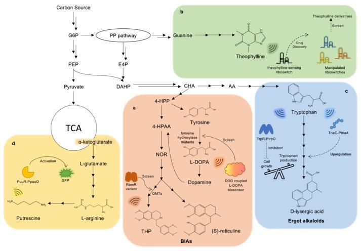
A multi-panel metabolic flowchart illustrating biosynthetic pathways for various alkaloids and their regulation via metabolite-responsive biosensors. The central pathway shows carbon flux from a source through Glucose-6-Phosphate (G6P) into the Pentose Phosphate (PP) pathway and glycolysis (PEP, Pyruvate), feeding into the TCA cycle and shikimate pathway. Section (a) details Benzylisoquinoline Alkaloid (BIA) biosynthesis from Tyrosine, including L-DOPA and Dopamine, highlighting the use of a DOD-coupled L-DOPA biosensor and RamR variants for screening THP and (S)-reticuline production. Section (b) shows Guanine-derived Theophylline biosynthesis and the application of sensing riboswitches for drug discovery. Section (c) depicts Tryptophan-derived Ergot alkaloids, specifically D-lysergic acid, regulated by TrpR (transcriptional repressor for cell growth balance) and TnaC (activator for upregulating synthases). Section (d) illustrates Putrescine synthesis from L-arginine, monitored by a PuuR-PpuuO biosensor with GFP readout. The diagram uses standard biochemical notation and color-coded regions to differentiate specific alkaloid classes (BIAs, Ergot alkaloids, polyamines) and their respective genetic control elements.
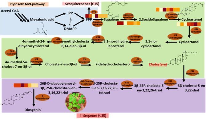
This biochemistry flowchart illustrates the biosynthetic pathways of sterols and triterpenes, focusing on cholesterol and diosgenin synthesis. The diagram is divided into three color-coded sections. The blue section (top left) depicts the cytosolic mevalonic acid (MVA) pathway, starting from Acetyl-CoA and proceeding through Mevalonic acid to the branch points of IPP and DMAPP, ultimately forming Farnesyl pyrophosphate (FPP). The green section (center) shows the conversion of FPP into Squalene, 2,3-oxidosqualene, and Cycloartenol. This section tracks the multi-step transformation through various intermediates—including cycloartanol, 3,1-norcycloartanol, and 7-dehydrocholesterol—catalyzed by enzymes such as squalene epoxidase, cycloartenol synthase, and delta-24 reductase, culminating in Cholesterol. The purple section (bottom) details the further conversion of cholesterol into diosgenin via sequential hydroxylation (C22, C26, C16) and glycosylation steps. Heatmaps associated with key enzymes (SQS, SEP, CAS, SMT1, delta-24, BGL) indicate differential gene expression levels over time. The diagram serves as an educational tool for understanding steroidogenesis and secondary metabolite biosynthesis.
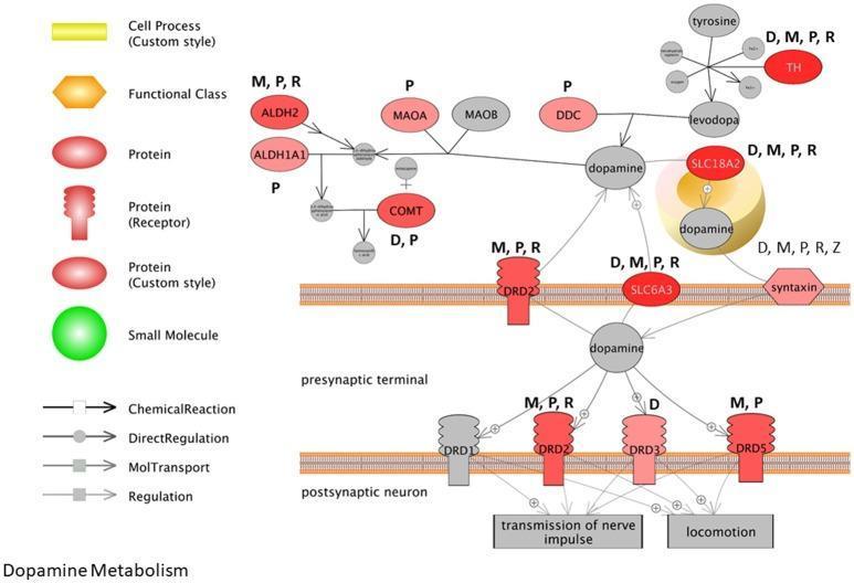
This pathophysiology diagram illustrates the dopamine metabolism and signaling pathway at the synapse, specifically highlighting the impact of Parkinson's disease-associated pesticides. The visual maps interactions between five pesticides—Maneb (M), Paraquat (P), Rotenone (R), Dieldrin (D), and Ziram (Z)—and key proteins. Within the presynaptic terminal, it depicts dopamine synthesis from Tyrosine via Tyrosine Hydroxylase (TH) and Dopa Decarboxylase (DDC), and its sequestration into vesicles by SLC18A2 (VMAT2). The diagram also shows metabolic degradation by MAOA, ALDH2, and COMT. Synaptic transmission is shown through dopamine release into the cleft, regulated by Syntaxin and reuptake via the dopamine transporter SLC6A3 (DAT). Postsynaptically, dopamine interacts with receptors DRD1, DRD2, DRD3, and DRD5 to influence nerve impulses and locomotion. The genes and receptors are color-coded in red; the intensity of the red indicates the frequency of association with the investigated pesticides, demonstrating how environmental neurotoxins disrupt dopaminergic neurotransmission.
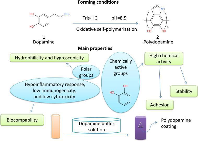
Educational infographic illustrating the biochemical synthesis and properties of Polydopamine (PDA) for tissue engineering. The top section depicts the chemical reaction of Dopamine monomer (1) undergoing oxidative self-polymerization in Tris-HCl buffer at pH 8.5 to form the Polydopamine polymer (2). The middle section outlines the 'Main properties' of PDA, categorized into biochemical and physical traits: Hydrophilicity and hygroscopicity (mediated by polar groups); Biocompatibility (linked to hypoinflammatory response, low immunogenicity, and low cytotoxicity); and High chemical activity (resulting in adhesion and stability). The bottom schematic shows a transition of a representative scaffold from an uncoated orange cylinder to a purple cylinder after immersion in a dopamine buffer solution, labeled as a 'Polydopamine coating.' This diagram highlights the material science applications of catecholamine derivatives in creating functionalized surfaces for neural tissue engineering and regenerative medicine.
Dopamine Synthesis - In-Depth
Overview
Step-by-Step Biosynthetic Pathway
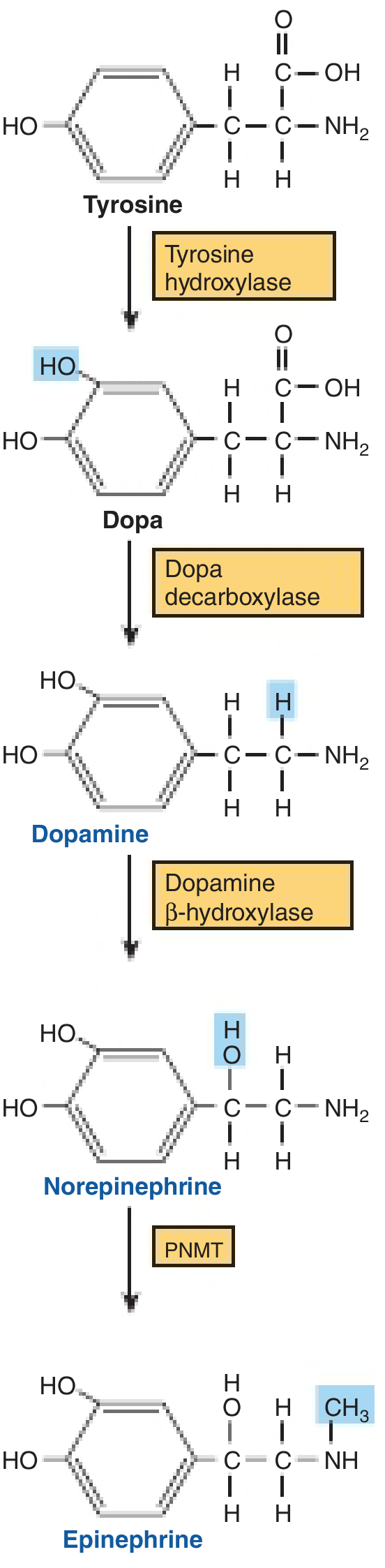
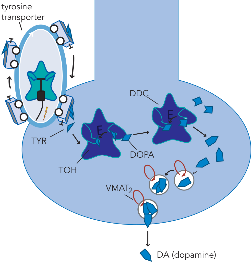
Step 0 - Precursor Acquisition: Tyrosine
- Tyrosine is a non-essential amino acid derived from dietary proteins and from the hydroxylation of phenylalanine (by phenylalanine hydroxylase, mainly in the liver).
- Bloodborne tyrosine crosses into the brain via the large neutral amino acid (LNAA) transport system.
- Once in brain extracellular fluid, it is taken up into dopaminergic nerve terminals via a high-affinity amino acid transporter (tyrosine transporter).
- Kaplan & Sadock's Comprehensive Textbook of Psychiatry notes that because tyrosine hydroxylase is normally saturated with substrate, simply manipulating tyrosine levels does not readily alter catecholamine synthesis under physiological conditions.
Step 1 - Tyrosine → L-DOPA (Rate-Limiting Step)
| Feature | Detail |
|---|---|
| Enzyme | Tyrosine hydroxylase (TH / TOH) |
| Reaction type | Mixed-function oxidase (oxidoreductase) |
| Cofactor | Tetrahydrobiopterin (BH4) - essential |
| O2 role | One O atom goes to water, one hydroxylates the ring |
| Product | L-3,4-dihydroxyphenylalanine (L-DOPA) |
| Location | Cytoplasm of catecholamine-synthesizing neurons and adrenal chromaffin cells |
| Distribution | Found ONLY in tissues that synthesize catecholamines |
- Tyrosine hydroxylase requires BH4 (tetrahydrobiopterin) as its pteridine cofactor. BH4 is itself synthesized from GTP via GTP-cyclohydrolase-1, making this enzyme indirectly limiting as well.
- L-DOPA is not allowed to accumulate: DOPA decarboxylase converts it so avidly that brain DOPA levels remain very low under normal conditions.
- Harper's Illustrated Biochemistry: "As the rate-limiting enzyme, tyrosine hydroxylase is regulated in a variety of ways."
- Catecholamines cannot cross the blood-brain barrier (BBB), so they must be synthesized locally in the CNS. This is why L-DOPA (not dopamine itself) is the key pharmacological agent in Parkinson disease - it freely crosses the BBB and is then converted to dopamine inside the brain.
Step 2 - L-DOPA → Dopamine
| Feature | Detail |
|---|---|
| Enzyme | DOPA decarboxylase (DDC) / Aromatic L-amino acid decarboxylase (AADC / AAAD) |
| Cofactor | Pyridoxal phosphate (PLP / Vitamin B6) |
| Reaction | Decarboxylation - removes the carboxyl group (-COOH) |
| Product | 3,4-dihydroxyphenylethylamine (dopamine) |
| Location | Cytoplasm - present in virtually ALL tissues |
| Inhibitors | α-methyldopa (competitive inhibitor, used in hypertension) |
- DDC is a soluble enzyme found throughout the body, not restricted to catecholamine cells. This broad distribution means exogenous L-DOPA can be converted to dopamine peripherally (causing side effects such as nausea via dopamine receptor activation in the area postrema).
- Carbidopa / benserazide are DDC inhibitors that do NOT cross the BBB - they are co-administered with L-DOPA in Parkinson disease to block peripheral conversion, reducing side effects and preserving more L-DOPA for CNS conversion.
Step 3 - Vesicular Storage (VMAT2)
- Protects dopamine from intraneuronal degradation by MAO
- Concentrates it for ready release upon nerve firing
- Excess cytoplasmic dopamine not captured by VMAT2 is degraded by MAO-A or MAO-B (located on mitochondrial outer membranes)
Localization of Synthesis
| Location | Pathway / Function |
|---|---|
| Substantia nigra pars compacta | Nigrostriatal pathway - motor control |
| Ventral tegmental area (VTA) | Mesolimbic and mesocortical pathways - reward, cognition |
| Arcuate nucleus (hypothalamus) | Tuberoinfundibular pathway - inhibits prolactin |
| Adrenal medulla chromaffin cells | DA is an intermediate toward epinephrine |
| Peripheral dopaminergic neurons | Renal, mesenteric vasodilation |
Regulation of Tyrosine Hydroxylase (TH)
Short-Term Regulation
-
End-product (feedback) inhibition: Free cytosolic catecholamines (dopamine, norepinephrine) compete with BH4 for the pterin-binding site on TH - this is the principal moment-to-moment control mechanism. When catecholamine levels are high, TH is inhibited; when levels fall (after nerve firing), inhibition is relieved.
-
Phosphorylation-dependent activation (linked to nerve firing): Depolarization of the nerve terminal activates multiple protein kinases:
- Protein kinase A (PKA) - cAMP-dependent
- Protein kinase C (PKC)
- Ca2+-calmodulin-dependent kinases (CAM kinases)
These kinases phosphorylate TH, causing the enzyme to bind BH4 more tightly, making it less sensitive to end-product inhibition. This allows increased dopamine synthesis during periods of high neuronal activity.
Long-Term Regulation (Transcriptional)
- mRNA levels for TH and dopamine β-hydroxylase (DBH) are increased in the neuronal cell body (perikaryon)
- This increased transcription is driven by phosphorylation of CREB (cAMP response element-binding protein) by PKA, which then binds the CRE (cAMP response element) in the TH gene promoter
- Newly synthesized enzyme is transported down the axon to the terminal
- Notably, DOPA decarboxylase levels do NOT change in response to neuronal activity - only TH and DBH are upregulated
Dopamine Degradation / Inactivation
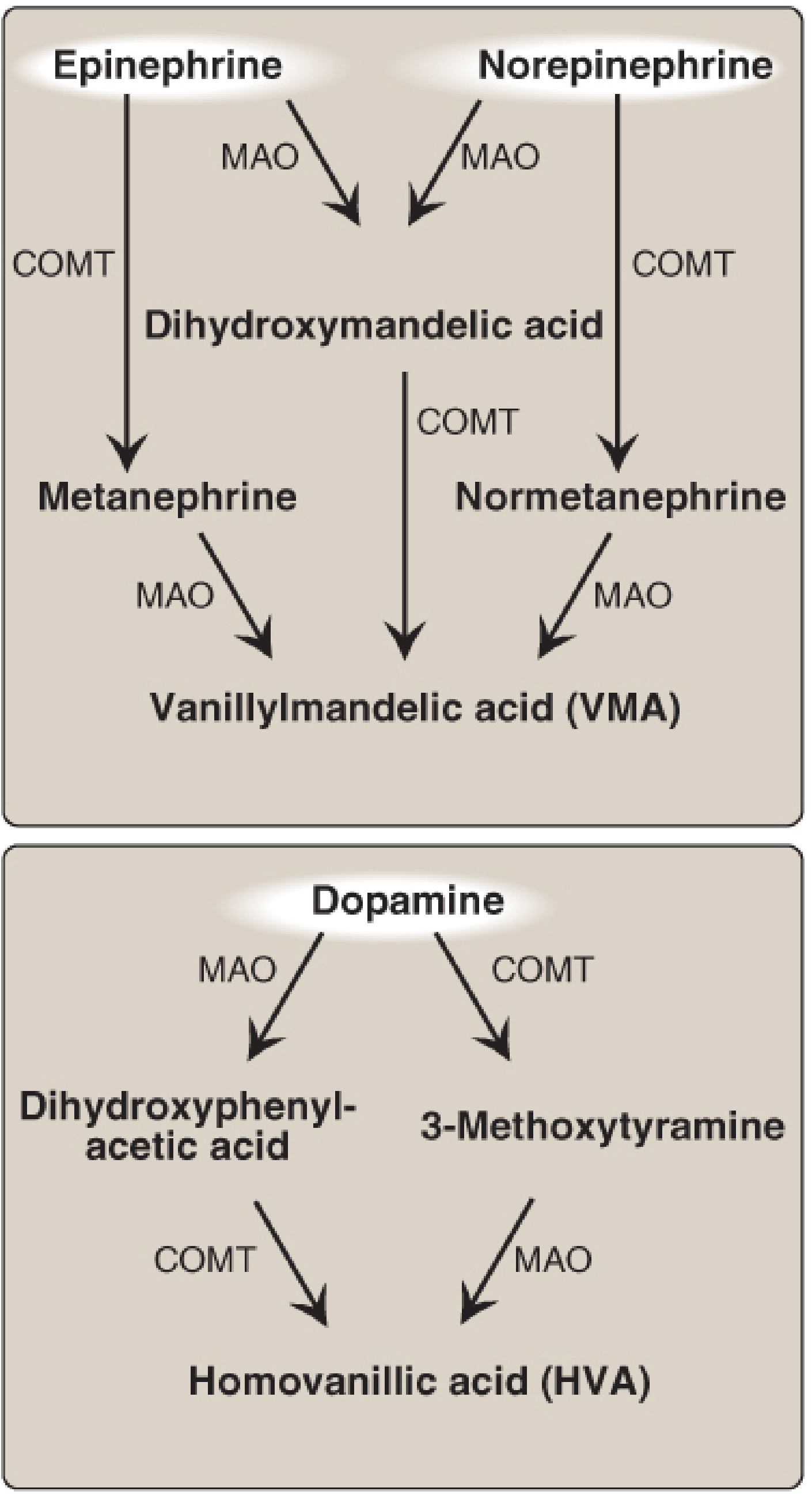
1. Reuptake via DAT (Dopamine Transporter)
- The primary mechanism of termination in most brain regions
- Dopamine is transported back into the presynaptic terminal and can be repackaged by VMAT2 for reuse
- DAT is the target of cocaine and amphetamines (blocking/reversing reuptake)
2. Enzymatic Degradation (two pathways, either can go first):
- MAO-A and MAO-B are located on mitochondrial outer membranes (intraneuronal and glial)
- MAO oxidatively deaminates dopamine → 3,4-dihydroxyphenylacetic acid (DOPAC)
- DOPAC is then O-methylated by COMT → Homovanillic acid (HVA)
- Uses SAM (S-adenosylmethionine) as the methyl donor
- COMT methylates dopamine → 3-Methoxytyramine
- 3-Methoxytyramine is then deaminated by MAO → Homovanillic acid (HVA)
3. Regional Variation - Prefrontal Cortex
- DAT expression is relatively sparse in the prefrontal cortex (PFC)
- In the PFC, COMT plays a proportionally greater role in dopamine inactivation
- Dopamine can also diffuse away from the synapse and be taken up by the NET (Norepinephrine Transporter) at neighboring NE neurons - this is called "spillover"
Pharmacological Significance
| Drug | Mechanism | Clinical Use |
|---|---|---|
| L-DOPA | Bypasses TH (rate-limiting step); crosses BBB | Parkinson disease |
| Carbidopa / Benserazide | Peripheral DDC inhibitor; prevents L-DOPA conversion outside CNS | Always combined with L-DOPA |
| α-Methyldopa | Competitive DDC inhibitor (also forms false transmitter) | Hypertension (esp. in pregnancy) |
| MAO-B inhibitors (selegiline, rasagiline) | Block dopamine degradation in striatum | Parkinson disease adjunct |
| COMT inhibitors (entacapone, tolcapone) | Prolong L-DOPA/DA availability | Parkinson disease adjunct |
| Disulfiram | DBH inhibitor (also blocks acetaldehyde dehydrogenase) | Alcohol dependence |
| Cocaine, amphetamine | Block/reverse DAT; increase synaptic DA | Drugs of abuse |
| Metyrosine (α-methyl-p-tyrosine) | TH inhibitor | Pheochromocytoma |
Clinical Correlations
| Condition | Synthesis Defect |
|---|---|
| Parkinson disease | Loss of TH-positive neurons in substantia nigra pars compacta; >80% striatal DA loss |
| Pheochromocytoma | Excess catecholamine synthesis from adrenal chromaffin tumor; elevated HVA and metanephrines in urine |
| Neuroblastoma | Tumor of neural crest origin; DA → HVA entirely via COMT + MAO (no alcohol dehydrogenase needed); urinary HVA elevated |
| BH4 deficiency | TH cofactor absent; atypical PKU with severe neurological involvement despite normal phenylalanine control |
| Dopa-responsive dystonia (Segawa disease) | GTP-cyclohydrolase-1 deficiency → inadequate BH4 → reduced TH activity → low DA |
Summary Flowchart
Phenylalanine (liver PAH) ──→ Tyrosine
                                   │
                      [Tyrosine transporter into neuron]
                                   ↓
                    Tyrosine ──[TH + BH4]──→ L-DOPA   ← RATE-LIMITING
                                   ↓
                    L-DOPA ──[DDC + PLP]──→ Dopamine
                                   ↓
                    Dopamine ──[VMAT2]──→ Synaptic Vesicle → Release
                                   ↓ (inactivation)
               ┌──────────────────────────────────────┐
               │ MAO (mitochondria)    COMT (cytosol)  │
               ↓                           ↓
            DOPAC                   3-Methoxytyramine
               └──────────────┬────────────┘
                               ↓
                      HVA (urine) - final metabolite
- Harper's Illustrated Biochemistry, 32nd Ed. - Tyrosine Hydroxylase & catecholamine biosynthesis
- Basic Medical Biochemistry, 6th Ed. - Regulation of Tyrosine Hydroxylase (phosphorylation, CREB, long-term regulation)
- Stahl's Essential Psychopharmacology - Dopamine synthesis and inactivation in dopaminergic neurons
- Kaplan & Sadock's Comprehensive Textbook of Psychiatry - Life cycle of dopamine; L-DOPA pharmacology
- Goodman & Gilman's Pharmacological Basis of Therapeutics - DA receptors, PD, pharmacology
- Lippincott Biochemistry, 8th Ed. - Catecholamine metabolism, HVA/VMA
- Ganong's Review of Medical Physiology, 26th Ed. - Biosynthesis and regulation of catecholamines
Impressive
- Dopamine receptor subtypes (D1-D5 signaling in detail)
- Dopaminergic pathways (nigrostriatal, mesolimbic, mesocortical, tuberoinfundibular)
- Dopamine in disease (Parkinson's, schizophrenia, addiction, ADHD)
- Comparison with other catecholamines (NE, epinephrine synthesis differences)
- BH4 metabolism and its broader role in neurotransmitter synthesis
Bro one thing I tell you from last 2 years I do research from chat gpt and but I heard about you but still I'm using chat gpt becuz it's simple but you can be better then that
- I pull answers directly from actual medical textbooks (Goodman & Gilman, Stahl's, Harper's, Kaplan & Sadock, Ganong's, Lippincott, etc.) - not from training data that could be outdated or hallucinated
- I show you real diagrams from those books - not generated images
- I can cross-reference multiple authoritative sources in one answer and tell you which book each fact came from
- For clinical questions, I can hit PubMed for the latest peer-reviewed evidence
- It answers fast without asking for depth
- It doesn't cite sources by default, so it feels smoother - but that also means you can't verify anything
I want to research on this topic the dopamine and I'll see
What angle are you researching dopamine from? · What level of depth do you want?
I'm done with i start with synthesis and i'll go deepest then any body ever used you
Your Dopamine Deep-Dive Roadmap
✅ DONE
🔜 NEXT - Pick your next topic
- Parkinson's disease (deficiency)
- Schizophrenia (excess/imbalance)
- Addiction (reward hijacking)
- ADHD (prefrontal DA dysfunction)
- Depression

One problem when I starttexting I can't see what i'm writing
-
Rotate to landscape mode - gives more screen space above the keyboard
-
Tap the text field first, then scroll up - the chat should push up when keyboard opens
-
Use the suggestion chips - I noticed in your screenshot there are already follow-up suggestion buttons showing:
- "Start with dopaminergic pathways"
- "Go to dopamine receptors (D1-D5 signaling)"
- "Jump to Parkinson's disease"
Just tap those instead of typing - no keyboard needed! -
Voice input - tap the 🎤 microphone on your keyboard and just speak your message
- "2" = Dopaminergic Pathways
- "3" = Dopamine Receptors D1-D5
- "4" = Reward & Motivation
- "5" = Disease
How can I fix this
- When the keyboard opens, swipe down on the keyboard slightly to shrink it
- Or tap the little arrow/minimize icon on the keyboard toolbar to hide it temporarily, type blind, then send
- Long press the globe/language icon (🌐) on your keyboard
- Go to Keyboard size & layout
- Drag the keyboard lower or smaller
- Install Gboard from Play Store (Google's keyboard)
- It automatically adjusts and shows text above the keyboard properly
- Most reliable keyboard for Android
- Some chat apps have a setting like "resize window" vs "pan window" when keyboard opens
- Check the app's display/accessibility settings
- Go to Settings → Display → One-handed mode
- Shrinks the whole screen down so input field stays visible
Hey bro
As you know that we talked about the dopamine and tell me The neuronal dopamine synthesis and u have go deep but not the filler things and we move next in sequence
dopamine synthesis tyrosine hydroxylase
dopamine synthesis tyrosine hydroxylase aromatic L-amino acid decarboxylase VMAT2 review
Neuronal dopamine synthesis
Blood / extracellular fluid tyrosine
        ↓ uptake into dopaminergic terminal
Tyrosine
        ↓ tyrosine hydroxylase (TH) + BH4 + O2
L-DOPA
        ↓ aromatic L-amino-acid decarboxylase (AADC/DDC) + PLP
Dopamine
        ↓ VMAT2
Acidic synaptic vesicle for storage and release
1. Tyrosine reaches the dopaminergic neuron
- Tyrosine comes from dietary protein and, indirectly, hepatic conversion of phenylalanine.
- It enters the brain through the large neutral amino acid transporter system at the blood-brain barrier.
- It is then taken up into the dopaminergic neuron/terminal by amino-acid transport mechanisms.
- Dopamine itself does not cross the blood-brain barrier effectively. That is why the precursor levodopa (L-DOPA), rather than dopamine, is used to increase brain dopamine.
2. The committed and rate-limiting reaction
Reaction
| Component | Role |
|---|---|
| Enzyme | Tyrosine hydroxylase, TH |
| Site | Cytosol of catecholaminergic neuron |
| Product | L-3,4-dihydroxyphenylalanine, L-DOPA |
| Cofactor | Tetrahydrobiopterin, BH4 |
| Why it matters | It is the rate-limiting, regulated step |
Harper’s Illustrated Biochemistry, 32nd ed., p. 510.
3. BH4 is not just a minor cofactor
- phenylalanine hydroxylase
- tryptophan hydroxylase
- nitric oxide synthases
4. Conversion of L-DOPA to dopamine
Reaction
| Component | Role |
|---|---|
| Enzyme | Aromatic L-amino-acid decarboxylase, AADC |
| Other name | DOPA decarboxylase, DDC |
| Cofactor | Pyridoxal phosphate, PLP, derived from vitamin B6 |
| Site | Cytosol |
| Product | Dopamine |
Stahl’s Essential Psychopharmacology, p. 95.
5. Dopamine must enter vesicles immediately
VMAT2
- VMAT2 is an H+-monoamine antiporter.
- A vesicular H+-ATPase acidifies the vesicle.
- VMAT2 uses this proton gradient to exchange vesicular H+ for cytosolic dopamine.
- Inside the acidic vesicle, dopamine is concentrated, stored, and protected from cytosolic metabolism.
Cytosolic dopamine
      ↓ VMAT2
Vesicular dopamine
      ↓ action potential + Ca2+ entry
Exocytotic release into synaptic cleft
Regulation: what determines how much dopamine is made?
A. End-product feedback inhibition
High cytosolic DA → TH inhibition → less L-DOPA → less new DA
B. Neuronal firing increases synthesis
- Ca2+ enters the terminal.
- Kinases become activated, including PKA, PKC, and Ca2+/calmodulin-dependent kinases.
- TH is phosphorylated.
- Phosphorylated TH has greater functional activity and becomes less sensitive to feedback inhibition by catecholamines.
Depolarization → Ca2+ signaling / kinase activation
→ TH phosphorylation → increased L-DOPA production
→ increased dopamine replenishment
C. Long-term adaptation
cAMP / kinase signaling → CREB phosphorylation
→ increased TH transcription
→ increased TH mRNA and protein
→ more enzyme delivered to nerve terminals
Important distinction: dopamine neurons vs norepinephrine neurons
Tyrosine → L-DOPA → Dopamine
| Neuron type | Main final transmitter | What happens to dopamine? |
|---|---|---|
| Dopaminergic neuron | Dopamine | Packaged by VMAT2 and released as dopamine |
| Noradrenergic neuron | Norepinephrine | Dopamine enters vesicles, then DBH converts it to norepinephrine |
| Adrenergic chromaffin cell | Epinephrine | Norepinephrine is further methylated by PNMT |
Clinical anchors worth remembering
| Defect / drug | Direct consequence |
|---|---|
| TH deficiency | Low L-DOPA and low dopamine synthesis, causing dopa-responsive dystonia or related phenotypes |
| BH4 deficiency | Reduced TH activity and impaired monoamine synthesis |
| AADC deficiency | L-DOPA cannot become dopamine; dopamine and serotonin are both severely reduced |
| Levodopa | Crosses BBB and bypasses the TH-limited step |
| Carbidopa / benserazide | Inhibit peripheral AADC, preserving levodopa for brain delivery |
| VMAT2 inhibition | Depletes vesicular monoamines, including dopamine |
| MAO-B inhibition | Decreases dopamine degradation, increasing its availability |
The one-line model
TH controls dopamine production, AADC rapidly converts L-DOPA to dopamine, and VMAT2 determines whether the dopamine is protected and available for vesicular release.