
What happen when T vwave repeat?
Reading File
The question is about T wave repeat on ECG - this likely refers to a "U wave" (which follows the T wave) or possibly "T wave alternans" or re-entrant T wave phenomena. Let me search the medical library for the correct content.
Quick answer: When a T wave appears to "repeat" on ECG, this is known as the U wave - a second, smaller deflection following the T wave. It is most visible in leads V2-V3. A prominent U wave can indicate hypokalemia, bradycardia, or drug effects. Alternatively, "T wave alternans" (alternating T wave morphology) is linked to ventricular arrhythmia risk.
Searching sources...
Finding Sources
Finding Sources
Now let me read the detailed Harrison's section on T wave and U wave, and fetch the relevant ECG images:
Reading File
Reading File
Loading Image
Loading Image
When a T Wave Appears to "Repeat" - The U Wave
When a second small positive deflection appears after the T wave on ECG, it is called the U wave. This is the "repeat" or extra bump you see following the T wave.
What is the U Wave?
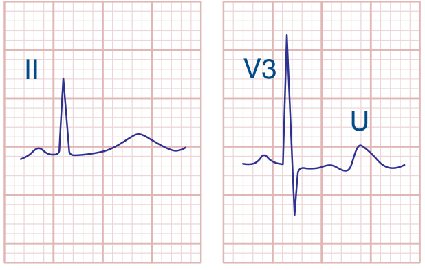
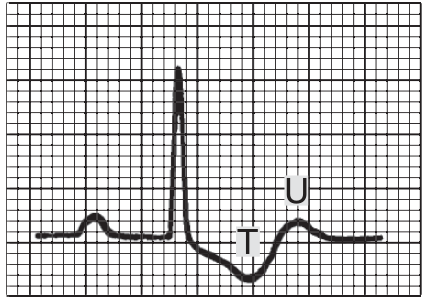
The U wave is a small, rounded deflection (≤1 mm) that follows the T wave and normally has the same polarity as the T wave (both upright in most leads). It is best seen in leads V2-V3.
Origin (still debated):
- Late repolarization of the Purkinje system
- Delayed repolarization of mid-myocardial M cells
(Guyton and Hall Textbook of Medical Physiology)
ECG showing a prominent U wave in hypokalemia (lead V3):
(Harrison's Principles of Internal Medicine, 22E)
Normal vs. Abnormal U Wave
| Feature | Normal U wave | Abnormal (Prominent) U wave |
|---|---|---|
| Amplitude | ≤1 mm | >1 mm (larger than T wave) |
| Polarity | Same as T wave | Can be inverted |
| Appearance | Barely visible | Clearly visible, sometimes merges with T |
Causes of a Prominent (Large) U Wave
-
Hypokalemia - most common cause
- As K+ drops, the U wave becomes progressively larger
- At K+ ~3.5 mEq/L: prominent U wave present, ST depression
- At K+ ~2.5 mEq/L: T wave inverts, U wave very prominent, PR interval lengthens
- Important trap: The U wave can be mistaken as part of the T wave, causing a falsely prolonged QT interval to be measured (e.g., true QT = 0.4s but appears as 0.6s)
-
Drugs - dofetilide, amiodarone, sotalol, quinidine
-
Bradycardia
-
Hypercalcemia, hypomagnesemia
(Ganong's Review of Medical Physiology; Harrison's 22E)
Why Prominent U Waves Are Clinically Important
Very prominent U waves indicate prolonged ventricular repolarization. This is a marker of increased susceptibility to Torsades de Pointes (TdP) - a dangerous polymorphic ventricular tachycardia that can degenerate into ventricular fibrillation and sudden cardiac death.
(Harrison's Principles of Internal Medicine, 22E - T Wave and U Wave section)
ECG diagram showing U wave following T wave:
(Ganong's Review of Medical Physiology - Hypokalemia ECG changes)
Key Clinical Takeaway
If you see what looks like a "repeated T wave" on an ECG:
- Check the patient's serum potassium (hypokalemia is #1 cause)
- Review medications (especially antiarrhythmics)
- Measure the true QT interval carefully - do not include the U wave in your QT measurement
- Be alert for risk of Torsades de Pointes
This is a shared conversation. Sign in to Orris to start your own chat.