
Give me all set of image related questions for breastfeeding week and breastmilk
breastfeeding technique and positions
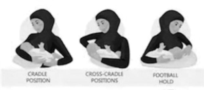
Summary : This illustration demonstrates three common breastfeeding positions used by mothers to hold their infants: cradle position, cross-cradle position, and football hold. photo: Scene Overview : • The image shows three side-by-side illustrations of a mother holding a baby in different breastfeeding positions. • Each position is labeled below the corresponding illustration: "CRADLE POSITION," "CROSS-CRADLE POSITION," and "FOOTBALL HOLD." • The mother is depicted wearing a dark, long-sleeved garment and a head covering. • The baby is held close to the mother's chest in each position. Technical Details : • The illustrations are simple, with clear outlines and minimal shading. • No scale bar, magnification, or technical imaging details are present. • The background is plain and uncluttered. Spatial Relationships : • Left: Cradle position – mother’s arm supports the baby’s head and body, baby lies horizontally across the front. • Center: Cross-cradle position – mother’s opposite arm supports the baby’s head, with the baby’s body across the front. • Right: Football hold – mother holds the baby under her arm, with the baby’s body tucked alongside her side. Analysis : • The figure visually compares three breastfeeding holds, highlighting differences in arm placement and baby orientation. • The cradle and cross-cradle positions both involve the baby lying across the mother’s front, but with different supporting arms. • The football hold positions the baby along the mother’s side, which may be useful for certain feeding situations or comfort preferences.
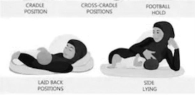
Summary : This illustration demonstrates four common breastfeeding positions for mothers and infants, each labeled with its respective name and depicted with simple, shaded figures. illustration: # Breastfeeding Positions : • Cradle Position: Mother reclined, baby held across her body in a traditional cradle hold. • Cross-Cradle Position: Mother reclined, baby held across her body with opposite arm supporting the baby. • Football Hold: Mother seated, baby tucked under her arm at her side, similar to holding a football. • Side Lying: Mother and baby both lying on their sides facing each other. # Labels & Layout : • Top row: Position names—Cradle Position, Cross-Cradle Positions, Football Hold. • Bottom row: Laid Back Positions (left), Side Lying (right). • Each position is visually separated and clearly labeled above or below the corresponding illustration. # Scene Overview : • Simple, monochrome drawing style. • Figures are shown on soft surfaces (pillows or mats). • No background details; focus is on body posture and baby placement. # Spatial Relationships : • Left side: Laid back positions (mother reclined, baby on chest). • Right side: Side lying (mother and baby parallel on their sides). • Each position demonstrates a different angle and arm placement for supporting the baby. Analysis : • The figure provides a clear visual comparison of four distinct breastfeeding positions, highlighting differences in body orientation and arm support. It serves as an educational guide for mothers to choose a comfortable and effective feeding posture.
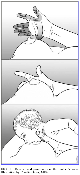
Summary : This illustration demonstrates the "dancer hand position" technique for breastfeeding, as viewed from the mother's perspective. The figure consists of three panels showing the step-by-step hand placement and support for effective latch and feeding. illustration: # Scene Overview : • The main subject is a mother’s hand supporting her breast and the infant’s head during breastfeeding. • The perspective is from the mother’s viewpoint, focusing on the hand, breast, and infant. • The illustration is in grayscale, with clear anatomical labeling in the top panel. # Technical Details : • No scale bar or magnification is present. • The top panel labels anatomical landmarks: "cheek," "chin," and "breast." • The hand is shown with fingers supporting the breast and the thumb positioned near the infant’s cheek and chin. • The middle panel shows the hand maintaining support as the infant approaches the breast. • The bottom panel depicts the infant latched onto the breast, with the mother’s hand still in the dancer position. # Spatial Relationships : • The hand is consistently placed under the breast, with the thumb and fingers providing support and gentle pressure. • The infant’s head is cradled by the mother’s hand, ensuring alignment of the chin and cheek with the breast for optimal latch. • The sequence moves from preparation (top), approach (middle), to active feeding (bottom). # Analysis : • The illustration visually instructs on the dancer hand position, emphasizing anatomical alignment and support for successful breastfeeding. • The stepwise depiction clarifies the technique, showing how the hand stabilizes both the breast and the infant’s head to facilitate feeding. • The labeled anatomical points guide correct finger placement for mothers learning this method.
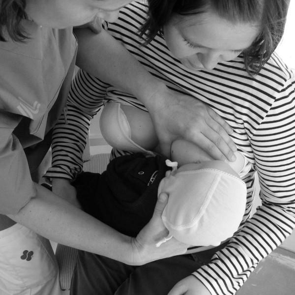
This clinical photograph captures a hands-on teaching interaction between a nurse and a mother within a neonatal or maternity care setting. The image demonstrates a breastfeeding support technique where the nurse is actively involved in positioning the infant for latching. The nurse, wearing professional medical scrubs, is positioned on the mother's left side. Her right hand is shown supporting the infant’s head and neck to maintain alignment, while her left hand is used to stabilize the mother’s breast to facilitate a proper latch. The mother is seated, wearing a striped shirt, and holds the infant close to her chest, focusing on the baby. The infant is wearing a bonnet and is swaddled, a common practice for maintaining warmth in neonatal care units. This visual illustrates the 'hands-on' approach to breastfeeding education, which is a clinical concept used to demonstrate physical positioning and latching techniques for new mothers, particularly in the context of preterm or sick infants requiring specialized support.

A black-and-white clinical photograph documenting an infant breastfeeding, serving as an educational example of maternal-child health and nutrition in global health settings. The image illustrates the 'cradle hold' positioning, with the infant's face turned toward the breast and a deep latch visible. The adult provides manual support to the infant's occiput to facilitate stable positioning during feeding. Notable cultural markers include stacked brass neck rings and wrist coils on the adult, which are characteristic of specific ethnic groups in Southeast Asia. This visual emphasizes the clinical significance of breastfeeding as a critical intervention for infant survival and immune support, particularly in resource-limited or emergency environments like refugee camps. Key educational concepts include lactation management, infant latch technique, and the sociocultural context of neonatal care.

A clinical photograph depicting postpartum lactation support in a healthcare setting. The image shows a mother in a hospital environment seated and breastfeeding a newborn infant. A lactation consultant or nurse, wearing a professional striped uniform, is positioned beside the mother, providing manual guidance to facilitate proper positioning and latch-on technique. The infant is supported by a patterned nursing pillow to optimize ergonomic alignment. This visual illustrates key concepts in neonatal care, maternal education, and the clinical management of breastfeeding. The background includes elements of a hospital bed, identifying the setting as a maternity ward or obstetric unit. This content is designed for medical education focusing on maternal-child health, nursing interventions, and the mechanics of early breastfeeding support.
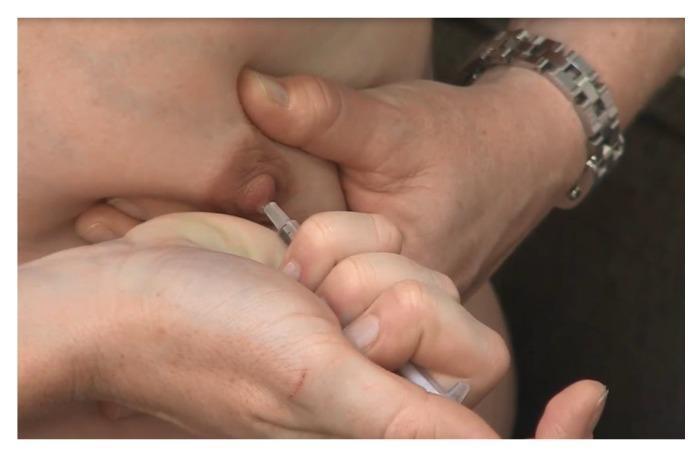
This clinical photograph captures a close-up procedural demonstration of Antenatal Colostrum Expression (ACE). The image illustrates the 'C-hold' hand technique, where the breast tissue is compressed between the thumb and fingers, positioned approximately 2-3 cm behind the nipple at the edge of the areola. A second hand is shown holding a small oral syringe directly at the nipple tip to collect the expressed droplets of colostrum. The procedural focus is on the manual stimulation and compression of the lactiferous sinuses to facilitate milk removal. This visual serves as an educational tool for lactation consultants and expectant mothers, highlighting the correct hand placement and the use of a syringe for hygienic colostrum harvesting and storage during late pregnancy. The setting is likely an antenatal clinic or educational session designed to improve breastfeeding confidence and knowledge.
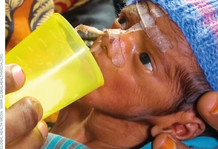
Clinical photograph of a preterm infant receiving nutritional support in a neonatal care setting. The image demonstrates a 'cup feeding' technique, an alternative oral feeding method for infants who cannot be directly breastfed. A healthcare provider or caregiver is shown holding a small, translucent yellow cup containing an opaque, off-white liquid (likely expressed breast milk or formula) to the infant's lips. The infant exhibits several medical devices and clinical features including a nasogastric or orogastric tube secured with adhesive medical tape across the cheek and nasal bridge for enteral feeding. Protective adhesive dressings are also visible on the forehead. The infant is wearing a knitted cap to assist with thermoregulation, a critical component of neonatal care. This visual illustrates neonatal nutritional management and the use of supplemental feeding implements in resource-limited or specific clinical scenarios where direct breastfeeding is not possible.
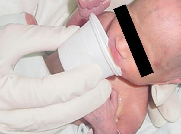
This clinical photograph demonstrates the alternative neonatal feeding method known as cup feeding. A caregiver, wearing white protective gloves, holds a small, white plastic medicine-style cup to the lips of an infant. The infant's identity is anonymized with a black bar over the eyes. The image captures the moment liquid (breast milk or formula) makes contact with the infant's lower lip, facilitating lapping or sipping movements rather than active suction used in bottle feeding. Visible fluid is noted trickling from the corner of the mouth onto the infant's chest, a common finding during the learning phase of this technique. The clinical context involves assessing masseter muscle electromyographic (EMG) activity post-electrode removal to compare oral motor patterns between breastfeeding, bottle feeding, and cup feeding. This image serves as an educational resource for neonatal nutrition, showing the proper positioning of the cup and the physical interaction between the caregiver and the neonate in a clinical or research setting.
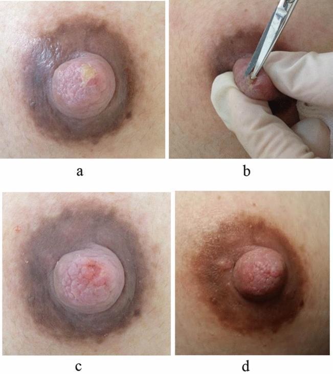
A series of four clinical photographs (a-d) documenting the management and healing of a chronic nipple injury in a breastfeeding patient. Image (a) shows the initial presentation of the nipple with visible white-matter keratinization and necrotic plaque on the superior surface, accompanied by hyperpigmentation of the areola. Image (b) captures the intraoperative debridement procedure where a clinician, using sterile technique, employs ophthalmic scissors to excise the keratinized necrotic tissue. Image (c) shows the immediate post-debridement state, revealing fresh, pink underlying tissue with the necrotic layer successfully removed. Image (d) illustrates the clinical outcome five days post-intervention, showing a completely healed nipple surface with resolved inflammation and restoration of normal tissue texture. This series demonstrates the effectiveness of mechanical debridement in treating breastfeeding-related nipple trauma and secondary hyperkeratosis to facilitate wound healing and pain relief.
breastmilk composition benefits nutrition
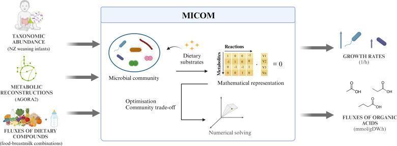
This pathophysiology diagram illustrates the workflow of the MICOM computational model used to predict the impact of infant nutrition on the gut microbiome. The flowchart is divided into three sections: inputs, model processing, and outputs. Inputs include taxonomic abundance data from weaning infants, metabolic reconstructions (using the AGORA2 database), and fluxes of dietary compounds from various food-breastmilk combinations. The central 'MICOM' box depicts the internal logic, showing a microbial community interacting with dietary substrates, represented mathematically via a metabolite-reaction matrix and community trade-off optimization. The model employs numerical solving to generate two primary outputs: microbial growth rates (measured in 1/h) and fluxes of organic acids (measured in mmol/gDW*h), including short-chain fatty acids like acetate, propionate, and butyrate. This educational visual explains the systems biology approach to neonatal nutrition and gastrointestinal microbiology, highlighting the relationship between diet, colonic microbiota metabolism, and the production of health-relevant metabolites.
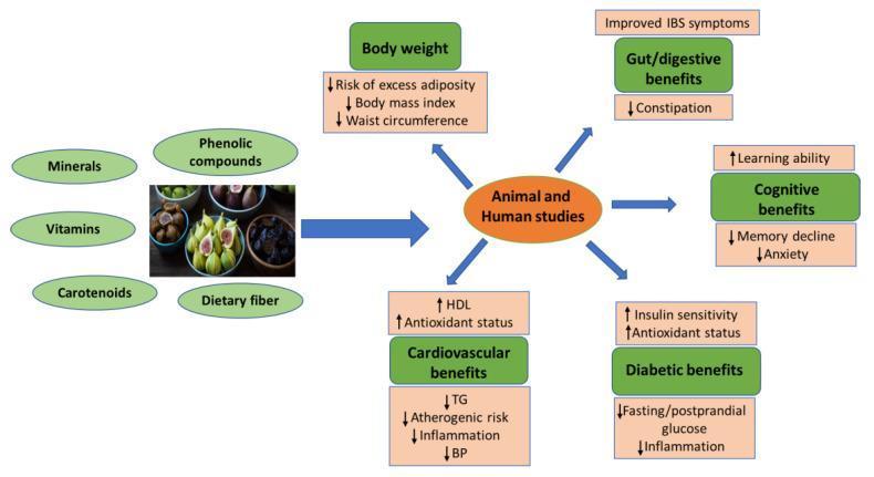
An educational infographic and flowchart summarizing the nutritional components and systemic health benefits of figs (Ficus carica) derived from animal and human studies. The left section identifies the primary bioactive phytochemicals and nutrients including minerals, vitamins, carotenoids, phenolic compounds, and dietary fiber. A central node representing 'Animal and Human studies' connects these components to five major clinical benefit categories. 1. Body weight: Reduced risk of adiposity, BMI, and waist circumference. 2. Gut/Digestive health: Improved IBS symptoms and decreased constipation. 3. Cognitive function: Increased learning ability and reduced memory decline/anxiety. 4. Cardiovascular health: Increased HDL and antioxidant status, with decreased triglycerides (TG), atherogenic risk, inflammation, and blood pressure (BP). 5. Diabetic management: Increased insulin sensitivity and antioxidant status, alongside decreased fasting/postprandial glucose and inflammation. The diagram uses color-coded nodes and directional arrows (up/down) to indicate physiological changes, serving as a comprehensive summary of fig-derived nutrition in preventive medicine and metabolic health.
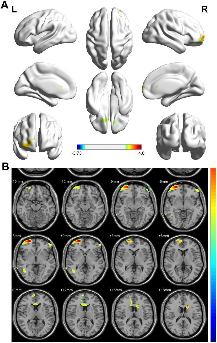
This diagnostic image displays a Voxel-Based Morphometry (VBM) comparison of regional gray matter volumes in infants. Panel A presents a 3D cortical surface mapping (brain render) from multiple orientations, including lateral, medial, superior, and inferior views. Yellow and red highlights indicate clusters of significantly higher gray matter volume in breastmilk-fed versus formula-fed infants, primarily localized to the bilateral frontal lobes, right temporal lobe, and focal areas of the occipital cortex. Panel B provides a complementary 2D visualization using a series of axial brain slices ranging from -15mm to +18mm in MNI space, following radiologic convention. These slices demonstrate significant volume differences in the prefrontal cortex (Brodmann areas 11 and 46), as well as subcortical involvement in the left caudate nucleus/basal ganglia region. A color-coded statistical scale represents T-values, with warmer colors indicating higher statistical significance. This neuroimaging study illustrates the impact of early nutrition on neonatal structural brain development and neuroanatomy.
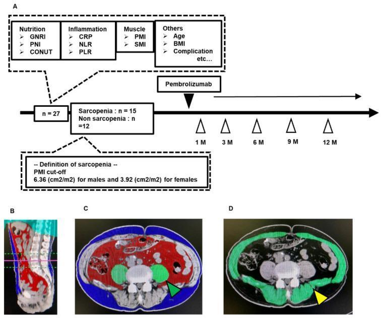
Educational figure illustrating a clinical study workflow and diagnostic imaging for body composition analysis in patients with urothelial carcinoma. Section A presents a flowchart detailing the assessment of 27 patients before and during Pembrolizumab treatment, highlighting biomarkers for nutrition (GNRI, PNI, CONUT), inflammation (CRP, NLR, PLR), and muscle mass (PMI, SMI). It defines sarcopenia based on sex-specific psoas muscle index (PMI) cut-offs. Sections B, C, and D showcase abdominal computed tomography (CT) images processed with the SYNAPSE VINCENT analysis system. Figure B provides a 3D sagittal reconstruction at the L3 level. Figure C is a color-coded axial/coronal slice differentiating abdominal subcutaneous adipose tissue (blue), visceral adipose tissue (red), and the psoas muscle (green, green arrow). Figure D identifies the total skeletal muscle area at the L3 level in green (yellow arrow). The visual demonstrates the quantitative radiological assessment of sarcopenia and body composition as prognostic factors in oncological management.
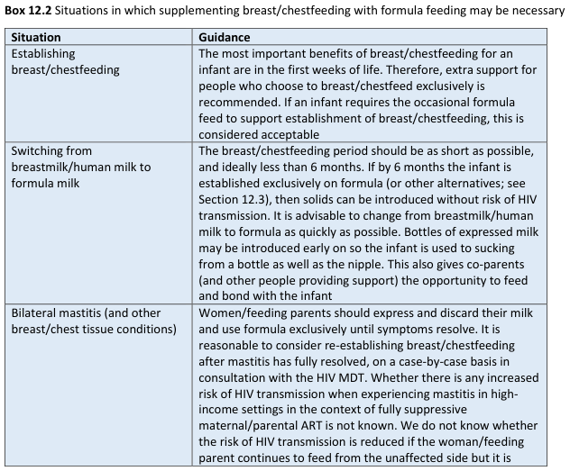
Box 12.2 Situations in which supplementing breast/chestfeeding with formula feeding may be necessary <table><thead><tr><th>Situation</th><th>Guidance</th></tr></thead><tbody><tr><td>Establishing breast/chestfeeding</td><td>The most important benefits of breast/chestfeeding for an infant are in the first weeks of life. Therefore, extra support for people who choose to breast/chestfeed exclusively is recommended. If an infant requires the occasional formula feed to support establishment of breast/chestfeeding, this is considered acceptable</td></tr><tr><td>Switching from breastmilk/human milk to formula milk</td><td>The breast/chestfeeding period should be as short as possible, and ideally less than 6 months. If by 6 months the infant is established exclusively on formula (or other alternatives; see Section 12.3), then solids can be introduced without risk of HIV transmission. It is advisable to change from breastmilk/human milk to formula as quickly as possible. Bottles of expressed milk may be introduced early on so the infant is used to sucking from a bottle as well as the nipple. This also gives co-parents (and other people providing support) the opportunity to feed and bond with the infant</td></tr><tr><td>Bilateral mastitis (and other breast/chest tissue conditions)</td><td>Women/feeding parents should express and discard their milk and use formula exclusively until symptoms resolve. It is reasonable to consider re-establishing breast/chestfeeding after mastitis has fully resolved, on a case-by-case basis in consultation with the HIV MDT. Whether there is any increased risk of HIV transmission when experiencing mastitis in high-income settings in the context of fully suppressive maternal/parental ART is not known. We do not know whether the risk of HIV transmission is reduced if the woman/feeding parent continues to feed from the unaffected side but it is</td></tr></tbody></table>
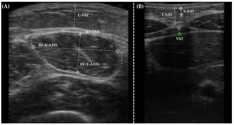
This diagnostic ultrasound image demonstrates 'Nutritional Ultrasound' techniques for assessing body composition through muscular and adipose tissue measurements. Image (A) shows a transverse view of the rectus femoris muscle. Key annotations include the rectus femoris cross-sectional area (RF-CSA) traced with a dotted white line, and its internal dimensions marked by the RF-X-AXIS and RF-Y-AXIS. The overlying leg subcutaneous adipose tissue (L-SAT) is measured with a vertical calliper. Image (B) displays an abdominal ultrasound assessment of adipose compartments. Measurements highlight the total subcutaneous abdominal adipose tissue (T-SAT), the superficial subcutaneous abdominal fat layer (S-SAT), and the deeper visceral adipose tissue (VAT) indicated by a green marker. These imaging modalities are used in clinical nutrition to objectively quantify muscle mass and fat distribution, providing more detailed data than traditional anthropometric measurements for monitoring metabolic health and nutritional status in hospital settings.
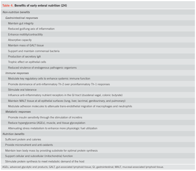
Table 4. Benefits of early enteral nutrition (24) <table><thead><tr><th colspan="2">Non-nutrition benefits</th></tr></thead><tbody><tr><td colspan="2">Gastrointestinal responses</td></tr><tr><td></td><td>Maintain gut integrity</td></tr><tr><td></td><td>Reduced gut/lung axis of inflammation</td></tr><tr><td></td><td>Enhance motility/contractility</td></tr><tr><td></td><td>Absorptive capacity</td></tr><tr><td></td><td>Maintain mass of GALT tissue</td></tr><tr><td></td><td>Support and maintain commensal bacteria</td></tr><tr><td></td><td>Production of secretory IgA</td></tr><tr><td></td><td>Trophic effect on epithelial cells</td></tr><tr><td></td><td>Reduced virulence of endogenous pathogenic organisms</td></tr><tr><td colspan="2">Immune responses</td></tr><tr><td></td><td>Modulate key regulatory cells to enhance systemic immune function</td></tr><tr><td></td><td>Promote dominance of anti-inflammatory Th-2 over proinflammatory Th-1 responses</td></tr><tr><td></td><td>Stimulate oral tolerance</td></tr><tr><td></td><td>Influence anti-inflammatory nutrient receptors in the GI tract (duodenal vagal, colonic butyrate)</td></tr><tr><td></td><td>Maintain MALT tissue at all epithelial surfaces (lung, liver, lacrimal, genitourinary, and pulmonary)</td></tr><tr><td></td><td>Modulate adhesion molecules to attenuate trans-endothelial migration of macrophages and neutrophils</td></tr><tr><td colspan="2">Metabolic responses</td></tr><tr><td></td><td>Promote insulin sensitivity through the stimulation of incretins</td></tr><tr><td></td><td>Reduce hyperglycemia (AGEs), muscle, and tissue glycosylation</td></tr><tr><td></td><td>Attenuating stress metabolism to enhance more physiologic fuel utilization</td></tr><tr><th colspan="2">Nutrition benefits</th></tr><tr><td></td><td>Sufficient protein and calories</td></tr><tr><td></td><td>Provide micronutrient and anti-oxidants</td></tr><tr><td></td><td>Maintain lean body mass by providing substrate for optimal protein synthesis</td></tr><tr><td></td><td>Support cellular and subcellular (mitochondria) function</td></tr><tr><td></td><td>Stimulate protein synthesis to meet metabolic demand of the host</td></tr></tbody></table> AGEs, advanced glycolytic end products; GALT, gut-associated lymphoid tissue; GI, gastrointestinal; MALT, mucosal-associated lymphoid tissue.

This clinical photograph illustrates the process of complementary feeding in an infant, typically occurring between 5 to 12 months of age. The image shows a caregiver (mother) introducing semi-solid food to an infant seated in a high chair. The mother is using a small, orange plastic spoon to deliver food from a green and orange bowl, demonstrating the transition from exclusive liquid nutrition (breastmilk or formula) to solid foods. The infant is actively engaged, holding a small object in one hand, which reflects normal developmental motor skills associated with this age group. This stage is medically significant as a critical window for establishing lifelong dietary patterns, promoting growth, and diversifying the gut microbiome. The visual emphasizes the manual coordination required during the weaning period and the role of caregiver-led feeding in ensuring adequate nutrient intake, particularly protein and fiber, to prevent undesired growth patterns or obesity later in life.
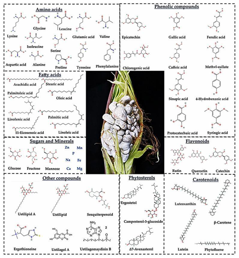
An educational infographic summarizing the nutritional and phytochemical composition of huitlacoche (corn smut), caused by the biotrophic fungus Ustilago maydis. The central image is a clinical photograph of an infected corn ear, demonstrating characteristic galls: swollen, irregularly shaped kernels transformed into grayish-white fungal masses. Surrounding the photograph are chemical diagrams (ball-and-stick models) categorized by molecular class, illustrating the fungus's biochemical profile. These include essential and non-essential amino acids (e.g., lysine, leucine, glutamic acid), long-chain fatty acids (e.g., oleic and linoleic acid), sugars, and essential minerals (Zn, Mn, Fe, Mg). The graphic also highlights diverse bioactive secondary metabolites such as phenolic acids (ferulic, chlorogenic), flavonoids (quercetin, rutin), phytosterols (ergosterol), and carotenoids (beta-carotene, lutein). Specialized fungal compounds like ustilipids and ergothioneine are also featured. This visual serves to educate on the interface of phytopathology and human nutrition, emphasizing the transformation of a crop pathogen into a nutrient-dense food source with potential therapeutic properties.
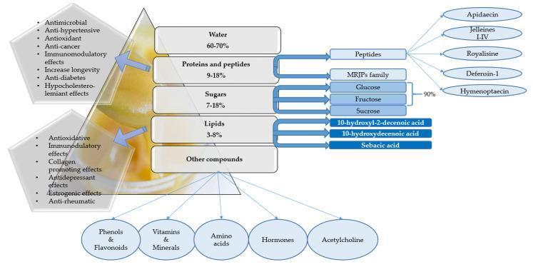
This infographic and infographic-style diagram illustrates the biochemical composition and biological properties of royal jelly, a substance used in complementary medicine and nutrition. The central hierarchical structure lists the primary macronutrients by percentage: water (60-70%), proteins and peptides (9-18%), sugars (7-18%), lipids (3-8%), and other minor compounds. The diagram specifically identifies key bioactive molecules including peptides such as Apidaecin, Jelleines I-IV, Royalisine, Defensin-1, Hymenoptaecin, and the Major Royal Jelly Protein (MRJP) family. Listed lipids include 10-hydroxyl-2-decenoic acid, 10-hydroxydecenoic acid, and sebacic acid. The 'other' category details minor constituents like phenols, flavonoids, vitamins, minerals, amino acids, hormones, and acetylcholine. Flanking the composition are clinical functional activities attributed to these components, highlighting antimicrobial, antioxidant, anti-hypertensive, immunomodulatory, estrogenic, anti-diabetic, and collagen-promoting effects. This diagram is designed for pharmacology and nutritional science education, summarizing the complex biochemical profile and potential therapeutic pathways of royal jelly in human health and aging-related contexts.
colostrum breastmilk newborn infant

This clinical photograph shows a newborn infant in a supine position, representing standard neonatal care and attire within a maternity ward setting. The infant is dressed in a light-colored, long-sleeved cotton shirt and matching leggings. The shirt is designed with a side-tie closure, currently open at the abdomen to allow for clinical observation or umbilical cord care. The infant appears in a relaxed, sleeping state with closed eyes and arms flexed toward the head, indicating normal neurological tone and comfort. The infant is placed on a flat, padded surface covered with a protective sheet, typical of a nursery changing station or bassinet. A plush toy is visible near the feet. This image serves to illustrate traditional postpartum care practices and the use of loose-fitting infant apparel as an alternative to swaddling in clinical research focused on early infant development and breastfeeding outcomes.

Clinical photograph of a newborn immediately following delivery, likely via cesarean section as indicated by the surgical setting. The infant presents with a healthy reddish skin tone (erythema neonatorum) and is partially covered in vernix caseosa, a protective white, creamy biofilm visible on the face, scalp, and limbs. The newborn is lying in a supine position on a green sterile surgical drape. Anatomical features include a flexed posture of the lower extremities and a blue plastic umbilical cord clamp applied to the umbilical stump. The face shows mild physiological edema common in neonates. This image serves as an educational example of immediate neonatal appearance and the outcome of obstetric anesthetic management. For patient privacy, the eyes are obscured by a black bar.

This clinical photograph depicts a newborn infant in a state of distress, being held by a caregiver. The infant exhibits a facial expression of crying or discomfort, characterized by tightly closed eyelids, a furrowed brow, and a slightly open, downturned mouth. The infant's skin shows a faint, physiologic mottling (cutis marmorata) on the upper extremities, which is a common finding in neonates due to vasomotor instability. The image serves as a visual reference for pediatric behavioral health and neonatal clinical assessment, specifically regarding infant temperament, soothability, and neurodevelopmental observation. It illustrates the physical manifestations of early emotional reactivity in a clinical or developmental psychology context.

Clinical photograph of a newborn in a supine position, demonstrating asymmetrical upper limb posturing and signs of acute distress (crying). The infant's left arm is flexed at the elbow with the hand clenched in a tight fist, while the right arm is extended. A small adhesive bandage is present on the lateral aspect of the right arm just above the elbow joint. On the left forearm, a small, dark skin lesion or mark is visible. The image illustrates a clinical examination of neonatal motor function and limb symmetry, potentially related to neonatal radial nerve palsy or subcutaneous fat necrosis following a traumatic birth. The facial expression and open mouth indicate irritability or pain, which is clinically relevant when assessing painful subcutaneous nodules or nerve injuries in neonates.

This clinical photograph depicts a newborn infant, likely a preterm newborn (PTNB), in a neonatal intensive care unit (NICU) setting. The infant is supine on white hospital linens and is monitored with various medical devices. A non-invasive heart rate variability (HRV) monitor or similar telemetry device is secured to the chest with white medical adhesives. An orogastric or nasogastric tube is visible, taped to the infant's cheek and entering the mouth, used for enteral feeding or gastric decompression. Additional medical tubing or leads are positioned near the lower torso and legs, indicating continuous physiological monitoring. The image illustrates standard neonatal monitoring protocols used during clinical assessments, such as visual acuity testing or baseline physiological data collection. The environment includes colored hospital equipment and padded bedding to support the infant's positioning and thermal regulation.
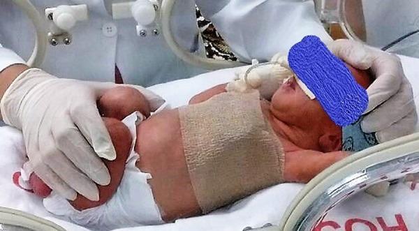
Clinical photograph of a newborn infant in a neonatal intensive care unit (NICU) setting, illustrating methods of manual and mechanical restraint for physiological monitoring. The infant is positioned in a dorsal decubitus (supine) posture within an incubator. Key clinical features include a tan elastic bandage wrapped circumferentially around the mid-torso to provide gentle abdominal and thoracic restraint, and a blue phototherapy eye shield protecting the infant's eyes. A healthcare professional wearing white nitrile gloves is seen performing manual restraint at the infant's feet. The infant is wearing a standard disposable diaper and is lying on a white sterile sheet. In the background, NICU infrastructure is visible, including incubator access ports, transparent plastic shielding, and medical monitoring tubing. This image demonstrates standard neonatal care positioning and restraint techniques used during clinical evaluations, such as heart rate variability (HRV) or vital sign monitoring in a controlled environment.

Clinical photograph of a newborn infant in a supine position, demonstrating several craniofacial dysmorphic features and congenital anomalies. Key findings include right-sided anophthalmia (complete absence of the eyeball and orbital cavity), a prominent beaked nose, severe micrognathia (undersized jaw), and posteriorly rotated, low-set ears. The infant exhibits generalized skin mottling (cutis marmorata appearance) and a blue umbilical cord clamp is visible on the abdomen. The overall posture shows moderate flexion of the extremities. This image is used in neonatological and genetic contexts to illustrate syndromic presentations or midline defects. The photograph serves as a medical reference for identifying distinct morphological patterns associated with rare congenital disorders during initial newborn physical examination.
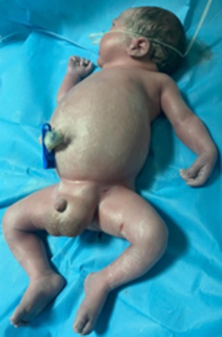
Clinical photograph of a newborn in the immediate postnatal period demonstrating hydrops fetalis with severe anasarca. The infant exhibits generalized, massive soft tissue edema. Key visual features include a markedly distended abdomen consistent with significant ascites, and severe swelling of the upper and lower extremities resulting in the loss of normal skin folds and obscured anatomical landmarks. The skin appears taut and shiny due to subcutaneous fluid accumulation. Pronounced scrotal edema is also visible. The infant is placed on a blue sterile field with a clamped umbilical cord and an orogastric tube in situ. This presentation is clinically significant for systemic fetal fluid overload, often secondary to cardiac failure (such as from a rhabdomyoma), severe anemia, or immune/non-immune hydrops. The image serves as a teaching example of the phenotypic manifestations of neonatal anasarca and its association with Mirror syndrome in the mother.
breast anatomy milk ducts lactation physiology
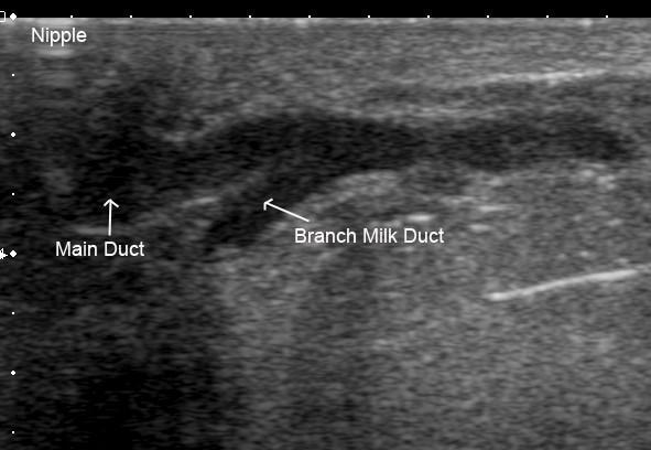
This diagnostic ultrasound image demonstrates the ductal anatomy of a human lactating breast. The image displays a longitudinal view of a main milk duct and a smaller branch milk duct. Both structures appear as hypoechoic (dark), fluid-filled tubular lumens contrasted against the more echogenic (brighter) surrounding glandular parenchyma and connective tissue. The main duct is shown with a diameter of approximately 2.4 mm, while the secondary branch milk duct measures 1.7 mm. The branching point is visible, showing the smaller duct draining glandular tissue and merging into the main collecting duct near the nipple region, which is labeled at the top left of the frame. This image illustrates the normal physiological state of milk ducts during lactation, emphasizing the lack of a prominent lactiferous sinus and the characteristic branching pattern close to the nipple. The clear differentiation between the hypoechoic ductal fluid and the hyperechoic breast tissue highlights the utility of high-resolution ultrasound in assessing mammary gland anatomy and potential obstructions.
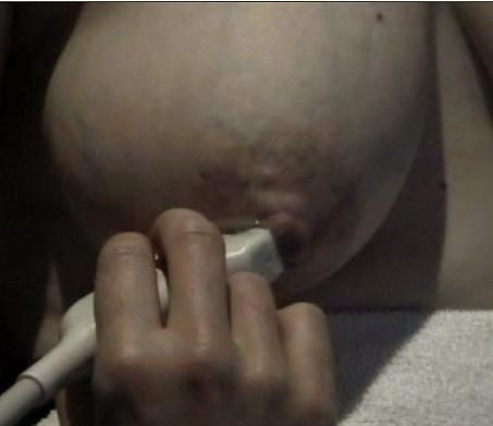
This clinical photograph and procedural image captures the right areola of a lactating woman during an ultrasound examination. The image demonstrates the anatomy of a full, lactating breast prior to milk ejection. Specifically, the areola region appears tense and engorged. Notable visual features include superficial bulging structures directly superior to the nipple, which represent dilated lactiferous ducts located just beneath the skin. A hand is seen holding a linear ultrasound probe in direct contact with the superior areolar surface, positioned to monitor these superficial ducts in the long axis. The image illustrates the clinical application of ultrasound for non-invasive monitoring of milk ejection and ductal physiology. Key educational concepts include the identification of superficial mammary anatomy, the visual presentation of ductal engorgement during lactation, and the ergonomic positioning of a transducer for real-time breast imaging.
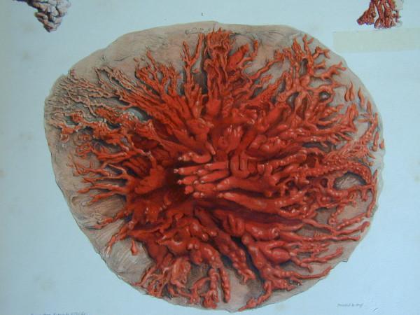
This historical anatomical illustration, derived from Sir Astley Cooper's 19th-century dissections, depicts the mammary ductal system of a lactating human breast. The image shows a wax-injected specimen where the lactiferous ducts are colored red to highlight their intricate architecture. The system exhibits a distinct radial symmetry, with major collecting ducts originating near the central nipple-areola complex and branching extensively as they move peripherally toward the glandular tissue. The ducts transition from thicker primary branches to increasingly fine, terminal arborizations, resembling a complex root or coral-like network. The illustration captures the high density of the ductal tree during lactation, demonstrating the interconnected pathways required for milk transport. This pedagogical material serves as a foundational reference for understanding breast anatomy, specifically the distribution of the lobulo-alveolar units and the patent ductal channels that accommodate milk ejection.
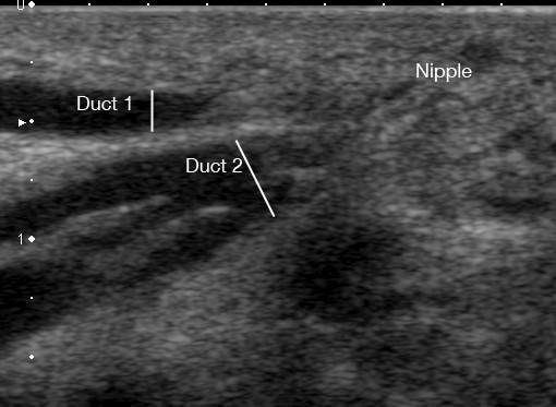
This diagnostic ultrasound image captures a sagittal view of milk ducts within a lactating human breast prior to milk ejection. The content demonstrates two distinct ductal structures, labeled Duct 1 and Duct 2, oriented toward the nipple region. Both ducts exhibit a characteristic sonographic appearance: hypoechoic (dark/black) internal lumens indicating fluid content (milk), bounded by echogenic (bright/white) walls. Duct 1 is positioned more superficially, measured at a depth of 1.95 mm from the skin surface, appearing as a single, linear tubular structure. Duct 2 is located deeper at 3.72 mm and displays a complex branching pattern where three separate tributary ducts are seen merging into a larger main channel. The surrounding breast parenchyma shows heterogeneous echogenicity typical of functional mammary tissue. This image serves as an educational example of maternal breast anatomy during lactation, highlighting the use of non-invasive ultrasonography to monitor ductal dilation and branching morphology in a clinical or research setting.
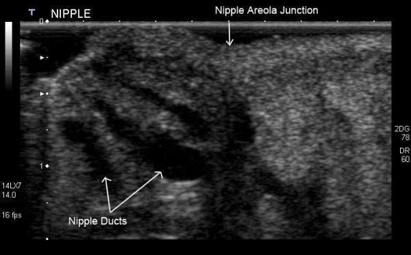
This diagnostic image is a high-resolution grayscale ultrasound scan of a lactating human breast, specifically focusing on the nipple-areola complex. The anatomy is labeled, showing the nipple and the nipple-areola junction. The surrounding nipple tissue exhibits a heterogeneous, primarily echogenic (bright) texture. Within the substance of the nipple, multiple milk ducts are clearly visualized as distinct hypoechoic (dark), tubular, and elongated structures. These ducts extend from the deeper mammary parenchyma toward the nipple surface. The educational focus of this image is the sonographic visualization of normal ductal anatomy during lactation, demonstrating how physiological distension allows these structures to be identified. The image highlights key diagnostic features such as the hypoechoic nature of fluid-filled ducts relative to the echogenic fibrous and glandular stromal tissue, which is clinically relevant for assessing ductal patency or identifying obstructive conditions like blocked ducts.
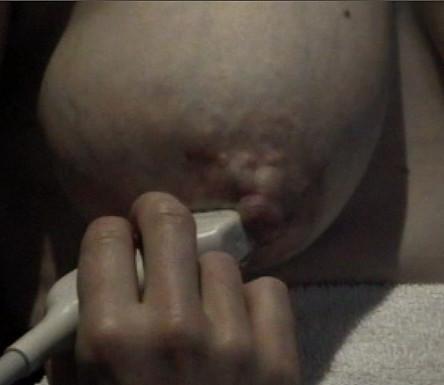
This clinical photograph displays a right lactating breast during the milk ejection reflex, alongside the application of a diagnostic ultrasound probe. The areola demonstrates significant physiological swelling and increased tension, resulting in a full, convex appearance. This visual change is indicative of the expansion of superficial milk ducts as oxytocin-induced myoepithelial contraction forces milk from the alveoli into the ductal system. A linear ultrasound transducer is positioned in direct contact with the inferior portion of the areola, stabilized by a hand to monitor ductal dilation and milk flow in real-time. The skin surface shows slight lobulation or prominence corresponding to the underlying distended ducts. This image illustrates the physical manifestations of the milk ejection reflex and the non-invasive clinical method of using ultrasound to study lactation physiology and breastfeeding dynamics.
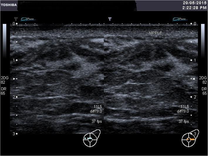
A grayscale diagnostic ultrasound image of the left breast in a postpartum patient, showing functional breast anatomy. The scan demonstrates a heterogeneous echotexture characteristic of lactating breast tissue, with a mix of hyperechoic glandular tissue and hypoechoic areas. Numerous dilated, hypoechoic linear and tubular structures are visible, representing lactiferous ducts. These ducts are oriented towards a focal point marked 'NIPPLE' at the superior aspect of the image, corresponding to the retroareolar region. The imaging highlights the presence of functional ductal systems even in clinical cases involving extensive external scarring (such as skin grafts). The ultrasound lacks the typical acoustic shadowing usually produced by a prominent nipple, reflecting the anatomical variation in this specific clinical case. Key educational features include the visualization of active milk-conducting ducts and the sonographic appearance of glandular tissue during the early postpartum/lactation phase.
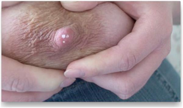
This clinical photograph shows a close-up view of the nipple-areola complex (NAC) during manual milk expression. The central focus is the nipple, a raised pinkish papilla mammaria, which exhibits several small, discrete white droplets of breast milk at the ductal orifices. The surrounding areola mammae displays a light tan or pinkish pigmentation with a textured, wrinkled surface marked by skin folds and creases. Fingers are visible at the upper and lower margins of the image, demonstrating the technique of manual compression to elicit milk flow. The image serves as an educational tool for identifying the functional anatomy of the mammary gland, specifically the location and distribution of milk ducts that transport milk to the surface during lactation. It illustrates clinical signs associated with breastfeeding and the physiological process of lactation in a female patient.
mastitis engorgement breast problems lactation
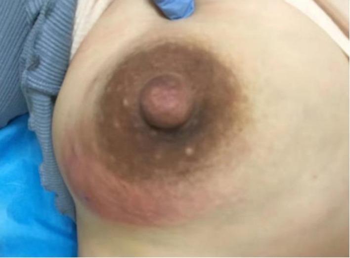
A clinical photograph of a right breast exhibiting signs of acute inflammation, specifically related to acute lactation mastitis (ALM). The lower-inner quadrant demonstrates a significant area of erythema (redness) and localized edema (swelling). This erythematous lesion extends toward the periareolar region. The areola appears hyperpigmented and displays prominent Montgomery tubercles (small, raised sebaceous glands). The nipple is slightly swollen with a reddish hue and an irregular surface texture. The clinical presentation is consistent with a breast abscess or inflammatory mastitis during the postpartum period. This image serves as an educational tool for identifying primary clinical manifestations of infectious breast disease, such as redness, swelling, and localized inflammatory changes in a lactating patient.
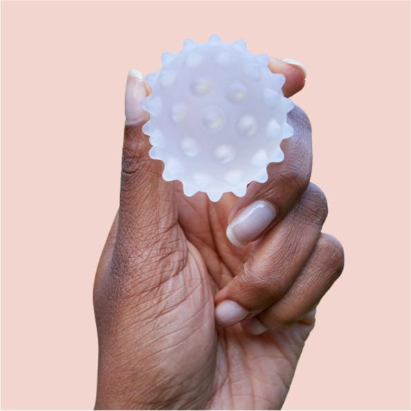
A clinical photograph of a Lactamo device, a specialized lactation aid designed for maternal health and breastfeeding support. The device is a small, handheld sphere, approximately 5 cm in diameter, constructed from a translucent, frosted medical-grade material. Its surface is characterized by multiple rounded, hollow protrusions or nodes distributed evenly across the sphere to provide varying levels of compression and tactile stimulation. The device is designed to be filled with a thermochromic gel, allowing for the delivery of hot or cold therapy in conjunction with breast massage and compression. It is used to address breastfeeding challenges such as engorgement, blocked ducts, and mastitis by facilitating milk flow through movement toward the nipple or axilla. The photograph shows the device held between fingers to demonstrate its scale and ergonomic design for self-application by postpartum patients.
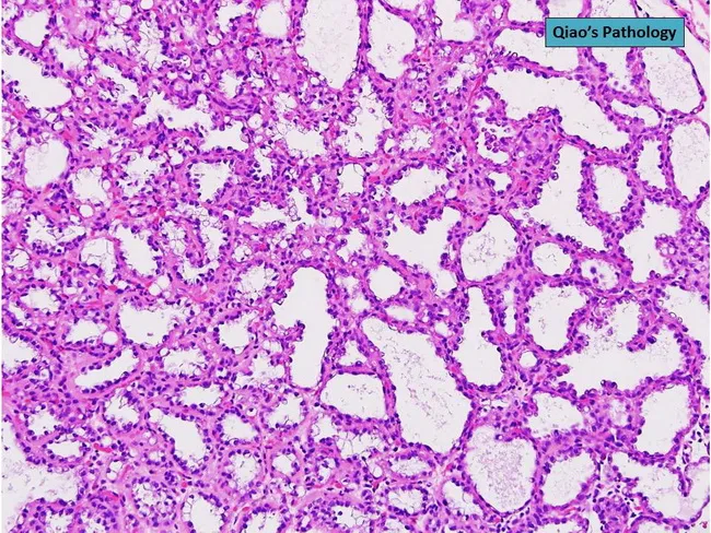
This is a histopathologic slide of the lactating breast illustrating a lactating adenoma. Imaging modality: Light microscopy of Hematoxylin and Eosin stained tissue section. The lesion shows multiple hyperplastic lobules within the breast parenchyma, with preserved lobular architecture. The luminal surfaces reveal actively secreting cuboidal to columnar epithelial cells, containing abundant eosinophilic cytoplasm and apical secretions, consistent with milk production. The stroma is mildly fibrous, with sparse inflammatory infiltrates and no significant atypia or mitotic activity. Ductal and acinar structures appear dilated, filled with secretory material, giving a benign, well-circumscribed, nodular appearance. Overall, features are classic for a lactating adenoma: a hormonally driven, benign lesion arising during pregnancy or lactation that mimics other secretory breast lesions but lacks malignant cytology. Diagnostic significance: distinguishes benign lactational changes from mastitis, fibroadenoma, or carcinoma; clinical correlation with recent pregnancy/breastfeeding supports diagnosis. Differential considerations include fibroadenoma with lactational change, secretory carcinoma (rare), and puerperal mastitis. This image is suitable for educational purposes in surgical pathology, breast disease, lactation-related breast lesions, and differential diagnosis of palpable breast masses in postpartum women. Potential clinical use cases: undergraduate/graduate medical education, residency training, and research on lactational breast pathology. These features confirm benign lactational physiology and correlate clinically.
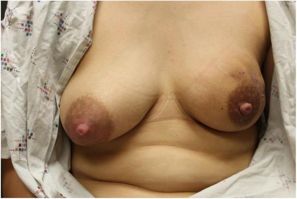
This clinical photograph shows a frontal view of a patient's chest at 10 weeks postpartum, illustrating significant breast asymmetry. The right breast (image left) appears full and engorged, consistent with active lactation. The left breast (image right) is notably smaller in volume with an elongated, flattened morphology, indicating a non-lactating state. The left breast exhibits sequelae of Idiopathic Granulomatous Mastitis (IGM), including mottled hyperpigmentation around the areola and several visible skin lesions or healing scars in the upper outer quadrant, where a prior fistula had been present. The left nipple is more prominent compared to the right. This image is used in medical education to demonstrate the physical manifestations of chronic inflammatory breast disease and the visual impact of unilateral cessation of lactation on breast tissue density and volume.

Content Type: Clinical Photograph. Anatomical Region: Anterior chest and bilateral breasts. Description: The image shows bilateral breast engorgement with diffuse erythema (redness) extending across the skin of both breasts. Visible features include linear striae (stretch marks) and prominent superficial vascular patterns (prominent veins) beneath the skin. The skin texture appears smooth and tense, consistent with significant swelling or engorgement. The areolae and nipples are digitally blurred for privacy. Clinical Significance: This image illustrates clinical signs associated with inflammatory breast conditions, severe engorgement, or postpartum mastitis. The educational focus is on identifying skin changes, vascular prominence, and the extent of inflammatory redness during therapeutic progression, specifically noted in the context of therapeutic ultrasound treatment for severe breast pain.

This clinical photograph set displays four distinct pathological conditions of the human breast and nipple in lactating patients across various skin tones. Panel A demonstrates breast engorgement featuring an inverted nipple with a light-colored central depression and taut, shiny skin. Panel B shows a breast abscess characterized by significant periareolar erythema, localized swelling, and a dark necrotic-appearing lesion at the nipple-areolar complex suggestive of nipple damage or a draining sinus. Panel C illustrates granulomatous mastitis with visible skin wrinkling, nipple inversion, and generalized breast inflammation. Panel D depicts acute nipple damage on dark-pigmented skin, highlighted by a prominent, raised, fluid-filled blood blister (hemorrhagic bleb) located on the lateral aspect of the nipple. This series serves as an educational comparison for differentiating mastitis, abscess, engorgement, and mechanical nipple trauma, while highlighting common visual pitfalls that can lead to clinical misclassification.
skin to skin kangaroo mother care bonding

A clinical photograph illustrating Kangaroo Mother Care (KMC), also known as skin-to-skin contact, between an adult and a newborn infant. The image depicts the neonate positioned vertically and prone against the adult's bare chest, a key practice in neonatal care for temperature regulation and physiological stability. The infant is wearing a light-colored soft cap to prevent heat loss through the scalp and is partially covered by a pink blanket to maintain thermal insulation. Both the adult and the infant appear relaxed with eyes closed, demonstrating the bonding and calming effects of this intervention. This clinical practice is commonly utilized in maternal and child health settings, particularly in neonatal intensive care units (NICUs) and for low-birth-weight infants, to improve clinical outcomes such as breastfeeding success, neurodevelopmental stability, and infection prevention. The photograph highlights the human-centric approach to neonatal medicine and the integration of psychosocial support in clinical protocols.

A clinical photograph illustrating Kangaroo Mother Care (KMC) performed by a male caregiver, demonstrating the practice of skin-to-skin contact for a preterm infant. The photograph shows an adult male of South Asian descent holding a small neonate against his bare chest, tucked inside a partially unbuttoned shirt to maintain warmth and bonding. The infant appears to be a premature newborn with dark hair, resting in a vertical position. This image serves as an educational example of neonatal supportive care, particularly relevant in the management of preterm births and the prevention of complications such as Retinopathy of Prematurity (ROP) through physiological stabilization. The setting appears to be a neonatal clinical environment, emphasizing the role of paternal involvement in evidence-based neonatal interventions in resource-limited or high-burden settings like India.

A clinical photograph depicting a row of adult males performing Kangaroo Mother Care (KMC), also known as skin-to-skin contact, within a Neonatal Intensive Care Unit (NICU). The men are seated in specialized chairs, wearing hospital-provided gowns designed to facilitate direct contact with newborn infants held against their chests. The newborns are positioned in a vertical or semi-vertical orientation. In the background, hospital infrastructure is visible, including neonatal cribs and wall-mounted electrical panels with medical-grade outlets. This image illustrates the involvement of fathers in developmental supportive care for preterm or low-birth-weight infants. The educational focus is on non-pharmacological interventions to stabilize neonatal physiology, promote bonding, and improve clinical outcomes in a NICU setting. Key concepts include neonatal nursing, paternal involvement in pediatrics, and the implementation of skin-to-skin contact protocols.
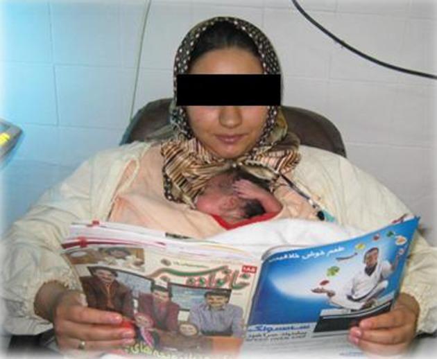
This clinical photograph illustrates Kangaroo Mother Care (KMC) being performed in a hospital setting, likely a Neonatal Intensive Care Unit (NICU). The image shows a mother seated in a supportive chair, holding a premature newborn in a skin-to-skin, vertical position against her chest. The infant is securely nestled within a specialized medical garment, often referred to as a 'Kanga Carrier' or 'Kangaroo Bag,' designed to provide support while keeping the mother's arms free. The newborn appears small with visible reddish skin tone and is positioned for optimal physiological stability. The mother is wearing a patterned headscarf and a light-colored top, demonstrating the implementation of KMC while engaged in a relaxing activity (reading). This visual highlights a key neonatology practice used to improve thermoregulation, facilitate breastfeeding, and enhance bonding for preterm infants. The setting emphasizes the clinical relevance of KMC as a routine, cost-effective intervention for high-risk neonates with stable respiratory and cardiovascular status.

This clinical photograph demonstrates the application of Kangaroo Mother Care (KMC) for a low-birth-weight neonate in a hospital setting. The image shows a preterm infant held in direct skin-to-skin contact against the mother's chest, a critical intervention for thermal regulation, bonding, and physiological stability. The neonate is wearing a white crocheted cap with a green trim to prevent heat loss through the head. The infant is secured in an upright position using a red-and-white striped cloth wrap, with the mother's hand providing additional support to the head. This visual illustrates key components of small and sick newborn care (SSNC), specifically addressing hypothermia prevention and essential newborn care in resource-limited environments. The background includes a neonatal cot, indicating the clinical environment of a Neonatal Intensive Care Unit (NICU). This image serves as an educational example of non-pharmacological interventions used to improve survival outcomes in premature or low-birth-weight infants.
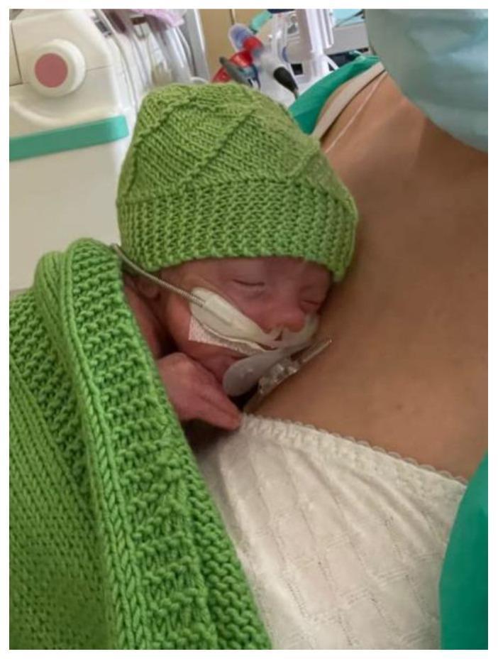
This clinical photograph illustrates a mother-infant dyad participating in a Skin-to-Skin Contact (SSC) session, also known as Kangaroo Care, within a Neonatal Intensive Care Unit (NICU) setting. The infant is positioned prone and upright in a slight 'sniffing position' against the mother’s bare chest, achieving sternum-to-sternum contact. To maintain thermoregulation, the neonate wears a light green knitted wool cap and is partially covered by a matching green knitted blanket. The infant appears to be receiving respiratory support via a nasal cannula or continuous positive airway pressure (CPAP) interface, secured with white medical tape and a stabilizing band across the face. The background shows clinical monitoring equipment and the mother wearing a white patterned top and a surgical mask, emphasizing the healthcare environment. This image serves as an educational tool for demonstrating proper positioning for SSC in stable preterm or ill infants and highlights the integration of family-centered care with medical technology for physiological stabilization.
breast pump expressing milk storage
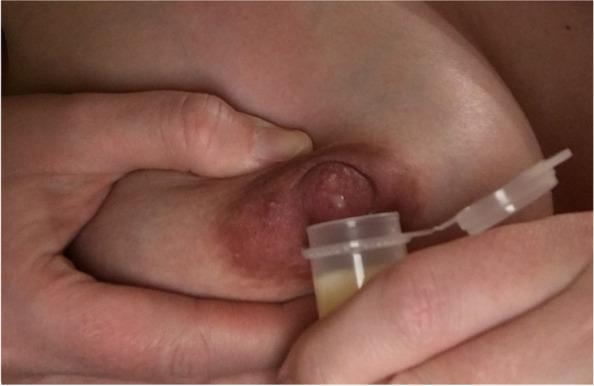
A close-up clinical photograph demonstrating the Marmet technique for hand-expressing antenatal milk or colostrum. The image shows a person's hands positioned on a lactating breast. One hand supports the breast tissue, with the thumb and index finger placed approximately 2-3 cm behind the nipple at the edge of the areola, applying rhythmic compression to the underlying milk ducts. The areola appears physiologically hyperpigmented and slightly engorged, typical of late pregnancy or the early postpartum period. A small, clear, flip-top collection vial is held directly beneath the nipple to capture the expressed fluid. The vial contains a small volume of yellowish, viscous fluid, characteristic of colostrum. This visual serves as an educational guide for lactation support, specifically teaching manual expression and collection techniques for individuals in the late third trimester (antenatal milk expression) or early postpartum phase.
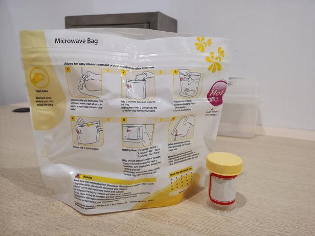
This clinical photograph displays a medical-grade disinfection system used for neonatal care accessories, specifically for the steam sterilization of breast milk collection equipment. The image features a specialized microwave steam sterilization bag alongside a sterile collection cup. The bag is a flexible, translucent white pouch with yellow instructional graphics and a 'Hold here' safety zone. It displays a six-step illustrated guide for cleaning, adding water (60ml), and microwave heating times based on wattage. A tracking grid at the bottom is included to monitor the bag's 20-use lifecycle. Next to the bag is a cylindrical, transparent collection cup with a yellow screw-top lid and a red-bordered label for patient identification. This visual represents standard protocols for infection control in a Neonatal Intensive Care Unit (NICU) or home care setting, demonstrating the process of ensuring the microbiological safety of colostrum and breast milk storage containers through rapid steam treatment.
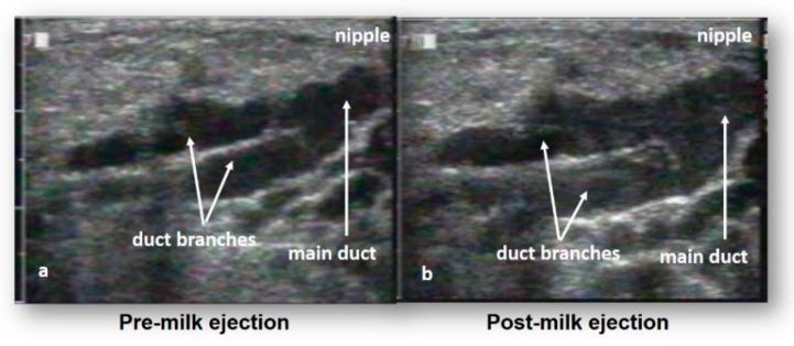
This diagnostic image consists of two side-by-side ultrasound frames (a and b) illustrating the functional anatomy of the lactating human breast during the milk ejection reflex. The imaging modality is B-mode ultrasound, used to visualize the mammary ductal system in real-time. In both images, the milk ducts are identified as prominent anechoic (black) tubular structures, providing high contrast against the surrounding hyperechoic, granular connective and adipose tissue. Figure (a), labeled 'Pre-milk ejection', shows the main duct and duct branches in a relatively narrow state. Figure (b), labeled 'Post-milk ejection', demonstrates significant ductal expansion and distention due to the presence of breast milk following the ejection reflex. Annotated labels indicate the 'nipple' position at the top right, with arrows highlighting the 'main duct' and 'duct branches'. This comparison serves as educational material to demonstrate that mammary ducts function primarily as transport vessels that dilate during milk flow rather than serving as large static storage reservoirs, contributing to contemporary understanding of breastfeeding physiology and mammary gland function.
This clinical photograph captures a close-up procedural demonstration of Antenatal Colostrum Expression (ACE). The image illustrates the 'C-hold' hand technique, where the breast tissue is compressed between the thumb and fingers, positioned approximately 2-3 cm behind the nipple at the edge of the areola. A second hand is shown holding a small oral syringe directly at the nipple tip to collect the expressed droplets of colostrum. The procedural focus is on the manual stimulation and compression of the lactiferous sinuses to facilitate milk removal. This visual serves as an educational tool for lactation consultants and expectant mothers, highlighting the correct hand placement and the use of a syringe for hygienic colostrum harvesting and storage during late pregnancy. The setting is likely an antenatal clinic or educational session designed to improve breastfeeding confidence and knowledge.
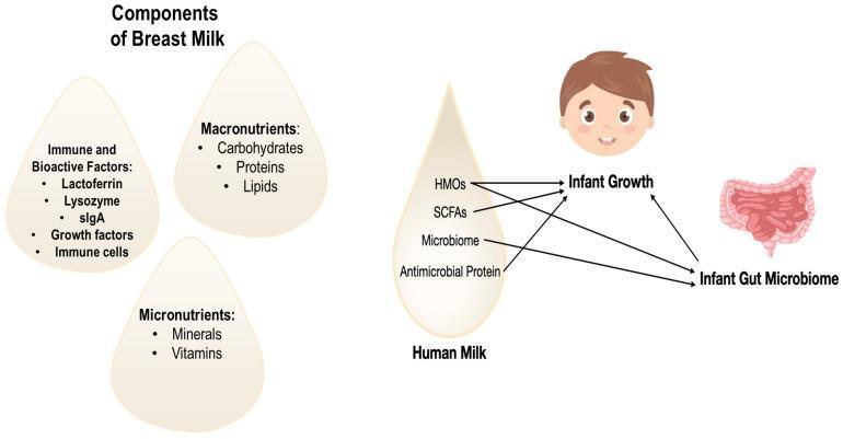
An educational infographic and signaling diagram illustrating the components of human breast milk and their physiological impact on neonatal development. The left side categorizes 'Components of Breast Milk' into three milk-droplet-shaped panels: 'Immune and Bioactive Factors' (including Lactoferrin, Lysozyme, sIgA, growth factors, and immune cells), 'Macronutrients' (Carbohydrates, Proteins, Lipids), and 'Micronutrients' (Minerals, Vitamins). The right side features a flow diagram detailing how specific human milk constituents influence clinical outcomes. Human Milk Oligosaccharides (HMOs), Short-Chain Fatty Acids (SCFAs), and Antimicrobial Proteins are shown to directly impact both 'Infant Growth' and the 'Infant Gut Microbiome.' The 'Microbiome' component within the milk is depicted as specifically influencing the infant's gut microbiome. The diagram highlights the bidirectional relationship between the developing gut microbiome and general infant growth, emphasizing the role of maternal milk in shaping metabolic and immunological health during early development.
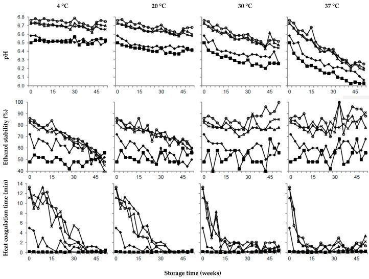
A multi-panel line graph assembly representing a biochemical and physicochemical stability study of ultra-high temperature (UHT) treated milk over a 52-week storage period. The data is organized into a grid with four columns representing storage temperatures (4°C, 20°C, 30°C, and 37°C) and three rows representing key stability metrics: pH, ethanol stability (%), and heat coagulation time (min). The study compares five distinct milk compositions: calcium-enriched (┐), citrate-enriched (○), urea-enriched (∆), calcium and citrate-enriched (♦), and an unmodified reference milk (x). Key findings illustrated include a linear decrease in pH at higher temperatures (30°C and 37°C), particularly in calcium-enriched samples. Ethanol stability generally declines at lower temperatures (4°C and 20°C) while fluctuating at higher temperatures. Heat coagulation time (HCT) shows a rapid decline across most samples within the first 15-30 weeks, especially at higher storage temperatures. This chart serves as an educational resource for food science and nutritional biochemistry, illustrating the impact of fortification and storage conditions on dairy product stability and shelf-life.
nipple latch correct attachment infant breastfeeding
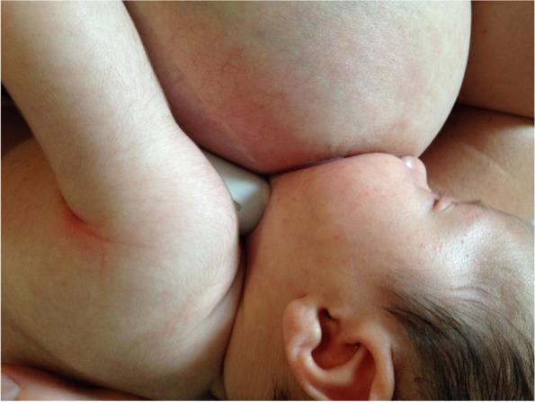
Clinical photograph of a 4-week-old infant breastfeeding using the 'gestalt' method, emphasizing optimal latching mechanics and positional stability. The image shows the infant in a cross-cradle or similar hold, with the head tilted slightly upward (asymmetric latch). The infant's mouth is wide open and deeply attached to the breast, though the nipple itself is not visible. Notably, the infant's nose is in close contact with the breast tissue, and the chin is tucked firmly into the breast. A white nipple shield is partially visible at the point of attachment. The infant's skin is fair with fine vellus hair, and the scalp has dark hair. The maternal breast appears full and rounded, with visible skin texture and faint vascular patterns. This image demonstrates post-intervention positioning intended to address nipple pain and infant fussiness associated with latch difficulties or oral connective tissue restrictions.
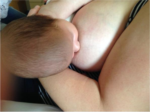
This clinical photograph demonstrates an infant breastfeeding, specifically illustrating an optimized latch following a gestalt intervention. The infant is positioned in close physical contact with the mother, demonstrating effective positioning and attachment. The infant's mouth is wide open and deeply latched onto the maternal breast, covering a significant portion of the areola. The infant's nose is in close proximity to the breast, and the chin is tucked into the breast tissue, supporting a stable, asymmetrical latch. The infant is supported by the mother's arm, ensuring a belly-to-belly orientation that facilitates proper alignment of the head, neck, and spine. This image serves as a clinical example of breastfeeding mechanics and positional stability, relevant for lactation consultancy and pediatric primary care. It highlights the visible contact points and body alignment necessary to maximize intra-oral breast tissue volume and potentially reduce maternal nipple pain associated with breastfeeding difficulties like tongue-tie or shallow latch.
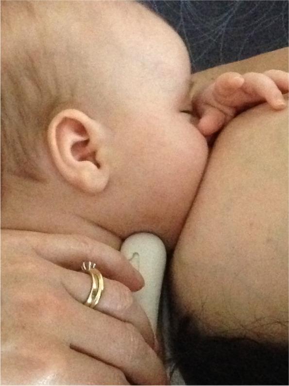
A clinical photograph depicting an infant latched onto a mother's breast during breastfeeding. The image provides a close-up, lateral view of the infant's head and neck in relation to the breast tissue. The infant is positioned with the chin pressed firmly into the breast, demonstrating an asymmetrical latch. The infant's upper body is oriented horizontally, and the nose appears clear of the breast tissue. A caregiver's hand is visible at the bottom of the frame, providing support to the infant's neck and base of the skull to maintain positioning. The photograph illustrates post-intervention breastfeeding mechanics following a 'gestalt' clinical approach, which focuses on optimizing maternal-infant positioning and attachment to address issues like nipple pain and poor milk transfer. The educational focus is on the visual assessment of latch quality, specifically the deep attachment and chin-breast contact required for effective suckling and maternal comfort.
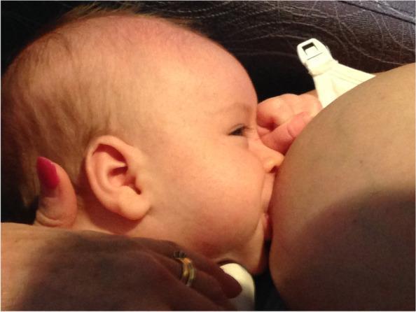
A clinical photograph depicting an 8-week-old infant breastfeeding, labeled as a 'pre-gestalt intervention' state. The image illustrates the physical dynamics of infant latching and maternal positioning. The infant is shown in a lateral profile with a relatively shallow latch; the mouth is open but primarily encompasses the nipple and only a minimal portion of the areola. The infant's chin is pressed against the breast tissue, while the nose remains close to the breast surface but clear for respiration. There is visible drawing of the breast tissue into the oral cavity. The mother's breast appears full, and her hand is visible supporting the infant's neck/head. A breastfeeding bra with a white plastic clasp is visible in the background. This clinical image is used in a medical educational context to demonstrate latch characteristics associated with maternal nipple pain and difficult attachment prior to corrective lactation support or positional interventions.
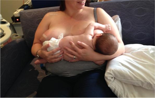
A clinical photograph depicting a mother and infant during a breastfeeding session, illustrating the 'gestalt' breastfeeding intervention for position and attachment. The infant is positioned in a modified cradle hold, latched onto the mother's left breast. The infant's body is aligned and supported by the mother's arms, with the head slightly tilted to facilitate a deep latch. The mother is seated on a dark blue couch, using a white pillow for additional lumbar or arm support. Notably, the infant appears to be breastfeeding with the assistance of a nipple shield, a common clinical tool used for managing nipple pain or latch difficulties. The mother's right breast is partially covered with white gauze, likely for milk collection or hygiene. The photograph serves as an educational resource in lactation consultancy and maternal-child health, demonstrating infant positioning, maternal support techniques, and the use of breastfeeding aids in cases involving neonatal oral connective tissue concerns or maternal nipple pain.
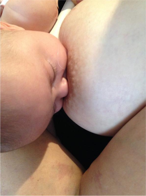
A clinical photograph depicting an infant latched onto a mother's breast, illustrating a post-gestalt intervention breastfeeding position. The image provides a lateral view of the infant's face in close proximity to the breast tissue. The infant's mouth is wide open and fully sealed around the nipple and a significant portion of the lower areola, demonstrating an asymmetrical, deep latch. The infant's nose is positioned lightly against the breast, and the chin is tucked firmly into the breast tissue. The cheeks appear rounded without visible dimpling, suggesting effective intra-oral vacuum and stability. The areola shows slight stretching and distortion consistent with active breastfeeding. This visual serves as an educational example of optimal breastfeeding mechanics and positioning designed to maximize milk transfer and minimize maternal nipple pain by increasing intra-oral breast tissue volume.
Image-Based Questions: Breastfeeding Week & Breastmilk
🤱 Category 1: Breastfeeding Positions & Holds
Answer hint: Football hold - avoids pressure on the abdominal incision site.
Answer hint: Side-lying position - reduces fatigue, suits post-operative mothers, promotes rest.
Answer hint: Dancer hand position - supports both the breast and baby's chin/cheek; used for hypotonic infants or those with low muscle tone.
🔬 Category 2: Latch Quality & Attachment
Answer hint: Only nipple in mouth (not areola), minimal jaw excursion - leads to nipple trauma, poor milk transfer, mastitis.
Answer hint: Wide-open mouth, lower areola covered more than upper, chin into breast, nose free, rounded cheeks, asymmetric latch.
Answer hint: Positioning infant's head and neck, supporting breast for latch. Lactation consultants guide positioning, assess milk transfer, manage complications.
🧪 Category 3: Breast Milk Composition & Components
Answer hint: Human Milk Oligosaccharides (HMOs) - prebiotic, unique to human milk, support gut microbiome development.
Answer hint: Bilateral frontal lobes, right temporal lobe, occipital cortex, prefrontal cortex (BA 11, 46), caudate nucleus. Suggests breastmilk promotes superior cognitive and executive function development.
🏥 Category 4: Breast Anatomy & Lactation Physiology
Answer hint: Main duct ~2.4 mm, branch duct ~1.7 mm. Ducts are hypoechoic (dark) against hyperechoic glandular tissue.
Answer hint: Milk ejection (let-down) reflex. Oxytocin causes myoepithelial contraction, dilating the ducts - visible as wider anechoic spaces in post-ejection image.
Answer hint: Radial branches from nipple-areola complex outward; multiple collecting ducts converge at the nipple from peripheral lobulo-alveolar units.
Answer hint: Areola becomes tense, swollen, convex. Superficial lactiferous ducts dilate due to oxytocin-induced milk ejection.
🤱 Category 5: Milk Expression & Antenatal Colostrum Collection

Answer hint: ACE (Antenatal Colostrum Expression) from 36 weeks gestation. Indications: gestational diabetes, expected neonatal hypoglycemia, planned separation from baby, cleft lip/palate.
Answer hint: Thumb and index finger 2-3 cm behind nipple, compress-roll motion toward nipple. Marmet emphasizes rhythmic compression of lactiferous sinuses.
🍼 Category 6: Alternative Feeding Methods
Answer hint: Cup feeding is used when direct breastfeeding is not possible but lapping/sipping reflexes are intact; it avoids nipple confusion and preserves oral motor patterns similar to breastfeeding.
🦘 Category 7: Kangaroo Mother Care (KMC)
Answer hint: KMC / Skin-to-skin care. Benefits: thermoregulation, breastfeeding initiation & success, neurodevelopmental support, infection reduction, improved weight gain, bonding.

Answer hint: Paternal KMC allows mother to rest and express milk; maintains infant warmth; supports maternal breastfeeding motivation; fosters parental bonding.
⚠️ Category 8: Breastfeeding Complications
Answer hint: Acute lactation mastitis. Signs: localized erythema, warmth, swelling + systemic fever. Management: Continue breastfeeding/expression, flucloxacillin/dicloxacillin, analgesia.
Answer hint: A = Engorgement with inverted nipple, B = Breast abscess (requires drainage), C = Granulomatous mastitis, D = Acute nipple trauma/hemorrhagic bleb.
Answer hint: Lactating adenoma - secretory lobular hyperplasia, benign cuboidal/columnar cells with apical secretions, no atypia or mitotic activity. Carcinoma shows cytological atypia, loss of architecture.
🤱 Category 9: Breastfeeding Support in Special Situations
Answer hint: Breastfeeding period should ideally be less than 6 months; switch from breastmilk to formula as quickly as possible; both bottle expression and direct feeding acceptable to ease transition.
💡 Quick-Reference Summary Table
| Category | No. of Image Questions |
|---|---|
| Positions & Holds | 3 |
| Latch Quality & Attachment | 3 |
| Breastmilk Composition | 2 |
| Breast Anatomy & Physiology | 4 |
| Milk Expression & ACE | 2 |
| Alternative Feeding Methods | 1 |
| Kangaroo Mother Care | 2 |
| Breastfeeding Complications | 3 |
| Special Situations | 1 |
| Total | 21 Questions |
Answers
✅ Complete Answers — Breastfeeding Week Image Questions
📌 Category 1: Breastfeeding Positions & Holds
Q1. Identify the breastfeeding positions. Which is best after cesarean section?
| Position | Description |
|---|---|
| Cradle | Baby's head rests in the crook of the mother's arm on the same side as the feeding breast; body lies across her front |
| Cross-Cradle | Opposite arm supports the baby's head; gives the mother more control over the baby's head — ideal for newborns learning to latch |
| Football Hold | Baby tucked under the mother's arm at her side, body along her forearm, feet pointing behind |
- The baby's body is tucked to the side, completely avoiding pressure on the abdominal incision
- Also useful for mothers with large breasts, flat nipples, or twins
Q2. Name all four positions. Which is the side-lying position and what are its advantages?
- Mother and baby lie on their sides facing each other
- Baby's mouth is level with the nipple
- Reduces maternal fatigue - no need to hold baby's weight
- Ideal for nighttime and early morning feeds
- Best for mothers recovering from perineal tears or episiotomy
- Reduces risk of postpartum back/shoulder pain
- Supports continuation of breastfeeding by making it less tiring
Q3. What hand technique is shown? Purpose and uses?
- Hand placed under the breast with 3 fingers supporting the breast
- Thumb positioned along the baby's cheek
- Index finger placed under the baby's chin (forming a "U" shape)
- Baby's head is guided into the breast
- Simultaneously supports the breast AND stabilizes the baby's jaw and chin
- Provides jaw support during feeding
- Infants with hypotonia (Down syndrome, premature babies)
- Infants with weak suck or poor jaw control
- Neurologically compromised infants
- Cleft palate infants (to maintain seal)
- Preterm infants with poor muscle coordination
📌 Category 2: Latch Quality & Attachment
Q4. What features indicate a SHALLOW latch? What problems arise?
- Only the nipple (not the areola) is in the baby's mouth
- Baby's mouth not opened wide enough
- Lower lip not sufficiently flanged outward
- Chin not pressed deeply into the breast
- Cheeks may appear sunken (dimpling visible during suckling)
- Nipple pain and trauma (cracking, blistering, bleeding)
- Poor milk transfer - baby doesn't get enough milk
- Insufficient breast emptying - leading to engorgement and blocked ducts
- Mastitis and breast abscess
- Poor infant weight gain / failure to thrive
- Early cessation of breastfeeding
- Nipple confusion if bottle supplementation started
Q5. Four features of an OPTIMAL latch
- Wide-open mouth - baby's mouth open at ~140° angle or more
- Asymmetric latch - more of the lower areola is covered than the upper areola
- Chin pressed firmly into the breast - buried in breast tissue
- Nose free/lightly touching breast - nostrils not obstructed
- Lower lip flanged outward (everted)
- Rounded cheeks - no dimpling, indicating good intraoral vacuum
- Rhythmic deep jaw movements (nutritive suckling)
- Swallowing sounds audible during feeding
Q6. What is the nurse providing? 3 key roles of a lactation consultant
- Supporting the infant's head and neck to achieve proper alignment
- Using her hand to stabilize the maternal breast and guide the nipple toward the baby's mouth
- Demonstrating positioning for an optimal latch - a "hands-on" education approach
- Assessment - evaluate maternal breast anatomy (flat/inverted nipples, engorgement), infant oral anatomy (tongue-tie, palate), and latch quality
- Education - teach correct positioning, attachment technique, feeding frequency, and signs of adequate intake
- Problem management - address pain, low supply, mastitis, weight concerns, and support transition back to exclusive breastfeeding after supplementation
📌 Category 3: Breast Milk Composition & Components
Q7. Classify breastmilk components. Which is unique and cannot be replicated?
| Category | Components |
|---|---|
| Macronutrients | Carbohydrates (lactose, HMOs), Proteins (whey/casein, lactoferrin), Lipids (triglycerides, DHA, ARA) |
| Immune/Bioactive Factors | sIgA, Lactoferrin, Lysozyme, Leukocytes, Cytokines, Growth factors (EGF, IGF-1), Hormones (leptin, adiponectin) |
| Micronutrients | Vitamins (A, D, E, K, B12), Minerals (calcium, zinc, iron, phosphorus) |
- 3rd most abundant component after lactose and lipids
- Over 200 structurally distinct oligosaccharides identified
- Serve as prebiotics - feed Bifidobacterium in the infant gut
- Act as decoy receptors - bind pathogens to prevent gut epithelium attachment
- Promote maturation of the infant gut barrier
- Current formulas can only partially replicate 2-3 HMO types
Q8. Brain regions with greater volume in breastfed infants. What does this mean?
- Bilateral frontal lobes
- Right temporal lobe
- Focal areas of occipital cortex
- Prefrontal cortex (Brodmann areas 11 and 46)
- Left caudate nucleus / basal ganglia
- Prefrontal cortex (BA 11, 46): Critical for executive function, working memory, decision-making, and social cognition
- Frontal lobes: Language development, motor planning, impulse control
- Caudate nucleus: Reward learning, motor control, procedural memory
- Temporal lobe: Auditory processing, language comprehension
- Breastmilk's DHA, ARA, and bioactive growth factors (EGF, IGF-1) promote structural brain development
- Breastfed infants demonstrate better cognitive outcomes, higher IQ scores, and better executive function in later childhood
- Duration of breastfeeding correlates with degree of structural brain advantage (dose-response relationship)
📌 Category 4: Breast Anatomy & Lactation Physiology
Q9. Label the ducts. Normal diameter on ultrasound?
- Main (collecting) duct: Larger, hypoechoic (dark/black) tubular structure - measured at ~2.4 mm
- Branch milk duct: Smaller tributary draining into the main duct - measured at ~1.7 mm
- Both appear as dark, fluid-filled tubes against bright (hyperechoic) glandular parenchyma
- Main ducts: 1.5 - 4.4 mm (mean ~2.4 mm)
- Ducts are NOT prominent reservoirs - they dilate transiently during the milk ejection reflex
- The old concept of "lactiferous sinuses" as large storage reservoirs has been disproved by modern ultrasound research
Q10. What physiological event occurred between panels (a) and (b)? What hormone triggers it?
- Sensory stimulation (infant suckling, sight/sound of baby) → hypothalamic oxytocin release from posterior pituitary
- Oxytocin reaches the breast via bloodstream
- Binds receptors on myoepithelial cells surrounding the alveoli
- Myoepithelial cells contract → milk expelled from alveoli into ductal system
- Ducts dilate as milk flows toward the nipple
- Multiple let-downs occur per feed (3-5 times)
Q11. Describe the ductal architecture. How does it facilitate milk delivery?
- Radial symmetry - 15-25 major collecting ducts radiate outward from the nipple-areola complex
- Each major duct branches progressively into smaller secondary and tertiary ducts
- Terminal ducts connect to lobulo-alveolar units (the milk-secreting acini)
- Resembles a complex tree/coral branching network
- Wax injection (red) shows the ducts are patent, interconnected, and highly branched during lactation
- All peripheral milk-secreting alveoli are connected to the nipple via this converging ductal tree
- Milk flows centripetally (outward to inward) from alveoli → terminal ducts → collecting ducts → nipple orifices
- The radiating architecture means milk from all quadrants of the breast drains efficiently through a compact nipple
- Multiple independent duct systems allow continued milk flow even if one duct is blocked
Q12. What visible change occurs at the areola? What underlying structure causes it?
- The areola becomes visibly tense, swollen, and takes on a full/convex shape
- The skin appears stretched and lobulated with small bulgings visible just under the surface
- This occurs during or immediately after a feed or let-down
- Superficial lactiferous ducts just beneath the areolar skin dilate dramatically during milk ejection
- These ducts, located only 2-3 mm beneath the skin surface, expand as oxytocin-driven milk flow enters them
- The ultrasound probe in the image is positioned to capture this dilation in real time
- This areolar engorgement can make it harder for the infant to latch during full engorgement
- Reverse pressure softening (gentle areolar compression) before feeding can help the baby latch on an engorged areola
📌 Category 5: Milk Expression & Antenatal Colostrum Collection
Q13. What technique is shown? Gestational age & indications?
- Thumb above, fingers below - forming a "C" approximately 2-3 cm behind the nipple at the edge of the areola
- Press back toward chest wall, then compress forward
- Small oral syringe held at nipple tip to collect golden/yellow colostrum droplets
- Generally from 36 weeks gestation (term)
- Some guidelines suggest from 34-36 weeks in low-risk pregnancies
- Gestational diabetes mellitus (GDM) - high risk of neonatal hypoglycemia requiring early colostrum feeds
- Expected neonatal hypoglycemia (large for gestational age, small for gestational age)
- Planned neonatal separation (planned NICU admission, cardiac surgery)
- Cleft lip/palate - infant may have difficulty latching
- Down syndrome (Trisomy 21) - poor initial suck reflex
- Maternal confidence/anxiety - having stored colostrum provides reassurance
Q14. Describe Marmet finger placement. How does it differ from the C-hold?
- Thumb placed above the areola at 12 o'clock position
- Index and middle fingers below at 6 o'clock
- All fingers placed 2-3 cm behind the nipple (not on the nipple itself)
- Step 1: Push back toward chest wall
- Step 2: Roll forward - thumb and fingers roll toward the nipple (not sliding - a rolling motion)
- Step 3: Release - return to starting position
- Repeat in a rhythmic pattern; rotate finger position around the areola to empty all sectors
| Feature | C-Hold (ACE) | Marmet Technique |
|---|---|---|
| Finger position | C-shape (thumb + 3 fingers) | Thumb + index/middle finger only |
| Motion | Compress and release | Push back → Roll → Release (3 steps) |
| Purpose | Colostrum harvesting in pregnancy | Ongoing milk expression postpartum |
| Yield | Small volumes (drops) of colostrum | Larger volumes of mature milk |
| Learning curve | Simpler | More technique-specific |
📌 Category 6: Alternative Feeding Methods
Q15. When is cup feeding preferred? What oral motor pattern does it preserve?
- Baby cannot latch directly to the breast but breastfeeding is still the goal
- Preterm infants with immature suck-swallow-breathe coordination
- Short-term supplementation to avoid nipple confusion (bottle teat vs. breast)
- Mother has sore/cracked nipples - temporary rest from direct feeding
- After NICU discharge - transition period back to breastfeeding
- Resource-limited settings where sterile bottles are unavailable
- Infants with cleft palate (can sip from cup edge)
- Cup feeding uses lapping and sipping movements
- The tongue moves in a wave-like forward motion (similar to breastfeeding)
- This is distinct from bottle feeding which requires negative pressure (suction) and tongue retraction
- Cup feeding does NOT cause nipple confusion because it does not train the tongue-retraction/peristaltic pattern of bottle teat sucking
- Maintains the infant's active participation and control of milk flow rate (pacing)
📌 Category 7: Kangaroo Mother Care (KMC)
Q16. What practice is shown? 5 evidence-based benefits?
- Preterm/low-birth-weight infant placed vertically, prone against the mother's bare chest
- Continuous or prolonged contact; infant wears only nappy and hat
| Benefit | Mechanism |
|---|---|
| 1. Thermoregulation | Maternal chest acts as a biological incubator; mother's body temperature adjusts dynamically to infant's needs (thermal synchrony) |
| 2. Breastfeeding success | Promotes oxytocin release, increases prolactin levels, improves milk supply; infant has immediate access to breast for cue-based feeding |
| 3. Neurodevelopmental outcomes | Reduces cortisol/stress hormones; promotes better sleep architecture, brain maturation, and later cognitive scores |
| 4. Infection prevention | Infant colonized with mother's skin flora (including beneficial Staphylococcus epidermidis); reduces nosocomial infection risk in NICU |
| 5. Improved weight gain | Better caloric intake from increased breastfeeding; reduced energy expenditure from thermoregulation; releases growth-promoting hormones |
Q17. Clinical significance of paternal KMC. How does it support breastfeeding?
- Fathers can provide continuous KMC while the mother rests and recovers
- Biologically, skin-to-skin contact releases oxytocin in fathers too, promoting bonding and paternal caregiving behaviors
- Provides identical thermoregulatory benefits to maternal KMC
- Studies show paternal KMC is equally effective for neonatal stability (heart rate, oxygen saturation, temperature)
- Mother can rest and express milk while the baby is in skin-to-skin with the father
- Rested mothers have better milk supply - fatigue is a major cause of early cessation
- Father witnesses infant cues firsthand → becomes a breastfeeding advocate and support person
- Creates family-centered care model - father is an active participant, not a bystander
- Increases maternal breastfeeding confidence and motivation - shared responsibility reduces burden
- Studies show higher rates of exclusive breastfeeding at discharge when fathers participated in NICU KMC
📌 Category 8: Breastfeeding Complications
Q18. Identify the condition. Cardinal signs & first-line management?
- Localized breast erythema - redness confined to one segment/quadrant
- Localized swelling, warmth, and tenderness - hard, warm, wedge-shaped area
- Systemic symptoms - fever >38.5°C, flu-like symptoms, myalgia, malaise
| Step | Action |
|---|---|
| 1. Continue breastfeeding | Most important - frequent, effective milk removal is the primary treatment; DO NOT stop breastfeeding |
| 2. Ensure effective drainage | Correct latch and positioning; start feed on affected side; massage toward nipple |
| 3. Analgesia | Ibuprofen (anti-inflammatory + analgesic) preferred; paracetamol if NSAID contraindicated |
| 4. Antibiotics | If no improvement in 12-24 hours: Flucloxacillin 500 mg QDS for 10-14 days (Staphylococcus aureus coverage); Dicloxacillin or Cefalexin alternatives; Clindamycin if MRSA risk |
| 5. Supportive | Warm compress before feeding (promotes drainage), cold compress after (reduces inflammation), rest, hydration |
Q19. Label panels A-D. Which requires immediate drainage?
| Panel | Diagnosis | Key Visual Features |
|---|---|---|
| A | Breast Engorgement with Inverted Nipple | Taut, shiny skin; central nipple depression (inversion); no erythema or systemic signs |
| B | Breast Abscess | Periareolar erythema, localized swelling, necrotic/draining lesion at nipple-areolar complex; fluctuant mass |
| C | Granulomatous Mastitis (IGM) | Skin wrinkling, nipple inversion, chronic inflammation; non-infectious in most cases |
| D | Acute Nipple Trauma / Hemorrhagic Bleb | Blood-filled blister (hemorrhagic bulla) on nipple surface; common on darker skin; painful |
- Ultrasound-guided needle aspiration - preferred (can be repeated; better cosmetic outcome; less disruption to breastfeeding)
- Surgical incision and drainage (I&D) if aspiration fails or abscess >5 cm
- Continue antibiotics (IV if severe: flucloxacillin 1-2g IV QDS)
- Continue breastfeeding from the unaffected breast; may resume from affected side after drainage if comfortable
- Do NOT stop breastfeeding - cessation worsens abscess by increasing milk stasis
Q20. Diagnosis from histopathology? How does it differ from carcinoma?
- Multiple hyperplastic lobules with preserved lobular architecture
- Cuboidal to columnar epithelial cells lining acini
- Cells have abundant eosinophilic cytoplasm
- Apical secretions (apocrine-pattern) visible - milk production actively occurring
- Dilated acinar and ductal structures filled with secretory material (milk)
- Mildly fibrous stroma
- No cytological atypia
- No mitotic activity or pleomorphism
- Well-circumscribed, benign appearance
| Feature | Lactating Adenoma | Breast Carcinoma |
|---|---|---|
| Architecture | Preserved lobular pattern | Loss of normal architecture |
| Nuclear atypia | Absent | Present (nuclear enlargement, irregular chromatin) |
| Mitotic figures | Rare/absent | Frequently present (atypical mitoses in high grade) |
| Cell polarity | Maintained | Lost |
| Invasiveness | Non-invasive, well-circumscribed | Invasive stromal infiltration |
| Myoepithelial layer | Intact | Absent (in invasive carcinoma) |
| Clinical context | Postpartum/lactating woman | Any age; not necessarily lactating |
| Prognosis | Benign; regresses after cessation | Requires staging and treatment |
📌 Category 9: Breastfeeding in Special Situations
Q21. HIV-positive mother on ART - breastfeeding duration and switch guidance
| Aspect | Guidance |
|---|---|
| Duration of breastfeeding | As short as possible; ideally less than 6 months |
| Goal | Establish infant on exclusive formula by 6 months so that solids can be introduced without risk of HIV transmission |
| Switching approach | Switch from breastmilk to formula as quickly as possible - abrupt rather than gradual weaning is preferred |
| Bottle introduction | Introduce bottles of expressed milk early so the infant adapts to bottle feeding before the switch to formula |
| Bilateral mastitis | Express and discard milk from BOTH breasts; use formula exclusively until mastitis resolves; then consider re-establishing breastfeeding on a case-by-case basis with HIV MDT |
| Supplementation | Occasional formula feeds during establishment of breastfeeding are acceptable |
- In high-income settings, formula feeding is safe (clean water, sanitation available)
- Maternal viral suppression reduces but does not eliminate HIV transmission via breastmilk to near zero
- Mastitis increases HIV viral load in breastmilk, potentially increasing transmission risk
- The 6-month cutoff aligns with when solids introduction begins, eliminating the need for breastmilk continuation
📊 Quick Answer Reference
| Q | Diagnosis/Topic | Key Answer |
|---|---|---|
| 1 | Breastfeeding positions | Football hold best after C-section |
| 2 | Side-lying position | Reduces fatigue; ideal for nighttime feeding |
| 3 | Dancer hand | For hypotonic/preterm infants |
| 4 | Shallow latch | Nipple only in mouth → trauma, poor transfer |
| 5 | Optimal latch | Wide mouth, asymmetric, chin in breast |
| 6 | Lactation nurse role | Assess, educate, manage complications |
| 7 | Breastmilk components | HMOs unique to human milk |
| 8 | Brain development | Greater gray matter in frontal, temporal, prefrontal cortex |
| 9 | Milk duct US | Main duct 2.4 mm; hypoechoic |
| 10 | Let-down reflex | Oxytocin → myoepithelial contraction → ductal dilation |
| 11 | Ductal architecture | Radial pattern; centripetal milk flow |
| 12 | Areolar engorgement | Superficial ductal dilation during let-down |
| 13 | ACE technique | From 36 weeks; GDM, anticipated separation |
| 14 | Marmet vs C-hold | 3-step roll motion vs simple compress |
| 15 | Cup feeding | Preserves lapping motion; avoids nipple confusion |
| 16 | KMC | Thermoregulation, breastfeeding, neurodevelopment, infection prevention, weight gain |
| 17 | Paternal KMC | Allows maternal rest, milk expression, breastfeeding support |
| 18 | Mastitis | Erythema, warmth, fever; flucloxacillin + continue breastfeeding |
| 19 | 4 breast conditions | B = Abscess requires immediate drainage |
| 20 | Lactating adenoma | Benign; no atypia, no invasion |
| 21 | HIV + breastfeeding | <6 months; switch to formula as quickly as possible |